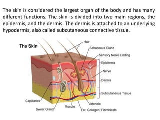
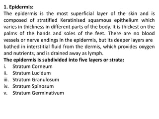
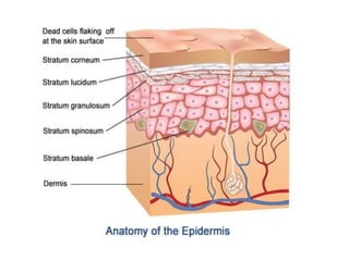
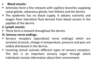
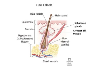
Limit test of sulphate is based on the reaction of soluble sulphate with barium chloride in presence of dilute hydrochloric acid to form barium sulphate which appears as solid particles (turbidity) in the solution.
this presentation on limit test for sulphates is very helpful for the diploma and bachelor pharmacy students. this includes principle procedure reaction and observation of limit test.
The presentation describes medical classification and importance of Integumentary System and working of the system. Moreover, it also highlights different parts of the system.
INTEGUMENTARY SYSTEM
THE INTEGUMENT
PROTECTION
First line of defense against
Bacteria
Viruses
Protects underlying structures from
Ultraviolet (UV) radiation
Dehydration
BODY TEMPERATURE REGULATION
If too hot
Dermal blood vessels dilate
Vessels carry more blood to surface so heat can escape
If too cold
Dermal blood vessels constrict
Prevents heat from escaping
EXCRETION
Small amounts of waste products are lost through perspiration
VITAMIN D PRODUCTION
Needed for calcium absorption
STRUCTURE OF SKIN
SKIN STRUCTURE : EPIDERMIS
Structures of the Epidermis
The five strata of keratinocytes in thick skin
From basal lamina to free surface
Stratum basale
Stratum spinosum
Stratum granulosum
Stratum lucidum
Stratum corneum
Second major layer of the skin
Provides mechanical strength, flexibility, and protection for underlying tissues
Highly vascular and contains a variety of sensory receptors that provide information about the external environment
Has two layers
The papillary layer
The reticular layer
Flexure lines - creases on palms
The Hypodermis (Subcutaneous Layer)
Lies below the integument
Stabilizes the skin
Allows separate movement
Made of elastic areolar and adipose tissues
Connected to the reticular layer of integument by connective tissue fibers
Deposits of Subcutaneous Fat
Distribution patterns determined by hormones
Reduced by cosmetic liposuction (lipoplasty)
STRUCTURE OF HAIR
The haIR FOLLICLE
Hair follicles are the organs that form the hairs.
Located deep in dermis.
Produces nonliving hairs.
Wrapped in a dense connective tissue sheath.
Base is surrounded by sensory nerves (root hair plexus).
Control bacteria
ACCESSORY STRUCTURES OF HAIR
Arrector pili
Involuntary smooth muscle
Causes hairs to stand up
Produces “goose bumps”
Sebaceous glands
Lubricate the hair
REGIONS OF HAIR
Hair root
Lower part of the hair
Attached to the integument
Hair shaft
Upper part of the hair
Not attached to the integument
HAIR FUNCTION
STRUCTURE AND FUNCTION OF NAIL
Structure and function of nail
Nails
Protect fingers and toes
Made of dead cells packed with keratin
Metabolic disorders can change nail structure
Nail Production
Occurs in a deep epidermal fold near the bone called the nail root
Structure and function of nail
The integumentary system comprises the skin and its appendages. Skin + derivatives= Integument.
It aims to protect the body from various kinds of damage, such as loss of water or damages from outside.
The integumentary system in chordates includes hair, scales, feathers, hooves, and nails.
It may serve to water proof, and protect the deeper tissues.
Excrete wastes, and regulate body temperature.
It is the attachment site for sensory receptors to detect pain, sensation, pressure, and temperature.
The branch of science concerned with the bodily structure of humans, animals, and other living organisms, especially as revealed by dissection and the separation of parts.
The integumentary system is an organ system consisting of the skin, hair, nails, and exocrine glands. The skin is only a few millimeters thick yet is by far the largest organ in the body. The average person's skin weighs 10 pounds and has a surface area of almost 20 square feet.
Integumentary system
-The organs of the integumentary system include the skin and its accessory structures including hair, nails, and glands, as well as blood vessels, muscles and nerves.
-Dermatology is the medical specialty for the diagnosis and treatment of disorders of the integumentary system.
Structure Of The Skin
The skin (cutaneous membrane) covers the body and is the largest organ of the body by surface area and weight.
Its area is about 2 square meters (22 square feet) and weighs 4.5-5kg (10-11 lb), about 7% of body weight.
It is 0.5 – 4 mm thick, thinnest on the eyelids, thickest on the heels, the average thickness is 1 – 2 mm.
It consists of two major layers:
Outer, thinner layer called the epidermis, consists of epithelial tissue. Inner, thicker layer called the dermis.
Beneath the dermis is a subcutaneous layer (also called hypodermis) which attaches the skin to the underlying tissues and organs.
1. EPIDERMIS
-Covers, protects and waterproofs.
Contains five main layers:
- Stratum Basale
- Stratum Spinosum
- Stratum Granulosum
- Stratum lucidum
- Stratum corneum
The epidermis has a number of important characteristics:
The epidermis is composed of keratinized stratified squamous epithelium.
-It contains four major types of cells:
Keratinocytes (90% of the cells): It produce keratin which is a tough fibrous protein that provides protection.
-Melanocytes: which produce the pigment melanin that protects against damage by ultraviolet radiation.
-Langerhans cells: involved in immune responses, arise from red bone marrow.
-Merkel cells: which function in the sensation of touch along with the adjacent tactile discs.
2. DERMIS
-It is a deeper layer of skin, composed of connective tissue containing collagen and elastic fibers.
-It contains blood and lymph vessels, nerves, and other structures, such as hair follicles and sweat glands.
-The epidermis is avascular and cells of this layer get their oxygen and nutrients from capillaries in the dermis.
-The dermis can be divided into papillary layer and reticular layer.
Hypodermis :
- The hypodermis (also called the subcutaneous layer) is a layer directly below the dermis and serves to connect the skin to the underlying fascia (fibrous tissue) of the bones and muscles.
--Two types of glands are present in the skin over most of the body
These are sweat glands and sebaceous glands.
--Function Of Skin
Protection:
Sensation:
Heat regulation:
Storage and synthesis:
Synthesis of vitamin D:
Excretion and homeostasis:
Secretion:
Absorption:
Water resistance:
Colour :
Healing of wounds:
Aids in the diagnosis:
Read| The latest issue of The Challenger is here! We are thrilled to announce that our school paper has qualified for the NATIONAL SCHOOLS PRESS CONFERENCE (NSPC) 2024. Thank you for your unwavering support and trust. Dive into the stories that made us stand out!
Introduction to AI for Nonprofits with Tapp NetworkTechSoup
Dive into the world of AI! Experts Jon Hill and Tareq Monaur will guide you through AI's role in enhancing nonprofit websites and basic marketing strategies, making it easy to understand and apply.
How to Make a Field invisible in Odoo 17Celine George
It is possible to hide or invisible some fields in odoo. Commonly using “invisible” attribute in the field definition to invisible the fields. This slide will show how to make a field invisible in odoo 17.
Synthetic Fiber Construction in lab .pptxPavel ( NSTU)
Synthetic fiber production is a fascinating and complex field that blends chemistry, engineering, and environmental science. By understanding these aspects, students can gain a comprehensive view of synthetic fiber production, its impact on society and the environment, and the potential for future innovations. Synthetic fibers play a crucial role in modern society, impacting various aspects of daily life, industry, and the environment. ynthetic fibers are integral to modern life, offering a range of benefits from cost-effectiveness and versatility to innovative applications and performance characteristics. While they pose environmental challenges, ongoing research and development aim to create more sustainable and eco-friendly alternatives. Understanding the importance of synthetic fibers helps in appreciating their role in the economy, industry, and daily life, while also emphasizing the need for sustainable practices and innovation.
Francesca Gottschalk - How can education support child empowerment.pptxEduSkills OECD
Francesca Gottschalk from the OECD’s Centre for Educational Research and Innovation presents at the Ask an Expert Webinar: How can education support child empowerment?
2024.06.01 Introducing a competency framework for languag learning materials ...Sandy Millin
http://sandymillin.wordpress.com/iateflwebinar2024
Published classroom materials form the basis of syllabuses, drive teacher professional development, and have a potentially huge influence on learners, teachers and education systems. All teachers also create their own materials, whether a few sentences on a blackboard, a highly-structured fully-realised online course, or anything in between. Despite this, the knowledge and skills needed to create effective language learning materials are rarely part of teacher training, and are mostly learnt by trial and error.
Knowledge and skills frameworks, generally called competency frameworks, for ELT teachers, trainers and managers have existed for a few years now. However, until I created one for my MA dissertation, there wasn’t one drawing together what we need to know and do to be able to effectively produce language learning materials.
This webinar will introduce you to my framework, highlighting the key competencies I identified from my research. It will also show how anybody involved in language teaching (any language, not just English!), teacher training, managing schools or developing language learning materials can benefit from using the framework.
June 3, 2024 Anti-Semitism Letter Sent to MIT President Kornbluth and MIT Cor...Levi Shapiro
Letter from the Congress of the United States regarding Anti-Semitism sent June 3rd to MIT President Sally Kornbluth, MIT Corp Chair, Mark Gorenberg
Dear Dr. Kornbluth and Mr. Gorenberg,
The US House of Representatives is deeply concerned by ongoing and pervasive acts of antisemitic
harassment and intimidation at the Massachusetts Institute of Technology (MIT). Failing to act decisively to ensure a safe learning environment for all students would be a grave dereliction of your responsibilities as President of MIT and Chair of the MIT Corporation.
This Congress will not stand idly by and allow an environment hostile to Jewish students to persist. The House believes that your institution is in violation of Title VI of the Civil Rights Act, and the inability or
unwillingness to rectify this violation through action requires accountability.
Postsecondary education is a unique opportunity for students to learn and have their ideas and beliefs challenged. However, universities receiving hundreds of millions of federal funds annually have denied
students that opportunity and have been hijacked to become venues for the promotion of terrorism, antisemitic harassment and intimidation, unlawful encampments, and in some cases, assaults and riots.
The House of Representatives will not countenance the use of federal funds to indoctrinate students into hateful, antisemitic, anti-American supporters of terrorism. Investigations into campus antisemitism by the Committee on Education and the Workforce and the Committee on Ways and Means have been expanded into a Congress-wide probe across all relevant jurisdictions to address this national crisis. The undersigned Committees will conduct oversight into the use of federal funds at MIT and its learning environment under authorities granted to each Committee.
• The Committee on Education and the Workforce has been investigating your institution since December 7, 2023. The Committee has broad jurisdiction over postsecondary education, including its compliance with Title VI of the Civil Rights Act, campus safety concerns over disruptions to the learning environment, and the awarding of federal student aid under the Higher Education Act.
• The Committee on Oversight and Accountability is investigating the sources of funding and other support flowing to groups espousing pro-Hamas propaganda and engaged in antisemitic harassment and intimidation of students. The Committee on Oversight and Accountability is the principal oversight committee of the US House of Representatives and has broad authority to investigate “any matter” at “any time” under House Rule X.
• The Committee on Ways and Means has been investigating several universities since November 15, 2023, when the Committee held a hearing entitled From Ivory Towers to Dark Corners: Investigating the Nexus Between Antisemitism, Tax-Exempt Universities, and Terror Financing. The Committee followed the hearing with letters to those institutions on January 10, 202
A Strategic Approach: GenAI in EducationPeter Windle
Artificial Intelligence (AI) technologies such as Generative AI, Image Generators and Large Language Models have had a dramatic impact on teaching, learning and assessment over the past 18 months. The most immediate threat AI posed was to Academic Integrity with Higher Education Institutes (HEIs) focusing their efforts on combating the use of GenAI in assessment. Guidelines were developed for staff and students, policies put in place too. Innovative educators have forged paths in the use of Generative AI for teaching, learning and assessments leading to pockets of transformation springing up across HEIs, often with little or no top-down guidance, support or direction.
This Gasta posits a strategic approach to integrating AI into HEIs to prepare staff, students and the curriculum for an evolving world and workplace. We will highlight the advantages of working with these technologies beyond the realm of teaching, learning and assessment by considering prompt engineering skills, industry impact, curriculum changes, and the need for staff upskilling. In contrast, not engaging strategically with Generative AI poses risks, including falling behind peers, missed opportunities and failing to ensure our graduates remain employable. The rapid evolution of AI technologies necessitates a proactive and strategic approach if we are to remain relevant.
This slide is special for master students (MIBS & MIFB) in UUM. Also useful for readers who are interested in the topic of contemporary Islamic banking.
A workshop hosted by the South African Journal of Science aimed at postgraduate students and early career researchers with little or no experience in writing and publishing journal articles.
1. P r e s e n t e d b y : A b h i s h e k D a b ra ( A s s t t . P r o f. )
B. PHARMACY-IST SEMESTER,
UNIT-II, CHAPTER-I
INTEGUMENTARY SYSTEM (SKIN)
Guru Gobind Singh College of Pharmacy,
Yamunanagar
2. Integumentary system (Skin)
The skin completely covers the body and is continuous with the
membranes lining the body orifices. It:
protects the underlying structures from injury and from invasion
by microbes
contains sensory (somatic) nerve endings of pain, temperature
and touch
is involved in the regulation of body temperature
Structure of Skin:
The skin has a surface area of about 1.5 to 2 m2 in adults and it
contains glands, hair and nails. There are two main layers:
1. Epidermis
2. Dermis.
3. Hypodermis (Subcutaneous)
Between the skin and underlying structures there is a layer of
subcutaneous fat.
3. The skin is considered the largest organ of the body and has many
different functions. The skin is divided into two main regions, the
epidermis, and the dermis. The dermis is attached to an underlying
hypodermis, also called subcutaneous connective tissue.
4. 1. Epidermis:
The epidermis is the most superficial layer of the skin and is
composed of stratified Keratinised squamous epithelium which
varies in thickness in different parts of the body. It is thickest on the
palms of the hands and soles of the feet. There are no blood
vessels or nerve endings in the epidermis, but its deeper layers are
bathed in interstitial fluid from the dermis, which provides oxygen
and nutrients, and is drained away as lymph.
The epidermis is subdivided into five layers or strata:
i. Stratum Corneum
ii. Stratum Lucidum
iii. Stratum Granulosum
iv. Stratum Spinosum
v. Stratum Germinativum
5.
6. i. Stratum corneum:
It is the uppermost layer, made up of keratin and horny scales
made up of dead keratinocytes. This is the layer which varies
most in thickness.
Within this layer, the dead keratinocytes secrete defensins which
are part of our first immune defense.
ii. Stratum Lucidum:
It is present in thicker skin found in the palms and soles. It is a
thin clear layer consisting of eleidin (clear intracellular protein)
which is a transformation product of keratohyalin.
iii. Stratum granulosum:
It contains diamond shaped cells with keratohyalin granules and
lamellar granules.
Keratohyalin granules contain keratin precursors that eventually
aggregate, crosslink, and form bundles.
7. The lamellar granules contain the glycolipids that get secreted
to the surface of the cells and function as a glue, keeping the
cells stuck together.
iv. Stratum Spinosum:
also known as the prickle cell layer contains irregular,
polyhedral cells with cytoplasmic processes, sometimes called
“spines”, that extend outward and contact neighboring cells by
desmosomes. Dendritic cells can be found in this layer.
v. Stratum germinativum/basale:
It is the deepest layer, separated from the dermis by the
basement membrane (basal lamina) and attached to the
basement membrane by hemidesmosomes.
The cells found in this layer are cuboidal to columnar mitotically
active stem cells that are constantly producing keratinocytes.
This layer also contains melanocytes.
8. 2. Dermis:
The dermis is tough and elastic. It is formed from connective
tissue and the matrix contains collagen fibres interlaced with
elastic fibres.
Rupture of elastic fibres occurs when the skin is overstretched,
resulting in permanent striae, or stretch marks, that may be
found in pregnancy and obesity.
Collagen fibres bind water and give the skin its tensile strength,
but as this ability declines with age wrinkles develop.
Fibroblasts, macrophages and mast cells are the main cells
found in the dermis.
The structures in the dermis are:
i. blood vessels
ii. lymph vessels
iii. sensory (somatic) nerve endings
iv. sweat glands and their ducts
v. hairs, arrector pili muscles and sebaceous glands
9. i. Blood vessels:
Arterioles form a fine network with capillary branches supplying
sweat glands, sebaceous glands, hair follicles and the dermis.
The epidermis has no blood supply. It obtains nutrients and
oxygen from interstitial fluid derived from blood vessels in the
papillae of the dermis.
ii. Lymph vessels:
These form a network throughout the dermis.
iii. Sensory nerve endings:
Sensory receptors (specialised nerve endings) which are
sensitive to touch, change in temperature, pressure and pain are
widely distributed in the dermis.
Incoming stimuli activate different types of sensory receptors.
The skin is an important sensory organ through which
individuals receive information about their environment.
10. iv. Sweat glands:
Sweat glands are found widely distributed throughout the skin
and are most numerous in the palms of the hands, soles of the
feet, axillae and groins.
They are composed of epithelial cells. The bodies of the glands
lie coiled in the subcutaneous tissue.
Some ducts open onto the skin surface at tiny depressions, or
pores, and others open into hair follicles. Glands opening into
hair follicles
11. 3. Hypodermis/ Subcutaneous:
The hypodermis is deep to the dermis and is also called
subcutaneous fascia.
It is the deepest layer of skin and contains adipose lobules along
with some skin appendages like the hair follicles, sensory
neurons, and blood vessels.
12. Skin Appendages:
1. Hair:
These are formed by a down-growth of epidermal cells into the
dermis or subcutaneous tissue, called hair follicles.
At the base of the follicle is a cluster of cells called the bulb. The
hair is formed by multiplication of cells of the bulb and as they
are pushed upwards, away from their source of nutrition, the
cells die and become Keratinised.
The part of the hair above the skin is the shaft and the
remainder, the root.
The color of the hair is genetically determined and depends on
the amount of melanin present.
White hair is the result of the replacement of melanin by tiny air
bubbles.
14. 2. The arrector pili:
These are little bundles of smooth muscle fibres attached to the
hair follicles. Contraction makes the hair stand erect and raises
the skin around the hair, causing 'goose flesh'.
The muscles are stimulated by sympathetic nerve fibres in
response to fear and cold. Erect hairs trap air, which acts as an
insulating layer.
This is an efficient warming mechanism especially when
accompanied by shivering, i.e. involuntary contraction of the
skeletal muscles.
15. 3. The sebaceous glands:
These consist of secretory epithelial cells derived from the same
tissue as the hair follicles.
They secrete an oily substance, sebum, into the hair follicles and
are therefore present in the skin of all parts of the body except
the palms of the hands and the soles of the feet.
They are most numerous in the skin of the scalp, face, axillae
and groins.
Sebum keeps the hair soft and pliable and gives it a shiny
appearance.
On the skin it provides some waterproofing and acts as a
bactericidal and fungicidal agent, preventing the successful
invasion of microbes.
It also prevents drying and cracking of skin, especially on
exposure to heat and sunshine.
16. 4. Nails:
The nails in human beings are equivalent to the claws, horns and
hoofs of animals.
They are derived from the same cells as epidermis and hair and
consist of a hard, horny keratin plate.
They protect the tips of the fingers and toes. The root of the nail
is embedded in the skin, is covered by the cuticle and forms the
hemispherical pale area called the lunula.
The nail plate of the nail is the exposed part that has grown out
from the germinative zone of the epidermis called the nail bed.
Finger nails grow more quickly than toe nails and growth is
quicker when the environmental temperature is high
18. Function of Skin:
1. Provides a protective barrier against mechanical, thermal and
physical injury and hazardous substances.
2. Prevent loss of moisture
3. Acts as a sensory organ (touch, detect temperature)
4. Helps regulate temperature
5. An immune organ to detect infections
6. Absorption
7. Excretion
19. References
1. Waugh A., Grant A., “ Ross and Wilson Anatomy & Physiology in health and illness,
Churchill Livingstone, 12th ed., 2014, Page no. 35-42.