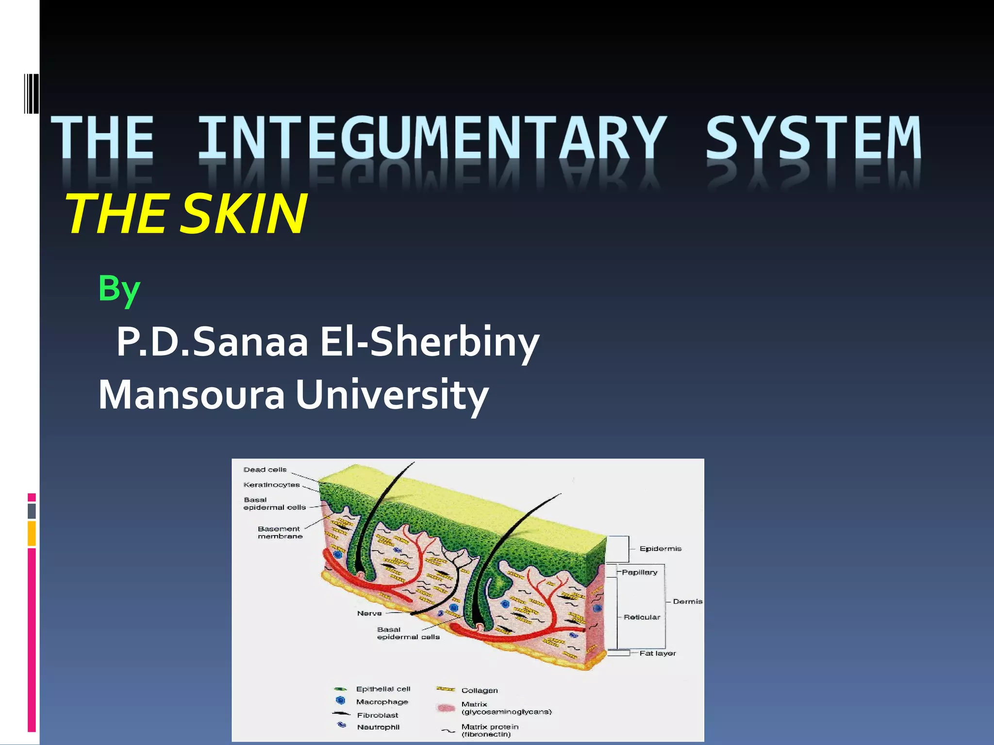
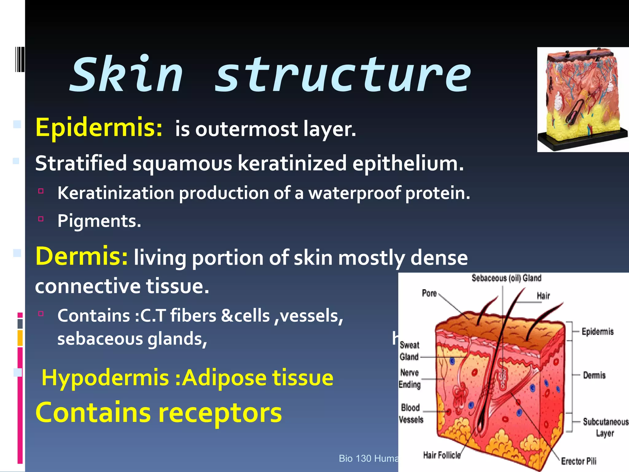
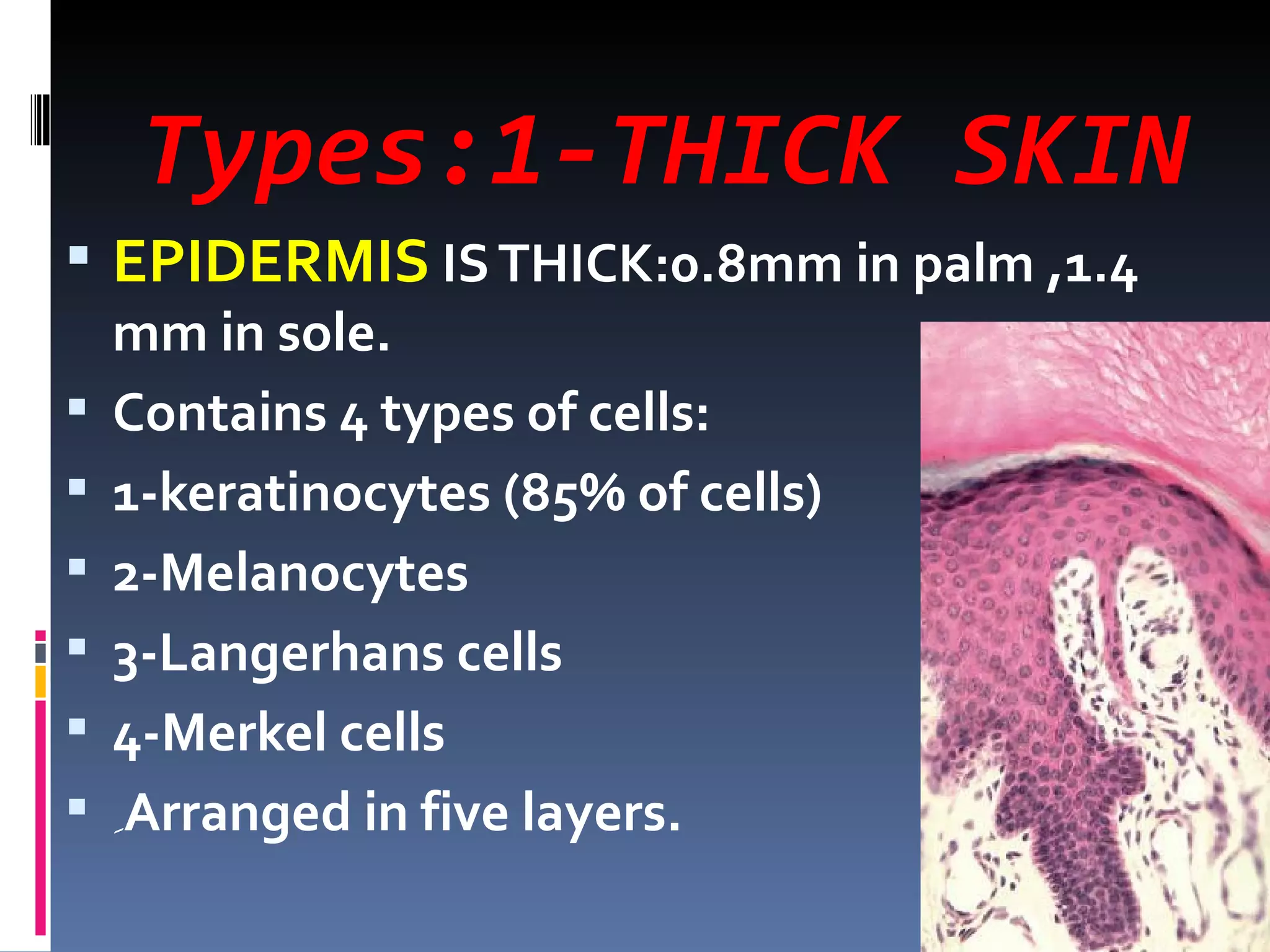
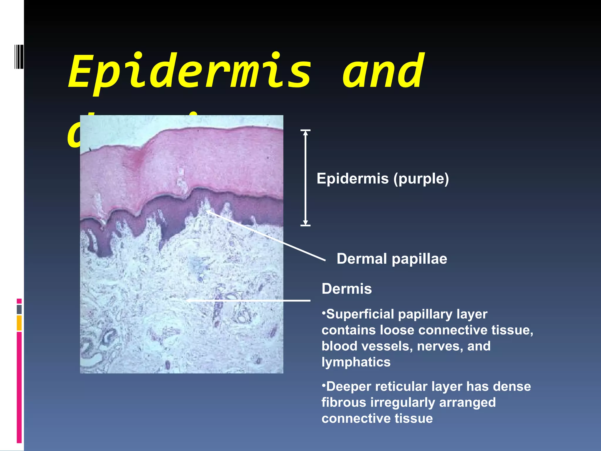
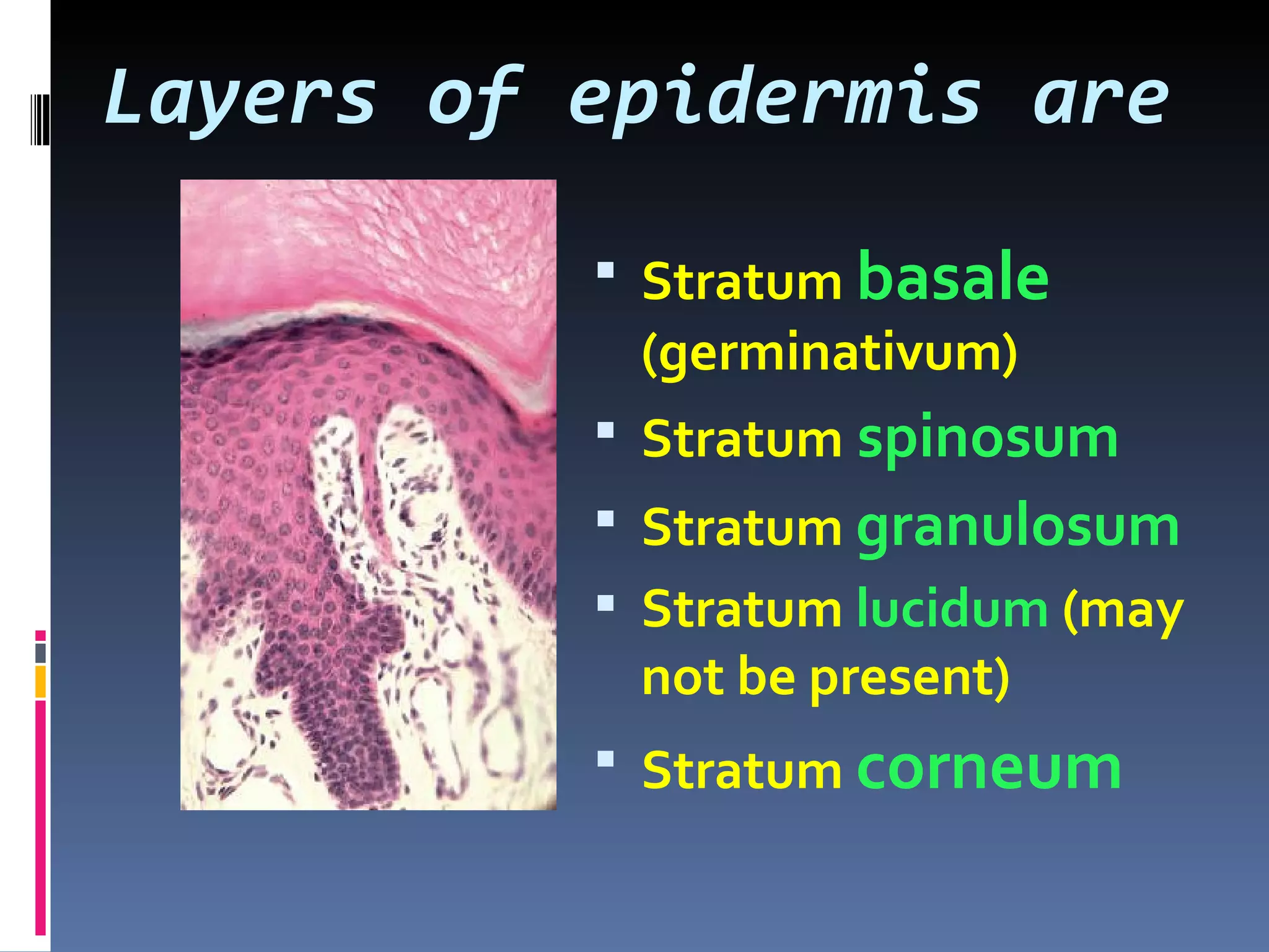
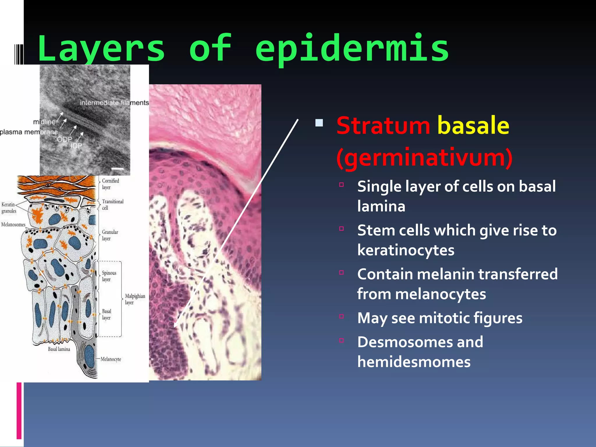
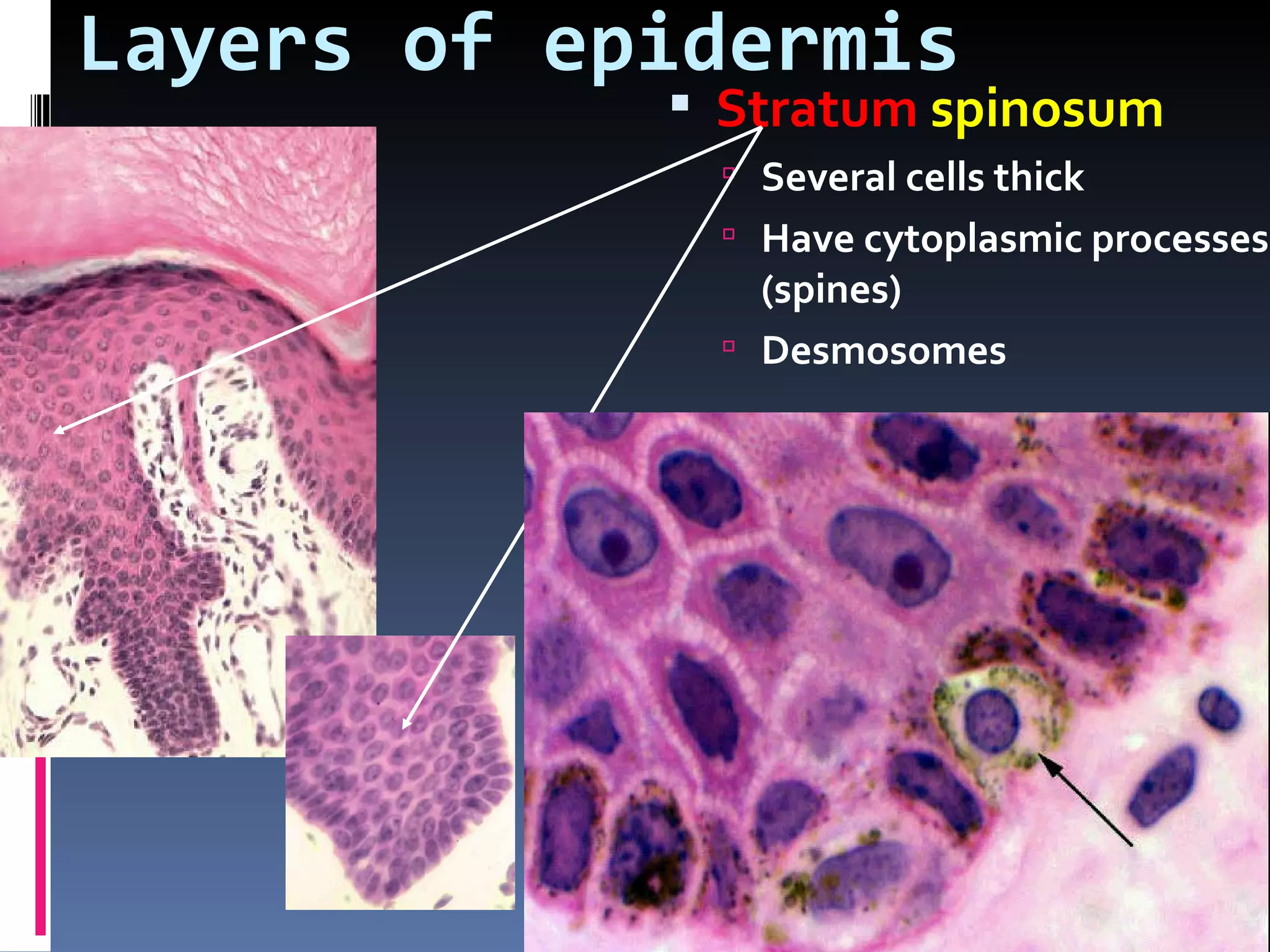
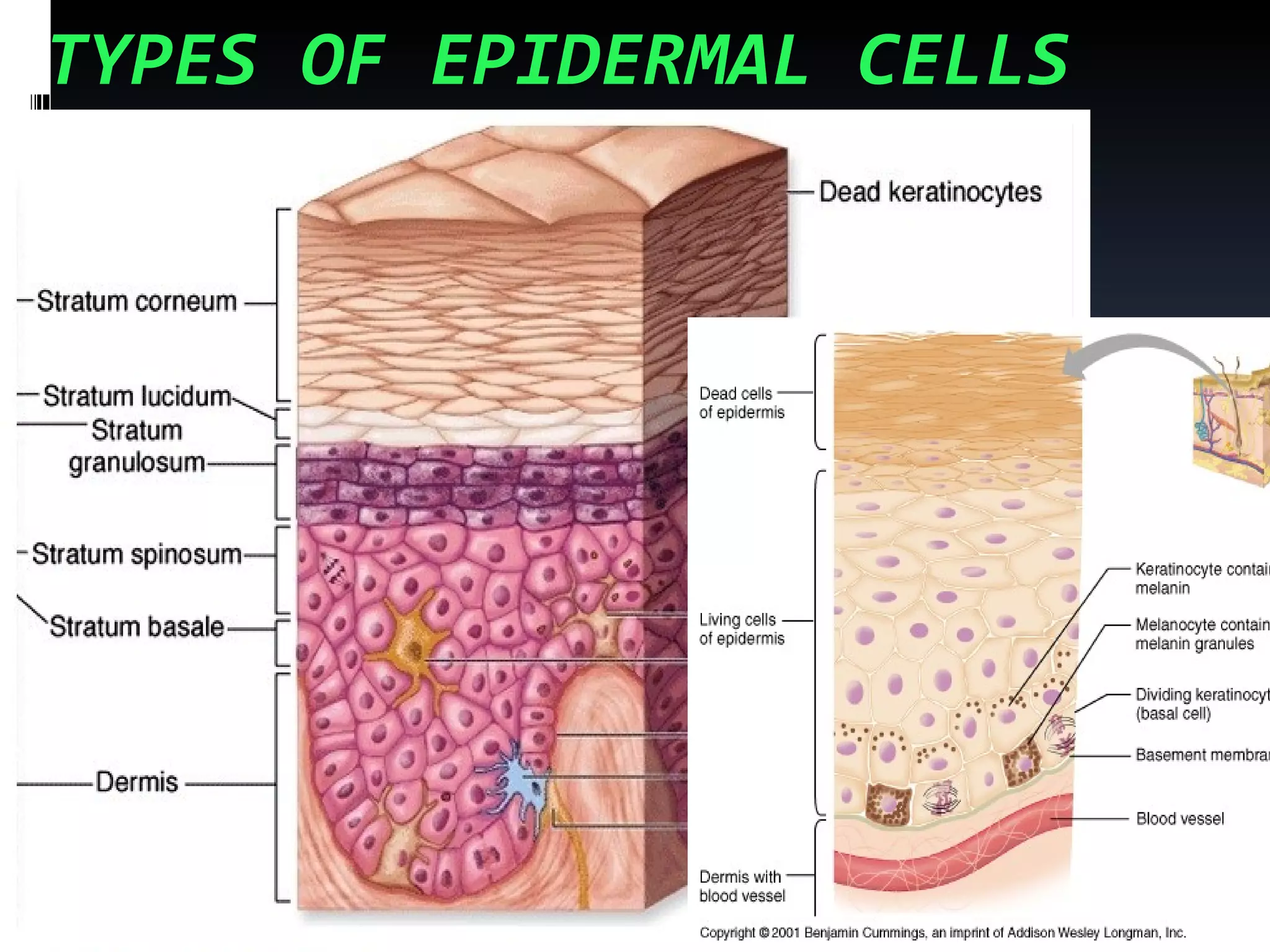
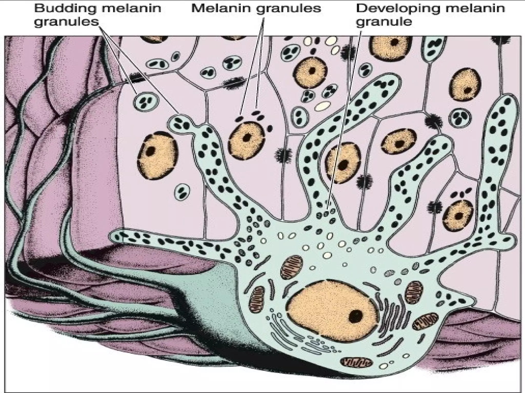
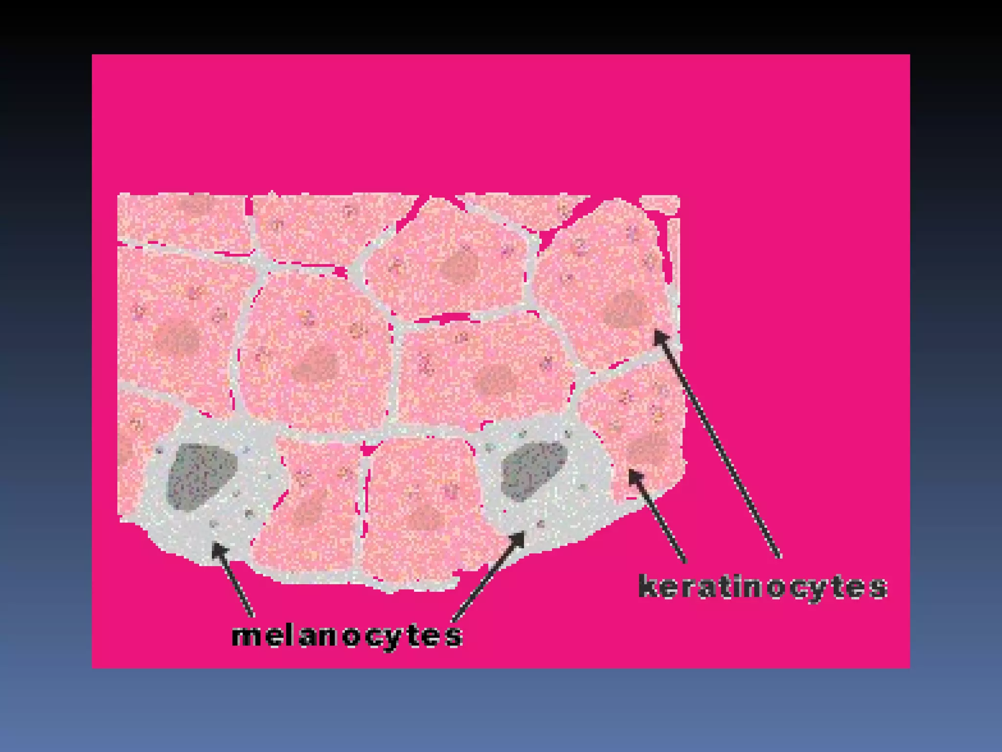
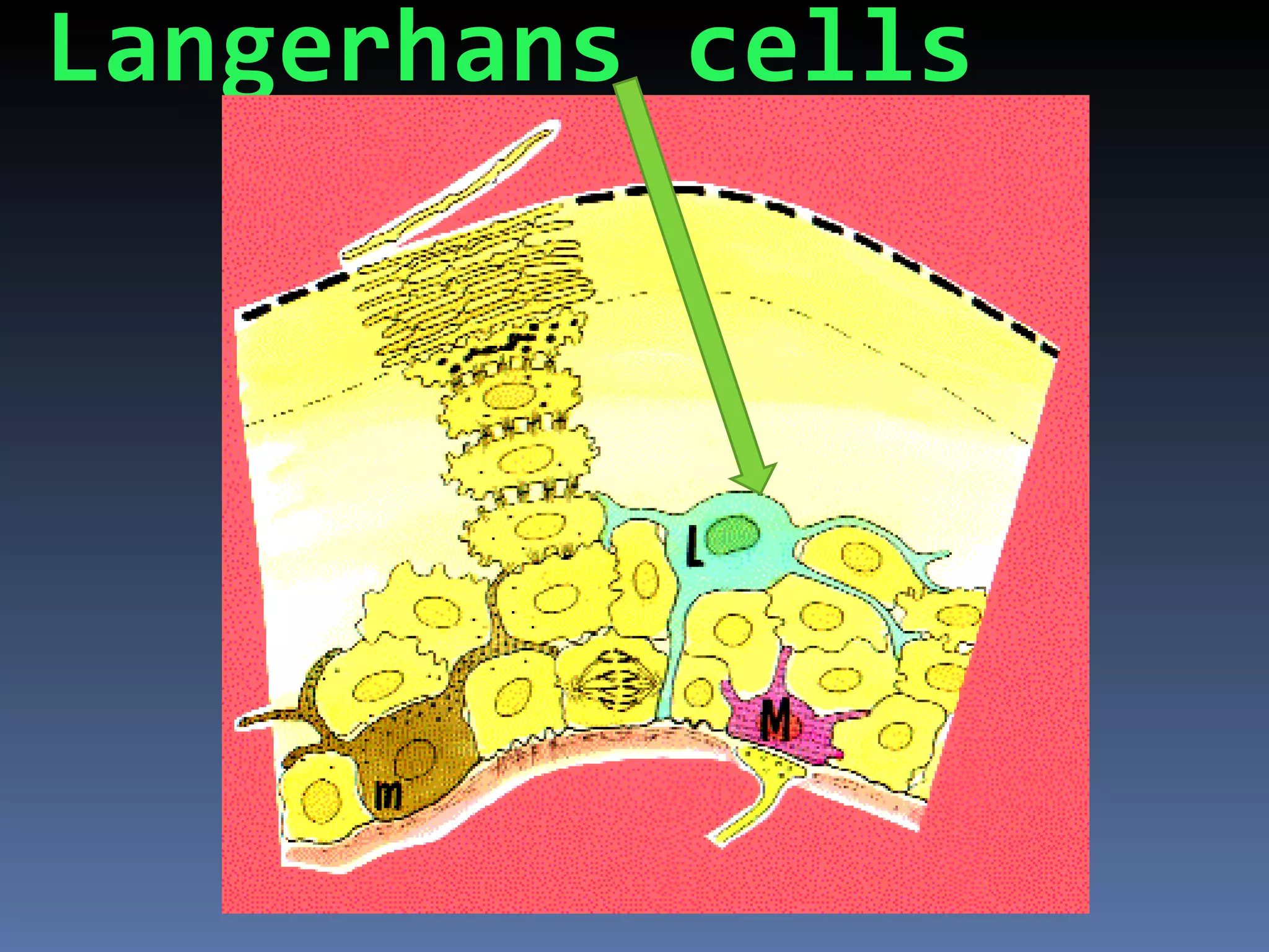
The skin has three layers - the epidermis, dermis, and hypodermis. The epidermis is made of stratified squamous epithelium and contains keratinocytes, melanocytes, Langerhans cells, and Merkel cells. It provides protection, regulates temperature and moisture, and produces vitamin D. The dermis contains dense connective tissue, hair follicles, sweat and sebaceous glands. It provides strength and elasticity to the skin. The hypodermis is a layer of adipose tissue that acts as insulation for the body. The skin comes in thick and thin varieties and has eccrine and apocrine sweat glands that help regulate temperature and release scent compounds.
Research Methods in Psychology | Cambridge AS Level | Cambridge Assessment In...
Histology (Skin) - Part 1
1.
2. THE SKIN
By
P.D.Sanaa El-Sherbiny
Mansoura University
3. Function of the
skinand protects the body.
Covers
Control internal temperature.
Produces vitamin D.
Receptors to detect environmental stimuli.
Regulates the movement of substances
into and out of the body
Forms the largest organ of the body
forms 16%of body weight
Bio 130 Human Biology
4. Skin structure
Epidermis: is outermost layer.
Stratified squamous keratinized epithelium.
Keratinization production of a waterproof protein.
Pigments.
Dermis: living portion of skin mostly dense
connective tissue.
Contains :C.T fibers &cells ,vessels, sweat glands,
sebaceous glands, hairs.
Hypodermis :Adipose tissue
Contains receptors
Bio 130 Human Biology
5. Types:1-THICK SKIN
EPIDERMIS IS THICK:0.8mm in palm ,1.4
mm in sole.
Contains 4 types of cells:
1-keratinocytes (85% of cells)
2-Melanocytes
3-Langerhans cells
4-Merkel cells
ِArranged in five layers.
7. Layers of epidermis are
Stratum basale
(germinativum)
Stratum spinosum
Stratum granulosum
Stratum lucidum (may
not be present)
Stratum corneum
8. Layers of epidermis
Stratum basale
(germinativum)
Single layer of cells on basal
lamina
Stem cells which give rise to
keratinocytes
Contain melanin transferred
from melanocytes
May see mitotic figures
Desmosomes and
hemidesmomes
9. Layers of epidermis
Stratum spinosum
Several cells thick
Have cytoplasmic processes
(spines)
Desmosomes
10. Layers of epidermis
Stratum
granulosum
1 to 3 layers of fusiform
shaped basophilic cells
Keratohyalin
granules contain
cystine-rich and
histidine-rich proteins
that associate with
keratin filaments
11. Layers of epidermis
Stratum
lucidum
Present only in
thick skin
Cells in which
keratinization
is advanced
12. Layers of epidermis
Stratum corneum
Superifical keratinized layer
Cells
Almost filled with keratin
Flattened, non nucleate
Coated with extra-cellular
lipids that form water barrier
of skin
Layer that varies most in
thickness
14. The epidermal cells
:1-keratinocytes
They are responsible
For keratin
formation
Formed of many
layers that
continuously shed
And regenerate
every 2-4 weeks
They are arranged
In many layers.
15. :Melanocytes
Found inbetween cells of the basal layer &
At the basal part of the hair follicles.
Branched cells with centeral nuclei
By EM contains organells for protein
synthesizes (rER, Golgi, mitochondria
&melanosomes).
They form melanin by tyrosinase from
tyrosine amino acid
By converting it to dioxyphenyl alanine
DOPA
16.
17.
18.
19.
20. :Langerhans cells -3
Found in upper layers of st.spinosum
Have branched shape ¢ral nuclei
Represent 3-8%of epid. Cells
Mesodermal in origin.
EM not connected to keratinocytes &
contain
Birbeck granules
Stained with silver & vital stains
Phagocytic & antigen presenting cells
22. Merkels cells-4
Found in basal cell layer
Are modified epidermal cells
Sensory nerve fibers form
terminal disk under
Merkels cells
Function as touch
receptors
23.
24. THIN SKIN
ALLOVER THE BODY EXCEPT SOLE &
PALMS.
EPIDERMIS
ISTHINNER,ST.spinosum,2-4LAYERS,ST.GRA.
ONE INCOMPLETE
LAYER:NO ST LUCI. ,ST,COR.THINNER
NUMEROUS SWEAT GLANDS.
*CONTAIN HAIRE, SEBACEOUS
GLANDS
*AND ERECTOR PILLI MUSCLE
29. Reticular layer -2
The thicker deep layer
Formed of dense c.T rich in interlacing wavy
collage fibers
It is less cellular &less vascular
Contain many nerve receptors:
Krause end bulbs
Ruffini corpuscles
Pacinian corpuscles
30.
31. :Glands and skin appendages
Sebaceous glands
Clumps of epithelial tissue distributed within dermis
Secrete “sebum”—oily, fat-based substance that is
also anti-bacterial
Located all over body
Sweat glands
Microscopic clumps of epithelial tissue distributed
within dermis, duct extends out through dermis to
pore their secretion.
More than 2.5 million glands per person
Eccrine sweat glands, concentrated on hands and
soles of feet and forehead, secrete sweat to cool
body, also at conditions of fear and emotion.
Apocrine glands, concentrated in armpits and groin,
analogous with sexual scent glands of other
animals, odor comes from bacteria that concentrate
here.
Ceruminous glands: modified sweat glands in ear
canal produce ear wax
32. sweat glands
Two types of sweat glands
Eccrine
Not associated with hair follicle
Duct segment
less coiled, leads to epidermis
Stratified cuboidal epithelium
Secretory segment
in deep dermis or hypodermis
Secretory cells
Myoepithelial cells lie between
secretory cells, contraction
expels sweat
Apocrine
Found in limited areas
Empty into hair follicle
33. SWEAT GLANDS
Merocrine glands: Apocrine glands:
Allover the body Axilla,groin,pubic
Secretory cells 2types region
Clear cells cubical rich in Secretory part similar
glycogen granules.
To mero. With wider
Dark with narrow basal
part with apical Lumen
Glycoprotein granules Their ducts are lined
Myoepithelialt cells With 2 layers of cubical cells
Ducts lined by 2layers of but open into hair follicles
cubical cells Secretion stim.by sex
hormons