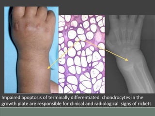
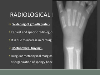
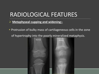
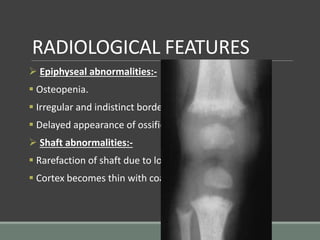
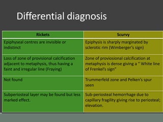
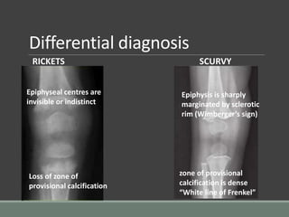
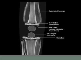
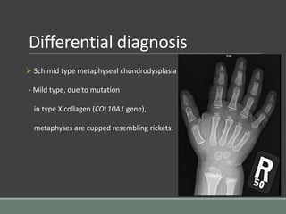
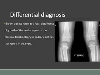
IMAGING
IN
RICKETS
PRESENTER: DR. YASHAS ULLAS L.
Contents
 Pathoradiological features
 Radiological features
 Differential diagnosis
Introduction
• Rickets is a metabolic disease of growing bone that is
unique to children.
• It caused by a failure of mineralization of osteoid tissue in a
developing skeleton, particularly at the growth plate.
• Imperfect calcification typically resulting in soft bones and
skeleton deformities.
Patho radiological features
 Radiological appearances truly portray the histological
changes occurring in the bones.
 Disordered proliferation of cartilage cells and defective
mineralization in the zone of provisional calcification.
 Decreased quantity of calcified osteoid.
Patho radiological features
 Owing to the lack of osteoid mineralization the
metaphyseal zone of provisional calcification will also be
absent radiographically.
 Bones appear more radiolucent with coarsened trabecular
pattern.
 Widened growth plate with irregular, frayed and cupped
metaphyseal margins (paintbrush metaphyses)
Apoptosis of hypertrophic
chondrocytes caused by
phosphate ions
Hypophosphatemia
No apoptosis of
hypertrophic
chondrocytes
Normal growth plate Rachitic growth plate
Impaired apoptosis of terminally differentiated chondrocytes in the
growth plate are responsible for clinical and radiological signs of rickets
RADIOLOGICAL FEATURES
 Widening of growth plate:-
 Earliest and specific radiological change.
 It is due to increase in cartilaginous cell mass.
 Metaphyseal fraying:-
 Irregular metaphyseal margins occurring due to fraying and
disorganization of spongy bone in the metaphyseal region.
RADIOLOGICAL FEATURES
 Metaphyseal cupping and widening:-
 Protrusion of bulky mass of cartilageneous cells in the zone
of hypertrophy into the poorly mineralized metaphysis.
Normal Rickets
RADIOLOGICAL FEATURES
 Epiphyseal abnormalities:-
 Osteopenia.
 Irregular and indistinct borders.
 Delayed appearance of ossification centres.
 Shaft abnormalities:-
 Rarefaction of shaft due to loss of mineral content.
 Cortex becomes thin with coarse echotexture.
RADIOLOGICAL FEATURES
 Growth plate abnormalities:-
 Generalised osteopenia.
 Coarse trabecular pattern.
 Frayed paintbrush metaphyseal margins.
 Lack of the zone of provisional calcification.
 Widening of the physis.
RADIOLOGICAL FEATURES
 Wrist:
 Widening of growth plates.
 Irregular margins of metaphysis with widening and
cupping.
 Osteopenia.
 Rarefraction in long bones.
RADIOLOGICAL FEATURES
 Bowing of long bones:
 Result of displacement of growth centres owing to
assymetrical musculotendinous pull on the weakened growth
plate.
RADIOLOGICAL FEATURES
 Rachitic rosary:
 Bulbous enlargement of costochondral junction.
 May indent on the pleural surface.
RADIOLOGICAL FEATURES
 Craniotabes:
 Excessive osteoid deposition in frontal and parietal regions
with posterior flattening of skull due to supine posture of
infant.
 Squared configuration of skull.
 Demineralisation of skull.
RADIOLOGICAL FEATURES
 Triradiate pelvis:-
 Protrusion of hip into the soft pelvis with protrusio acetabuli.
RADIOLOGICAL FEATURES OF
HEALING RICKETS
 Appearance of line of provisional calcification at the end of
metaphysis.
 Metaphysis show cupping and splaying but no fraying.
 Periosteum may show double periosteal line due to
subperiosteal deposition of osteoid tissue.
RADIOLOGICAL FEATURES OF
HEALED RICKETS
 The subperiosteal osteoid tissue is calcified (no double
periosteal line).
 Splaying and cupping is corrected.
Before After
Differential diagnosis
 Scurvy.
 Schimid type metaphyseal chondrodysplasia.
 Blount disease.
Differential diagnosis
Rickets Scurvy
Epiphyseal centres are invisible or
indistinct
Epiphysis is sharply marginated by
sclerotic rim (Wimberger’s sign)
Loss of zone of provisional calcification
adjacent to metaphysis, thus having a
faint and irregular line (Fraying)
Zone of proviosional calcification at
metaphysis is dense giving a “ White line
of Frenkel’s sign”
Not found Trummerfeld zone and Pelken’s spur
seen
Subperiosteal layer may be found but less
marked effect.
Sub-periosteal hemorrhage due to
capillary fragility giving rise to periosteal;
elevation.
Differential diagnosis
RICKETS SCURVY
Epiphyseal centres are
invisible or indistinct
Epiphysis is sharply
marginated by sclerotic
rim (Wimberger’s sign)
Loss of zone of
provisional calcification
zone of provisional
calcification is dense
“White line of Frenkel”
Differential diagnosis
 Schimid type metaphyseal chondrodysplasia
- Mild type, due to mutation
in type X collagen (COL10A1 gene),
metaphyses are cupped resembling rickets.
Differential diagnosis
Blount disease refers to a local disturbance
of growth of the medial aspect of the
proximal tibial metaphysis and/or epiphysis
that results in tibia vara.
Imaging in rickets

Imaging in rickets

  • 1.
  • 2.
    Contents  Pathoradiological features Radiological features  Differential diagnosis
  • 3.
    Introduction • Rickets isa metabolic disease of growing bone that is unique to children. • It caused by a failure of mineralization of osteoid tissue in a developing skeleton, particularly at the growth plate. • Imperfect calcification typically resulting in soft bones and skeleton deformities.
  • 4.
    Patho radiological features Radiological appearances truly portray the histological changes occurring in the bones.  Disordered proliferation of cartilage cells and defective mineralization in the zone of provisional calcification.  Decreased quantity of calcified osteoid.
  • 5.
    Patho radiological features Owing to the lack of osteoid mineralization the metaphyseal zone of provisional calcification will also be absent radiographically.  Bones appear more radiolucent with coarsened trabecular pattern.  Widened growth plate with irregular, frayed and cupped metaphyseal margins (paintbrush metaphyses)
  • 6.
    Apoptosis of hypertrophic chondrocytescaused by phosphate ions Hypophosphatemia No apoptosis of hypertrophic chondrocytes Normal growth plate Rachitic growth plate
  • 7.
    Impaired apoptosis ofterminally differentiated chondrocytes in the growth plate are responsible for clinical and radiological signs of rickets
  • 8.
    RADIOLOGICAL FEATURES  Wideningof growth plate:-  Earliest and specific radiological change.  It is due to increase in cartilaginous cell mass.  Metaphyseal fraying:-  Irregular metaphyseal margins occurring due to fraying and disorganization of spongy bone in the metaphyseal region.
  • 9.
    RADIOLOGICAL FEATURES  Metaphysealcupping and widening:-  Protrusion of bulky mass of cartilageneous cells in the zone of hypertrophy into the poorly mineralized metaphysis. Normal Rickets
  • 10.
    RADIOLOGICAL FEATURES  Epiphysealabnormalities:-  Osteopenia.  Irregular and indistinct borders.  Delayed appearance of ossification centres.  Shaft abnormalities:-  Rarefaction of shaft due to loss of mineral content.  Cortex becomes thin with coarse echotexture.
  • 11.
    RADIOLOGICAL FEATURES  Growthplate abnormalities:-  Generalised osteopenia.  Coarse trabecular pattern.  Frayed paintbrush metaphyseal margins.  Lack of the zone of provisional calcification.  Widening of the physis.
  • 12.
    RADIOLOGICAL FEATURES  Wrist: Widening of growth plates.  Irregular margins of metaphysis with widening and cupping.  Osteopenia.  Rarefraction in long bones.
  • 14.
    RADIOLOGICAL FEATURES  Bowingof long bones:  Result of displacement of growth centres owing to assymetrical musculotendinous pull on the weakened growth plate.
  • 15.
    RADIOLOGICAL FEATURES  Rachiticrosary:  Bulbous enlargement of costochondral junction.  May indent on the pleural surface.
  • 16.
    RADIOLOGICAL FEATURES  Craniotabes: Excessive osteoid deposition in frontal and parietal regions with posterior flattening of skull due to supine posture of infant.  Squared configuration of skull.  Demineralisation of skull.
  • 17.
    RADIOLOGICAL FEATURES  Triradiatepelvis:-  Protrusion of hip into the soft pelvis with protrusio acetabuli.
  • 18.
    RADIOLOGICAL FEATURES OF HEALINGRICKETS  Appearance of line of provisional calcification at the end of metaphysis.  Metaphysis show cupping and splaying but no fraying.  Periosteum may show double periosteal line due to subperiosteal deposition of osteoid tissue.
  • 19.
    RADIOLOGICAL FEATURES OF HEALEDRICKETS  The subperiosteal osteoid tissue is calcified (no double periosteal line).  Splaying and cupping is corrected.
  • 20.
  • 21.
    Differential diagnosis  Scurvy. Schimid type metaphyseal chondrodysplasia.  Blount disease.
  • 22.
    Differential diagnosis Rickets Scurvy Epiphysealcentres are invisible or indistinct Epiphysis is sharply marginated by sclerotic rim (Wimberger’s sign) Loss of zone of provisional calcification adjacent to metaphysis, thus having a faint and irregular line (Fraying) Zone of proviosional calcification at metaphysis is dense giving a “ White line of Frenkel’s sign” Not found Trummerfeld zone and Pelken’s spur seen Subperiosteal layer may be found but less marked effect. Sub-periosteal hemorrhage due to capillary fragility giving rise to periosteal; elevation.
  • 23.
    Differential diagnosis RICKETS SCURVY Epiphysealcentres are invisible or indistinct Epiphysis is sharply marginated by sclerotic rim (Wimberger’s sign) Loss of zone of provisional calcification zone of provisional calcification is dense “White line of Frenkel”
  • 25.
    Differential diagnosis  Schimidtype metaphyseal chondrodysplasia - Mild type, due to mutation in type X collagen (COL10A1 gene), metaphyses are cupped resembling rickets.
  • 26.
    Differential diagnosis Blount diseaserefers to a local disturbance of growth of the medial aspect of the proximal tibial metaphysis and/or epiphysis that results in tibia vara.

Editor's Notes

  • #7 Low serum phosphate is fundamental to pathogenesis of rickets. Impaired mineralization of growth plate and osteoid.
  • #14 Posteroanterior radiograph of both hands shows diffuse osteopenia, age-indeterminate fractures of several metacarpals (solid arrows), and cupped fragmented frayed metaphyses of the distal radii and ulnae (ovals). A peripheral rim of bone along the metaphysis (dashed arrow) occurs by membranous ossification.
  • #18 Square head appearance – caput quadratum
  • #23 Beneath the zone of provisional calcification, there is evidence of lucent zone related to lack of mineralized osteoid (Trummerfeld zone). Lateral projection of white line of frenkel may lead to formation of spur or marginal cleft.
  • #26 Widening of growth plate