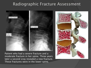
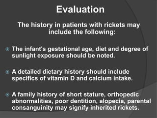
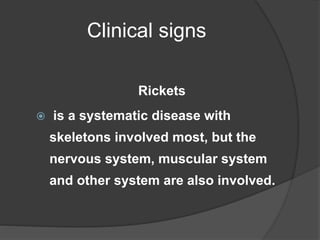
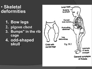
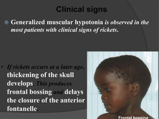
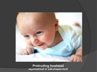
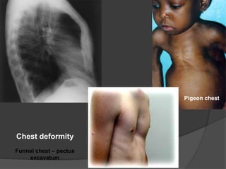
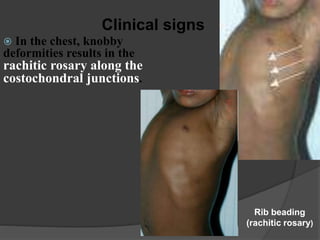
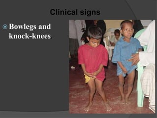
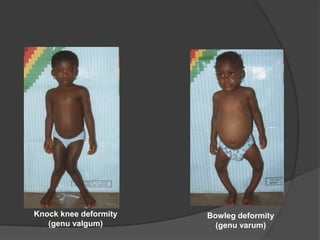
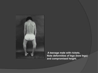
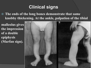
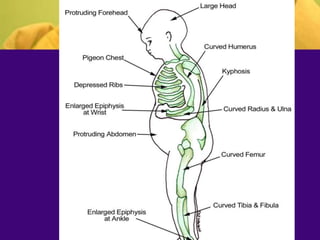
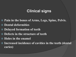
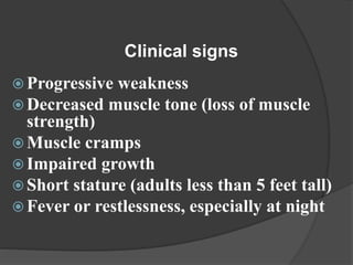
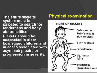
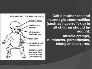
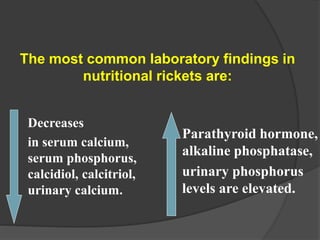
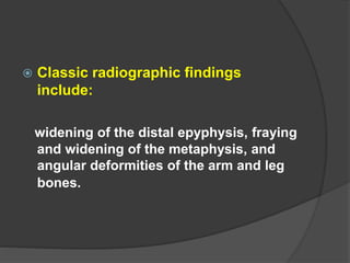
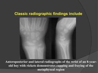
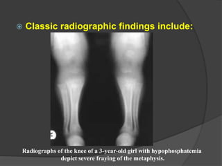
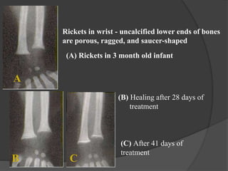
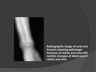
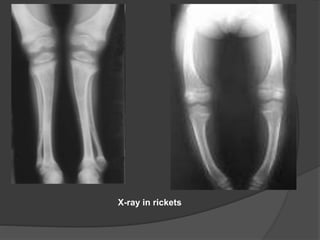
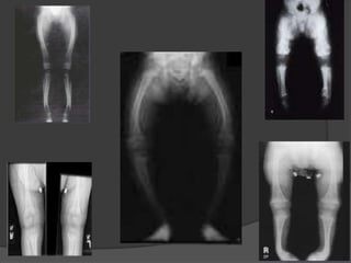
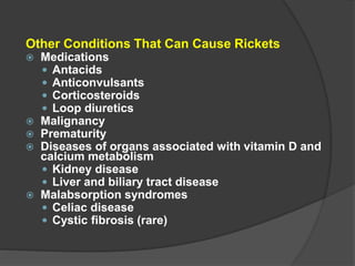
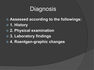
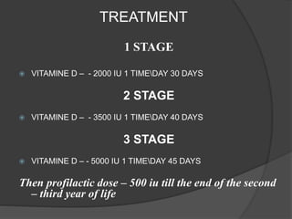
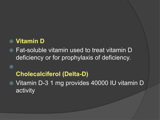
SlideShare a Scribd company logo
By
Dr Akkad Rafiq
Bone Structure
 Three main functions of bone
 support,
 protection
 leverage
 Composition
 Type I collagen fibers,
 mineral component
 Other non-collagenous proteins
 Osteopontin
 osteonectin
 Osteocalcin
 alkaline phosphatases
 Bone morphogenetic protein
Bone Minerals
 Almost half the bone volume is mineral
matter
 mainly calcium and phosphate in the
form of crystalline hydroxyapatite
 ‘demineralization’ of bone occurs only by
resorption of the entire matrix
Bone Cells
 Osteoblasts - concerned with bone
formation and osteoclast activation
 Osteocytes -These cells can be
regarded as spent osteoblasts
 Osteoclasts- These cells are the
principal mediators of bone resorption
Bone structure
 The mature tissue is lamellar
bone, in which the collagen
fibres are arranged parallel to
each other to form multiple
layers with the osteocytes
lying between the lamellae.
Minerals of bone
 Calcium
 Magnesium
 Vit D
 Phosphorus
 Calcitonin
Osteoporosis
 clinical disorder characterized by an
abnormally low bone mass and effects
in bone structure, which renders the
bone fragile and at greater risk of
fracture in a person of that age, sex and
race.
Pathology:
• results from an unhealthy imbalance
between two normal activities of
bone: bone resorption and bone
formation.
• The combined processes of bone
resorption and bone formation allow
the healthy skeleton to be
maintained continually by the
removal of old bone and its
replacement with new bone.
Pathology
 the destruction of bone begins to
exceed the formation of bone; this
imbalance leads to a net loss of
bone, and the beginnings of
osteoporosis.
PRIMERY RISK FACTORS
 Caucasoid (white) or Asiatic ethnicity
 Family history of osteoporosis
 History of anorexia nervosa and/or
amenorrhoea
 Low peak bone mass in the third decade
 Early onset of menopause
 Unusually slim or emaciated build
 Early hysterectomy
 Nutritional insufficiency
 Chronic lack of exercise
Causes of secondary
osteoporosis
 Nutritional
 Malabsorption
 Malnutrition
 Scurvy
 Inflammatory disorders
 Rheumatoid disease
 Ankylosing spondylitis
 Tuberculosis
 Drug induced
 Corticosteroids
 Excessive alcohol
consumption
 Anticonvulsants
 Heparin
 Immunosuppressives
 Endocrine disorders
 Gonadal insufficiency
 Hyperparathyroidism
 Thyrotoxicosis
 Cushing’s disease
 Malignant disease
 Carcinomatosis
 Multiple myeloma
 Leukaemia
 Other
 Smoking
 Chronic obstructive
 pulmonary disease
 Osteogenesis imperfecta
 Chronic renal disease
Investigations
 X-ray findings are
generally insufficient
• cannot reliably
measure bone density
• useful to identify
spinal factures,
explains back pain,
height loss or
kyphosis.
• X-rays may detect
osteopenia only when
bone loss is > 30%.
Patient who had a severe fracture and a
moderate fracture in her spine. Three years
later a second xray revealed a new fracture.
These fractures were in the lower spine.
Radiographic Fracture Assessment
DEXA
Dual energy x-ray absorptiometry (DEXA)
• This is the most popular and accurate test to
date
• non-invasive
• involves no special preparation.
• Radiation exposure is minimal,
• Can be used to measure bone mineral density in
the spine, hip, wrist, or total body.
•expensive
•not portable.
Screening- Ultrasound Densitometry
inexpensive,
portable
no radiation
can be used only in peripheral
sites (eg, the heel),
Fracture Reduction
 Goal: prevent fracture, not just treat
BMD
 Osteoporosis treatment options
 Calcium and vitamin D
 Calcitonin
 Bisphosphonates
 Selective Estrogen Receptor Modulators
 Parathyroid Hormone
Osteoporosis Treatment: Calcium and Vit
D
 Calcium and Vit D supplementation shown
to decrease risk of hip fracture in older
adults
 1000 mg/day standard;
 1500 mg/day in postmenopausal
women/osteoporosis
 Vitamin D (25 and 1,25): 400 IU/Day
Osteoporosis Treatment:
Bisphosphonates
 Decrease bone resorption
 decrease hip and vertebral fractures
 Alendronate, risodronate PO
 IV: pamidronate, zolendronate
 Ibandronate : once/month
Calcetonin
 not as effective as Bisphosphonates
 200 IU nasally/day
Osteoporosis Treatment:
Selective Estrogen Receptor Modulators
 Raloxifene
 Decrease bone resorption like estrogen
 No increased risk cancer (decrease risk
breast cancer)
Osteoporosis Treatment:
PTH
 Teriparatide
 INTERMITTENT PTH: overall improvement
in bone density
Current Guidelines
 US Preventive Task Force
 Test Bone Mineral Density in all women over
age 65, younger postmenopausal women
with at least one risk factor, and
postmenopausal women with a history of
fracture
 Treat patients with T score <-2 and no risk
factors, T score <1.5 if any risk factors, and
anyone with prior vertebral/hip fracture
Rickets:
Etiology, pathogenesis, clinical
features, diagnostics, treatment and
prevention
Rickets is a childhood disorder
involving softening and
weakening of the bones.
It is primarily caused by lack of
vitamin D, calcium, or
phosphate.
Etiology
1. Lack of sunshine due to:
 1) Lack of outdoor activities
 2) Lack of ultraviolet light in fall and winter
 3) Too much cloud, dust, vapour and smoke
Etiology
2. Improper feeding:
1) Inadequate intake of Vitamin D
 Breast milk 0-10IU/100ml
 Cow’s milk 0.3-4IU/100ml
 Egg yolk 25IU/average yolk
 Herring 1500IU/100g
2) Improper Ca and P ratio
Etiology
3. Fast growth, increased requirement
(relative deficiency)
4. Diseases and drug:
 Liver diseases, renal diseases
 Gastrointestinal diseases
 Antiepileptic
 Glucocorticosteroid
The history in patients with rickets may
include the following:
 The infant's gestational age, diet and degree of
sunlight exposure should be noted.
 A detailed dietary history should include
specifics of vitamin D and calcium intake.
 A family history of short stature, orthopedic
abnormalities, poor dentition, alopecia, parental
consanguinity may signify inherited rickets.
Evaluation
Clinical signs
Rickets
 is a systematic disease with
skeletons involved most, but the
nervous system, muscular system
and other system are also involved.
• Skeletal
deformities
1. Bow legs
2. pigeon chest
3. Bumps" in the rib
cage
4. odd-shaped
skull;
 Generalized muscular hypotonia is observed in the
most patients with clinical signs of rickets.
Clinical signs
• If rickets occurs at a later age,
thickening of the skull
develops. This produces
frontal bossing and delays
the closure of the anterior
fontanelle.
Protruding forehead
asymmetrical or odd-shaped skull
Chest deformity
Funnel chest – pectus
excavatum
Pigeon chest
Clinical signs
 In the chest, knobby
deformities results in the
rachitic rosary along the
costochondral junctions.
Rib beading
(rachitic rosary)
 Bowlegs and
knock-knees.
Clinical signs
Knock knee deformity
(genu valgum)
Bowleg deformity
(genu varum)
A teenage male with rickets.
Note deformities of legs (bow legs)
and compromised height.
 The ends of the long bones demonstrate that same
knobby thickening. At the ankle, palpation of the tibial
Clinical signs
malleolus gives
the impression
of a double
epiphysis
(Marfan sign).
 Pain in the bones of Arms, Legs, Spine, Pelvis.
 Dental deformities
 Delayed formation of teeth
 Defects in the structure of teeth
 Holes in the enamel
 Increased incidence of cavities in the teeth (dental
caries)
Clinical signs
 Progressive weakness
 Decreased muscle tone (loss of muscle
strength)
 Muscle cramps
 Impaired growth
 Short stature (adults less than 5 feet tall)
 Fever or restlessness, especially at night
Clinical signs
The entire skeletal
system must be
palpated to search for
tenderness and bony
abnormalities.
Rickets should be
suspected in older
bowlegged children and
in cases associated with
asymmetry, pain, or
progression in severity.
Physical examination
Gait disturbances and
neurologic abnormalities
(such as hyperreflexia) in
all children should be
sought.
muscle cramps,
numbness, paresthesias,
tetany and seizures.
Decreases
in serum calcium,
serum phosphorus,
calcidiol, calcitriol,
urinary calcium.
The most common laboratory findings in
nutritional rickets are:
Parathyroid hormone,
alkaline phosphatase,
urinary phosphorus
levels are elevated.
 Classic radiographic findings
include:
widening of the distal epyphysis, fraying
and widening of the metaphysis, and
angular deformities of the arm and leg
bones.
Classic radiographic findings include
Anteroposterior and lateral radiographs of the wrist of an 8-year-
old boy with rickets demonstrates cupping and fraying of the
metaphyseal region
 Classic radiographic findings include:
Radiographs of the knee of a 3-year-old girl with hypophosphatemia
depict severe fraying of the metaphysis.
Rickets in wrist - uncalcified lower ends of bones
are porous, ragged, and saucer-shaped
(A) Rickets in 3 month old infant
(B) Healing after 28 days of
treatment
(C) After 41 days of
treatment
A
B C
Radiographic image of wrist and
forearm showing pathologic
fractures of radius and ulna with
rachitic changes of distal end of
radius and ulna.
X-ray in rickets
Clinical manifestation
Stages
 Early stage
 Usually begin at 3 months old
 Symptoms: mental psychiatric symptoms
 Irritability, sleepless, hidrosis
 Signs: occipital bald
 Laboratory findings: Serum Ca, P normal or
decreased slightly, AKP normal or elevated
slightly, 25(OH)D3 decreased
 Roentgen-graphic changes: normal or
slightly changed
Clinical manifestation
Advanced stage
 On the base of early rickets, osseous
changes become marked and motor
development becomes delayed.
1. Osseous changes:
1) Head: craniotabes, frontal bossing, boxlike
appearance of skull, delayed closure of
anterior fontanelle
2) Teeth: delayed dentition with abnormal order,
defects
3) Chest: rachitic rosary, Harrison’s groove,
pigeon chest, funnel-shaped chest, flaring of
ribs
Clinical manifestation
4) Spinal column: scoliosis, kyphosis, lordosis
5) Extremities: bowlegs, knock knee,
greenstick fracture
6) Rachitic dwarfism
2. Muscular system: potbelly, late in standing
and walking
3. Motor development: delayed
4. Other nervous and mental symptoms
Clinical manifestation
Laboratory findings:
 Serum Ca and P decreased
 Ca and P product decreased
 AKP elevated
Roentgen-graphic changes:
Wrist is the best site for watching the changes
Widening of the epiphyseal cartilage
Blurring of the cup-shape metaphyses of long bone
 I Mild form: small changes of nervous
system, changes of one part of the skeleton;
 II Moderate form: changes of all organs and
systems, changes of two parts of the
skeleton;
 III Severe form: damaging function of all
organs and systems, changes of three parts
of the skeleton;
Classification
Types of Rickets
Nutritional
Nutritional rickets results from inadequate
sunlight exposure or inadequate intake of
dietary vitamin D, calcium, or phosphorus.
Vitamin D dependent
 Vitamin D-dependent rickets, type I is secondary
to a defect in the gene that codes for the
production of renal 25(OH)D3-1-alpha-
hydroxylase.
 Vitamin D-dependent rickets, type II is a rare
autosomal disorder caused by mutations in the
vitamin D receptor. Type II does not respond to
vitamin D treatment; elevated levels of circulating
calcitriol differentiate this type from type I.
Vitamin D resistant
 Rickets refractory to vitamin D treatment may be
caused by the most common heritable form,
known as vitamin D-resistant rickets or familial
hypophosphatemic rickets.
Other Conditions That Can Cause Rickets
 Medications
 Antacids
 Anticonvulsants
 Corticosteroids
 Loop diuretics
 Malignancy
 Prematurity
 Diseases of organs associated with vitamin D and
calcium metabolism
 Kidney disease
 Liver and biliary tract disease
 Malabsorption syndromes
 Celiac disease
 Cystic fibrosis (rare)
Diagnosis
 Assessed according to the followings:
 1. History
 2. Physical examination
 3. Laboratory findings
 4. Roentgen-graphic changes
Treatment for rickets
 The replacement of Vitamin D may correct rickets
using these methods of
ultraviolet light and medicine.
4000 IU of oral vitamin D per day for one month.
 Parents are instructed to take their infants outdoors
for approximately 20 minutes per day with their faces
exposed.
 Foods that are good sources of vitamin D include
cod liver oil,
 egg yolks,
butter
oily fish.
Some foods, including milk and breakfast cereals, are also
fortified with synthetic vitamin D.
Treatment
1. Special therapy: Vitamin D therapy
 A. General method: Vitamin D 2000-4000 IU/day
for 2-4 weeks, then change to
preventive dosage – 400 IU.
TREATMENT
1 STAGE
 VITAMINE D – - 2000 IU 1 TIMEDAY 30 DAYS
2 STAGE
 VITAMINE D – - 3500 IU 1 TIMEDAY 40 DAYS
3 STAGE
 VITAMINE D – - 5000 IU 1 TIMEDAY 45 DAYS
Then profilactic dose – 500 iu till the end of the second
– third year of life
 Vitamin D
 Fat-soluble vitamin used to treat vitamin D
deficiency or for prophylaxis of deficiency.

Cholecalciferol (Delta-D)
 Vitamin D-3 1 mg provides 40000 IU vitamin D
activity
Treatment
4. Calcium supplementation: Dosage: 1-3
g/day
 only used for special cases, such as baby
fed mainly with cereal or infants under 3
months of age and those who have already
developed tetany.
5. Plastic therapy:
In children with bone deformities after 4
years old plastic surgery may be useful.
Prevention
Vitamin D supplements
 Because of human milk contains only a small amount
of vitamin D, the American Academy of Pediatrics
(AAP) recommends that all breast-fed infants receive
400 IU of oral vitamin D daily beginning during the
first two months of life and continuing until the daily
consumption of vitamin D-fortified formula or milk
is two to three glasses, or 500 mL.
 AAP also recommends that all children and
adolescents should receive 400 IU a day of vitamin D.
Prevention
Vitamin D supplementation:
In prematures, twins and weak babies,
give Vitamin D 800IU per day,
For term babies and infants the demand
of Vitamin D is 400IU per day,
For those babies who can’t maintain a
daily supplementation, inject
muscularly
Vitamin D3 100000-200000 IU.
Prevention
Calcium supplementation:
0.5-1gm/day, for premature, weak babies and babies fed mainly with
cereal
 Recommended daily intake of calcium is as follows:
 1 to 3 years of age. 500 mg (two servings of dairy products a day)
 4 to 8 years of age. 800 mg (two to three servings of dairy products a
day)
 9 to 18 years of age. 1,300 mg (four servings of dairy products a day)
 19 to 50 years of age. 1,000 mg a day (three servings of dairy products
a day)
Sources of Vitamin D
 Sunlight is the most important source
 Fish liver oil
 Fish & sea food (herring & salmon)
 Eggs
 Plants do not contain vitamin D3

More Related Content

What's hot

metabolic bone diseases
metabolic bone diseasesmetabolic bone diseases
metabolic bone diseases
ssn zhd
 
Osteomalacia
OsteomalaciaOsteomalacia
Osteomalacia
MONTHER ALKHAWLANY
 
Neuropathic (Charcots) joints
Neuropathic (Charcots) joints Neuropathic (Charcots) joints
Neuropathic (Charcots) joints
Subodh Pathak
 
Legg calve perthes
Legg calve perthesLegg calve perthes
Legg calve perthes
airwave12
 
Slipped capital femoral epiphysis
Slipped  capital femoral epiphysisSlipped  capital femoral epiphysis
Slipped capital femoral epiphysis
Madhukar Reddy
 
Osteotomies around the hip
Osteotomies around the hipOsteotomies around the hip
Osteotomies around the hip
Santoshi Tanabuddi
 
perthes disease
perthes diseaseperthes disease
perthes disease
Maulik Patel
 
SLAC & SNAC WRIST
SLAC & SNAC WRISTSLAC & SNAC WRIST
SLAC & SNAC WRIST
Benthungo Tungoe
 
Giant cell tumor
Giant cell tumorGiant cell tumor
Giant cell tumor
Saurabh Chahar
 
Seronegative spondyloarthropathies
Seronegative spondyloarthropathiesSeronegative spondyloarthropathies
Seronegative spondyloarthropathies
airwave12
 
Ppt Approach to Rickets and it's Management
Ppt Approach to Rickets and it's Management Ppt Approach to Rickets and it's Management
Ppt Approach to Rickets and it's Management
Shinjan Patra
 
Perthes disease
Perthes diseasePerthes disease
Perthes disease
Fadzlina Zabri
 
Pathological fractures
Pathological fracturesPathological fractures
Pathological fractures
Raunak Milton
 
Femoroacetabular impingement
Femoroacetabular impingementFemoroacetabular impingement
Femoroacetabular impingement
Advanced Physiotherapy
 
Rickets and osteomalacia,ppt
Rickets and osteomalacia,pptRickets and osteomalacia,ppt
Rickets and osteomalacia,ppt
DrSiddique H. Ranna
 
Sprengle shoulder (congenital elevation of scapula)
Sprengle shoulder (congenital elevation of scapula)Sprengle shoulder (congenital elevation of scapula)
Sprengle shoulder (congenital elevation of scapula)
Gaurav Singh
 
Osteogenesis imperfecta - By Dr. Lokesh Sharoff
Osteogenesis imperfecta - By Dr. Lokesh SharoffOsteogenesis imperfecta - By Dr. Lokesh Sharoff
Osteogenesis imperfecta - By Dr. Lokesh Sharoff
Lokesh Sharoff
 
Metabolic bone diseases by Dr. Bharat Kumar Goud. C
Metabolic bone diseases by Dr. Bharat Kumar Goud. CMetabolic bone diseases by Dr. Bharat Kumar Goud. C
Metabolic bone diseases by Dr. Bharat Kumar Goud. C
Bharath Kumar
 
Crystal deposition diseases
Crystal deposition diseasesCrystal deposition diseases
Crystal deposition diseases
Muhammad Eimaduddin
 

What's hot (20)

metabolic bone diseases
metabolic bone diseasesmetabolic bone diseases
metabolic bone diseases
 
Osteomalacia
OsteomalaciaOsteomalacia
Osteomalacia
 
Neuropathic (Charcots) joints
Neuropathic (Charcots) joints Neuropathic (Charcots) joints
Neuropathic (Charcots) joints
 
Legg calve perthes
Legg calve perthesLegg calve perthes
Legg calve perthes
 
Slipped capital femoral epiphysis
Slipped  capital femoral epiphysisSlipped  capital femoral epiphysis
Slipped capital femoral epiphysis
 
Osteotomies around the hip
Osteotomies around the hipOsteotomies around the hip
Osteotomies around the hip
 
perthes disease
perthes diseaseperthes disease
perthes disease
 
Osteoporosis
OsteoporosisOsteoporosis
Osteoporosis
 
SLAC & SNAC WRIST
SLAC & SNAC WRISTSLAC & SNAC WRIST
SLAC & SNAC WRIST
 
Giant cell tumor
Giant cell tumorGiant cell tumor
Giant cell tumor
 
Seronegative spondyloarthropathies
Seronegative spondyloarthropathiesSeronegative spondyloarthropathies
Seronegative spondyloarthropathies
 
Ppt Approach to Rickets and it's Management
Ppt Approach to Rickets and it's Management Ppt Approach to Rickets and it's Management
Ppt Approach to Rickets and it's Management
 
Perthes disease
Perthes diseasePerthes disease
Perthes disease
 
Pathological fractures
Pathological fracturesPathological fractures
Pathological fractures
 
Femoroacetabular impingement
Femoroacetabular impingementFemoroacetabular impingement
Femoroacetabular impingement
 
Rickets and osteomalacia,ppt
Rickets and osteomalacia,pptRickets and osteomalacia,ppt
Rickets and osteomalacia,ppt
 
Sprengle shoulder (congenital elevation of scapula)
Sprengle shoulder (congenital elevation of scapula)Sprengle shoulder (congenital elevation of scapula)
Sprengle shoulder (congenital elevation of scapula)
 
Osteogenesis imperfecta - By Dr. Lokesh Sharoff
Osteogenesis imperfecta - By Dr. Lokesh SharoffOsteogenesis imperfecta - By Dr. Lokesh Sharoff
Osteogenesis imperfecta - By Dr. Lokesh Sharoff
 
Metabolic bone diseases by Dr. Bharat Kumar Goud. C
Metabolic bone diseases by Dr. Bharat Kumar Goud. CMetabolic bone diseases by Dr. Bharat Kumar Goud. C
Metabolic bone diseases by Dr. Bharat Kumar Goud. C
 
Crystal deposition diseases
Crystal deposition diseasesCrystal deposition diseases
Crystal deposition diseases
 

Viewers also liked

Imaging in rickets
Imaging in ricketsImaging in rickets
Imaging in rickets
Vikram Patil
 
Imaging in scurvy
Imaging in scurvyImaging in scurvy
Imaging in scurvy
Vikram Patil
 
Rickets
RicketsRickets
2 unilateral small, smooth kidney
2 unilateral small, smooth kidney2 unilateral small, smooth kidney
2 unilateral small, smooth kidney
Dr. Muhammad Bin Zulfiqar
 
DM Drugs
DM DrugsDM Drugs
DM Drugs
bajah423
 
Rickets in children
Rickets in childrenRickets in children
Rickets in children
Mohit Aggarwal
 
Metabolic (scurvy, gout, cppd, fluorosis) sayid
Metabolic (scurvy, gout, cppd, fluorosis) sayidMetabolic (scurvy, gout, cppd, fluorosis) sayid
Metabolic (scurvy, gout, cppd, fluorosis) sayidSayid Faisal
 
unilateral hyperlucent lung in children
unilateral hyperlucent lung in childrenunilateral hyperlucent lung in children
unilateral hyperlucent lung in children
Annie Agarwal
 
ACHONDROPLASIA -CASE REPORT & REVIEW OF LITERATURE/ dental implant courses
ACHONDROPLASIA -CASE REPORT & REVIEW OF LITERATURE/ dental implant coursesACHONDROPLASIA -CASE REPORT & REVIEW OF LITERATURE/ dental implant courses
ACHONDROPLASIA -CASE REPORT & REVIEW OF LITERATURE/ dental implant courses
Indian dental academy
 
Unilateral small kidney
Unilateral small kidneyUnilateral small kidney
Unilateral small kidney
maimusirdan
 
Nutrition and Bone Health
Nutrition and Bone HealthNutrition and Bone Health
Nutrition and Bone Health
Burke Rehabilitation
 
Agents that affect bone mineral homeostasis paul
Agents that affect bone mineral homeostasis paulAgents that affect bone mineral homeostasis paul
Agents that affect bone mineral homeostasis paul
Paul Ndung'u
 
Abd Pain and Drug Dependence
Abd Pain and Drug DependenceAbd Pain and Drug Dependence
Abd Pain and Drug Dependencejcm MD
 
Allergy Information
Allergy InformationAllergy Information
Allergy Information
allergy_information
 
Beacon Health Partners
Beacon Health PartnersBeacon Health Partners
Beacon Health Partners
Simon Prince
 
Sickle cell anemia
Sickle cell anemiaSickle cell anemia
Sickle cell anemia
Darshan Ranganath
 

Viewers also liked (20)

X rays
X raysX rays
X rays
 
Imaging in rickets
Imaging in ricketsImaging in rickets
Imaging in rickets
 
Imaging in scurvy
Imaging in scurvyImaging in scurvy
Imaging in scurvy
 
Rickets lecture
Rickets lectureRickets lecture
Rickets lecture
 
rickets
ricketsrickets
rickets
 
Rickets
RicketsRickets
Rickets
 
2 unilateral small, smooth kidney
2 unilateral small, smooth kidney2 unilateral small, smooth kidney
2 unilateral small, smooth kidney
 
DM Drugs
DM DrugsDM Drugs
DM Drugs
 
Rickets in children
Rickets in childrenRickets in children
Rickets in children
 
Metabolic (scurvy, gout, cppd, fluorosis) sayid
Metabolic (scurvy, gout, cppd, fluorosis) sayidMetabolic (scurvy, gout, cppd, fluorosis) sayid
Metabolic (scurvy, gout, cppd, fluorosis) sayid
 
unilateral hyperlucent lung in children
unilateral hyperlucent lung in childrenunilateral hyperlucent lung in children
unilateral hyperlucent lung in children
 
ACHONDROPLASIA -CASE REPORT & REVIEW OF LITERATURE/ dental implant courses
ACHONDROPLASIA -CASE REPORT & REVIEW OF LITERATURE/ dental implant coursesACHONDROPLASIA -CASE REPORT & REVIEW OF LITERATURE/ dental implant courses
ACHONDROPLASIA -CASE REPORT & REVIEW OF LITERATURE/ dental implant courses
 
Unilateral small kidney
Unilateral small kidneyUnilateral small kidney
Unilateral small kidney
 
Nutrition and Bone Health
Nutrition and Bone HealthNutrition and Bone Health
Nutrition and Bone Health
 
Agents that affect bone mineral homeostasis paul
Agents that affect bone mineral homeostasis paulAgents that affect bone mineral homeostasis paul
Agents that affect bone mineral homeostasis paul
 
Abd Pain and Drug Dependence
Abd Pain and Drug DependenceAbd Pain and Drug Dependence
Abd Pain and Drug Dependence
 
Allergy Information
Allergy InformationAllergy Information
Allergy Information
 
Beacon Health Partners
Beacon Health PartnersBeacon Health Partners
Beacon Health Partners
 
Sickle cell anemia
Sickle cell anemiaSickle cell anemia
Sickle cell anemia
 
Espiroq
EspiroqEspiroq
Espiroq
 

Similar to Metabolic disorders of bone

metabolicdisordersofbone-150219225723-conversion-gate02.pptx
metabolicdisordersofbone-150219225723-conversion-gate02.pptxmetabolicdisordersofbone-150219225723-conversion-gate02.pptx
metabolicdisordersofbone-150219225723-conversion-gate02.pptx
Yasiele897
 
metabolicendocrinedisordersaffectingbone-180222114735.pptx
metabolicendocrinedisordersaffectingbone-180222114735.pptxmetabolicendocrinedisordersaffectingbone-180222114735.pptx
metabolicendocrinedisordersaffectingbone-180222114735.pptx
Yasiele897
 
Metabolic & endocrine disorders affecting bone (Radiology)
Metabolic & endocrine disorders affecting bone (Radiology)Metabolic & endocrine disorders affecting bone (Radiology)
Metabolic & endocrine disorders affecting bone (Radiology)
Dr.Santosh Atreya
 
Metabolic & Endocrinal Diseases affecting bone
Metabolic & Endocrinal Diseases affecting boneMetabolic & Endocrinal Diseases affecting bone
Metabolic & Endocrinal Diseases affecting bone
Dr Sahidul
 
Radiograph of Skeletal System.pptmmmmmmmm
Radiograph of Skeletal System.pptmmmmmmmmRadiograph of Skeletal System.pptmmmmmmmm
Radiograph of Skeletal System.pptmmmmmmmm
IbrahemIssacGaied
 
Bones (applied physiology)
Bones (applied physiology)Bones (applied physiology)
Bones (applied physiology)
University of Sargodha Lahore Campus
 
Differential diagnosis of hip
Differential diagnosis of hipDifferential diagnosis of hip
Differential diagnosis of hip
Rutuja Patharkar
 
Bone matrix and its disorders
Bone matrix  and its disordersBone matrix  and its disorders
Bone matrix and its disorders
sri charan reddy
 
METABOLIC BONE DISORDER seminar presentation pptx
METABOLIC BONE DISORDER  seminar presentation pptxMETABOLIC BONE DISORDER  seminar presentation pptx
METABOLIC BONE DISORDER seminar presentation pptx
AyalewKomande1
 
Pathology Presentation Msc. Rebira Workineh.pptx
Pathology  Presentation Msc. Rebira Workineh.pptxPathology  Presentation Msc. Rebira Workineh.pptx
Pathology Presentation Msc. Rebira Workineh.pptx
RebiraWorkineh
 
Osteogenesis Imperfecta
Osteogenesis ImperfectaOsteogenesis Imperfecta
Osteogenesis ImperfectaPaudel Sushil
 
Osteoporosis surgical Spine tips and tricks
Osteoporosis surgical Spine tips and tricks Osteoporosis surgical Spine tips and tricks
Osteoporosis surgical Spine tips and tricks
Ghazwan Bayaty
 
Perthes disease in children
Perthes disease in childrenPerthes disease in children
Perthes disease in children
docortho Patel
 
Osteoporosis
OsteoporosisOsteoporosis
Osteoporosis
government hospital
 
Osteoporosis seminar final.pptx
Osteoporosis seminar final.pptxOsteoporosis seminar final.pptx
Osteoporosis seminar final.pptx
TechExcelLtd
 
Metabolic bone disease.pptx
Metabolic bone disease.pptxMetabolic bone disease.pptx
Metabolic bone disease.pptx
AbdUlrahman524508
 
Rickets
RicketsRickets
Rickets
Pro Faather
 
Osteoporosis
OsteoporosisOsteoporosis
Osteoporosis
Dr. Zubair Younis
 
Metabolic bone disease
Metabolic bone diseaseMetabolic bone disease
Metabolic bone disease
Radhika Chintamani
 
Osteoporosis my ppt
Osteoporosis my pptOsteoporosis my ppt
Osteoporosis my ppt
rajusvmc
 

Similar to Metabolic disorders of bone (20)

metabolicdisordersofbone-150219225723-conversion-gate02.pptx
metabolicdisordersofbone-150219225723-conversion-gate02.pptxmetabolicdisordersofbone-150219225723-conversion-gate02.pptx
metabolicdisordersofbone-150219225723-conversion-gate02.pptx
 
metabolicendocrinedisordersaffectingbone-180222114735.pptx
metabolicendocrinedisordersaffectingbone-180222114735.pptxmetabolicendocrinedisordersaffectingbone-180222114735.pptx
metabolicendocrinedisordersaffectingbone-180222114735.pptx
 
Metabolic & endocrine disorders affecting bone (Radiology)
Metabolic & endocrine disorders affecting bone (Radiology)Metabolic & endocrine disorders affecting bone (Radiology)
Metabolic & endocrine disorders affecting bone (Radiology)
 
Metabolic & Endocrinal Diseases affecting bone
Metabolic & Endocrinal Diseases affecting boneMetabolic & Endocrinal Diseases affecting bone
Metabolic & Endocrinal Diseases affecting bone
 
Radiograph of Skeletal System.pptmmmmmmmm
Radiograph of Skeletal System.pptmmmmmmmmRadiograph of Skeletal System.pptmmmmmmmm
Radiograph of Skeletal System.pptmmmmmmmm
 
Bones (applied physiology)
Bones (applied physiology)Bones (applied physiology)
Bones (applied physiology)
 
Differential diagnosis of hip
Differential diagnosis of hipDifferential diagnosis of hip
Differential diagnosis of hip
 
Bone matrix and its disorders
Bone matrix  and its disordersBone matrix  and its disorders
Bone matrix and its disorders
 
METABOLIC BONE DISORDER seminar presentation pptx
METABOLIC BONE DISORDER  seminar presentation pptxMETABOLIC BONE DISORDER  seminar presentation pptx
METABOLIC BONE DISORDER seminar presentation pptx
 
Pathology Presentation Msc. Rebira Workineh.pptx
Pathology  Presentation Msc. Rebira Workineh.pptxPathology  Presentation Msc. Rebira Workineh.pptx
Pathology Presentation Msc. Rebira Workineh.pptx
 
Osteogenesis Imperfecta
Osteogenesis ImperfectaOsteogenesis Imperfecta
Osteogenesis Imperfecta
 
Osteoporosis surgical Spine tips and tricks
Osteoporosis surgical Spine tips and tricks Osteoporosis surgical Spine tips and tricks
Osteoporosis surgical Spine tips and tricks
 
Perthes disease in children
Perthes disease in childrenPerthes disease in children
Perthes disease in children
 
Osteoporosis
OsteoporosisOsteoporosis
Osteoporosis
 
Osteoporosis seminar final.pptx
Osteoporosis seminar final.pptxOsteoporosis seminar final.pptx
Osteoporosis seminar final.pptx
 
Metabolic bone disease.pptx
Metabolic bone disease.pptxMetabolic bone disease.pptx
Metabolic bone disease.pptx
 
Rickets
RicketsRickets
Rickets
 
Osteoporosis
OsteoporosisOsteoporosis
Osteoporosis
 
Metabolic bone disease
Metabolic bone diseaseMetabolic bone disease
Metabolic bone disease
 
Osteoporosis my ppt
Osteoporosis my pptOsteoporosis my ppt
Osteoporosis my ppt
 

Recently uploaded

Pharynx and Clinical Correlations BY Dr.Rabia Inam Gandapore.pptx
Pharynx and Clinical Correlations BY Dr.Rabia Inam Gandapore.pptxPharynx and Clinical Correlations BY Dr.Rabia Inam Gandapore.pptx
Pharynx and Clinical Correlations BY Dr.Rabia Inam Gandapore.pptx
Dr. Rabia Inam Gandapore
 
Thyroid Gland- Gross Anatomy by Dr. Rabia Inam Gandapore.pptx
Thyroid Gland- Gross Anatomy by Dr. Rabia Inam Gandapore.pptxThyroid Gland- Gross Anatomy by Dr. Rabia Inam Gandapore.pptx
Thyroid Gland- Gross Anatomy by Dr. Rabia Inam Gandapore.pptx
Dr. Rabia Inam Gandapore
 
Ocular injury ppt Upendra pal optometrist upums saifai etawah
Ocular injury  ppt  Upendra pal  optometrist upums saifai etawahOcular injury  ppt  Upendra pal  optometrist upums saifai etawah
Ocular injury ppt Upendra pal optometrist upums saifai etawah
pal078100
 
Novas diretrizes da OMS para os cuidados perinatais de mais qualidade
Novas diretrizes da OMS para os cuidados perinatais de mais qualidadeNovas diretrizes da OMS para os cuidados perinatais de mais qualidade
Novas diretrizes da OMS para os cuidados perinatais de mais qualidade
Prof. Marcus Renato de Carvalho
 
Superficial & Deep Fascia of the NECK.pptx
Superficial & Deep Fascia of the NECK.pptxSuperficial & Deep Fascia of the NECK.pptx
Superficial & Deep Fascia of the NECK.pptx
Dr. Rabia Inam Gandapore
 
263778731218 Abortion Clinic /Pills In Harare ,
263778731218 Abortion Clinic /Pills In Harare ,263778731218 Abortion Clinic /Pills In Harare ,
263778731218 Abortion Clinic /Pills In Harare ,
sisternakatoto
 
A Classical Text Review on Basavarajeeyam
A Classical Text Review on BasavarajeeyamA Classical Text Review on Basavarajeeyam
A Classical Text Review on Basavarajeeyam
Dr. Jyothirmai Paindla
 
basicmodesofventilation2022-220313203758.pdf
basicmodesofventilation2022-220313203758.pdfbasicmodesofventilation2022-220313203758.pdf
basicmodesofventilation2022-220313203758.pdf
aljamhori teaching hospital
 
Lung Cancer: Artificial Intelligence, Synergetics, Complex System Analysis, S...
Lung Cancer: Artificial Intelligence, Synergetics, Complex System Analysis, S...Lung Cancer: Artificial Intelligence, Synergetics, Complex System Analysis, S...
Lung Cancer: Artificial Intelligence, Synergetics, Complex System Analysis, S...
Oleg Kshivets
 
KDIGO 2024 guidelines for diabetologists
KDIGO 2024 guidelines for diabetologistsKDIGO 2024 guidelines for diabetologists
KDIGO 2024 guidelines for diabetologists
د.محمود نجيب
 
Adv. biopharm. APPLICATION OF PHARMACOKINETICS : TARGETED DRUG DELIVERY SYSTEMS
Adv. biopharm. APPLICATION OF PHARMACOKINETICS : TARGETED DRUG DELIVERY SYSTEMSAdv. biopharm. APPLICATION OF PHARMACOKINETICS : TARGETED DRUG DELIVERY SYSTEMS
Adv. biopharm. APPLICATION OF PHARMACOKINETICS : TARGETED DRUG DELIVERY SYSTEMS
AkankshaAshtankar
 
Effective-Soaps-for-Fungal-Skin-Infections.pptx
Effective-Soaps-for-Fungal-Skin-Infections.pptxEffective-Soaps-for-Fungal-Skin-Infections.pptx
Effective-Soaps-for-Fungal-Skin-Infections.pptx
SwisschemDerma
 
ARTHROLOGY PPT NCISM SYLLABUS AYURVEDA STUDENTS
ARTHROLOGY PPT NCISM SYLLABUS AYURVEDA STUDENTSARTHROLOGY PPT NCISM SYLLABUS AYURVEDA STUDENTS
ARTHROLOGY PPT NCISM SYLLABUS AYURVEDA STUDENTS
Dr. Vinay Pareek
 
Light House Retreats: Plant Medicine Retreat Europe
Light House Retreats: Plant Medicine Retreat EuropeLight House Retreats: Plant Medicine Retreat Europe
Light House Retreats: Plant Medicine Retreat Europe
Lighthouse Retreat
 
TEST BANK for Operations Management, 14th Edition by William J. Stevenson, Ve...
TEST BANK for Operations Management, 14th Edition by William J. Stevenson, Ve...TEST BANK for Operations Management, 14th Edition by William J. Stevenson, Ve...
TEST BANK for Operations Management, 14th Edition by William J. Stevenson, Ve...
kevinkariuki227
 
Triangles of Neck and Clinical Correlation by Dr. RIG.pptx
Triangles of Neck and Clinical Correlation by Dr. RIG.pptxTriangles of Neck and Clinical Correlation by Dr. RIG.pptx
Triangles of Neck and Clinical Correlation by Dr. RIG.pptx
Dr. Rabia Inam Gandapore
 
Cervical & Brachial Plexus By Dr. RIG.pptx
Cervical & Brachial Plexus By Dr. RIG.pptxCervical & Brachial Plexus By Dr. RIG.pptx
Cervical & Brachial Plexus By Dr. RIG.pptx
Dr. Rabia Inam Gandapore
 
Physiology of Special Chemical Sensation of Taste
Physiology of Special Chemical Sensation of TastePhysiology of Special Chemical Sensation of Taste
Physiology of Special Chemical Sensation of Taste
MedicoseAcademics
 
BRACHYTHERAPY OVERVIEW AND APPLICATORS
BRACHYTHERAPY OVERVIEW  AND  APPLICATORSBRACHYTHERAPY OVERVIEW  AND  APPLICATORS
BRACHYTHERAPY OVERVIEW AND APPLICATORS
Krishan Murari
 
Ozempic: Preoperative Management of Patients on GLP-1 Receptor Agonists
Ozempic: Preoperative Management of Patients on GLP-1 Receptor Agonists  Ozempic: Preoperative Management of Patients on GLP-1 Receptor Agonists
Ozempic: Preoperative Management of Patients on GLP-1 Receptor Agonists
Saeid Safari
 

Recently uploaded (20)

Pharynx and Clinical Correlations BY Dr.Rabia Inam Gandapore.pptx
Pharynx and Clinical Correlations BY Dr.Rabia Inam Gandapore.pptxPharynx and Clinical Correlations BY Dr.Rabia Inam Gandapore.pptx
Pharynx and Clinical Correlations BY Dr.Rabia Inam Gandapore.pptx
 
Thyroid Gland- Gross Anatomy by Dr. Rabia Inam Gandapore.pptx
Thyroid Gland- Gross Anatomy by Dr. Rabia Inam Gandapore.pptxThyroid Gland- Gross Anatomy by Dr. Rabia Inam Gandapore.pptx
Thyroid Gland- Gross Anatomy by Dr. Rabia Inam Gandapore.pptx
 
Ocular injury ppt Upendra pal optometrist upums saifai etawah
Ocular injury  ppt  Upendra pal  optometrist upums saifai etawahOcular injury  ppt  Upendra pal  optometrist upums saifai etawah
Ocular injury ppt Upendra pal optometrist upums saifai etawah
 
Novas diretrizes da OMS para os cuidados perinatais de mais qualidade
Novas diretrizes da OMS para os cuidados perinatais de mais qualidadeNovas diretrizes da OMS para os cuidados perinatais de mais qualidade
Novas diretrizes da OMS para os cuidados perinatais de mais qualidade
 
Superficial & Deep Fascia of the NECK.pptx
Superficial & Deep Fascia of the NECK.pptxSuperficial & Deep Fascia of the NECK.pptx
Superficial & Deep Fascia of the NECK.pptx
 
263778731218 Abortion Clinic /Pills In Harare ,
263778731218 Abortion Clinic /Pills In Harare ,263778731218 Abortion Clinic /Pills In Harare ,
263778731218 Abortion Clinic /Pills In Harare ,
 
A Classical Text Review on Basavarajeeyam
A Classical Text Review on BasavarajeeyamA Classical Text Review on Basavarajeeyam
A Classical Text Review on Basavarajeeyam
 
basicmodesofventilation2022-220313203758.pdf
basicmodesofventilation2022-220313203758.pdfbasicmodesofventilation2022-220313203758.pdf
basicmodesofventilation2022-220313203758.pdf
 
Lung Cancer: Artificial Intelligence, Synergetics, Complex System Analysis, S...
Lung Cancer: Artificial Intelligence, Synergetics, Complex System Analysis, S...Lung Cancer: Artificial Intelligence, Synergetics, Complex System Analysis, S...
Lung Cancer: Artificial Intelligence, Synergetics, Complex System Analysis, S...
 
KDIGO 2024 guidelines for diabetologists
KDIGO 2024 guidelines for diabetologistsKDIGO 2024 guidelines for diabetologists
KDIGO 2024 guidelines for diabetologists
 
Adv. biopharm. APPLICATION OF PHARMACOKINETICS : TARGETED DRUG DELIVERY SYSTEMS
Adv. biopharm. APPLICATION OF PHARMACOKINETICS : TARGETED DRUG DELIVERY SYSTEMSAdv. biopharm. APPLICATION OF PHARMACOKINETICS : TARGETED DRUG DELIVERY SYSTEMS
Adv. biopharm. APPLICATION OF PHARMACOKINETICS : TARGETED DRUG DELIVERY SYSTEMS
 
Effective-Soaps-for-Fungal-Skin-Infections.pptx
Effective-Soaps-for-Fungal-Skin-Infections.pptxEffective-Soaps-for-Fungal-Skin-Infections.pptx
Effective-Soaps-for-Fungal-Skin-Infections.pptx
 
ARTHROLOGY PPT NCISM SYLLABUS AYURVEDA STUDENTS
ARTHROLOGY PPT NCISM SYLLABUS AYURVEDA STUDENTSARTHROLOGY PPT NCISM SYLLABUS AYURVEDA STUDENTS
ARTHROLOGY PPT NCISM SYLLABUS AYURVEDA STUDENTS
 
Light House Retreats: Plant Medicine Retreat Europe
Light House Retreats: Plant Medicine Retreat EuropeLight House Retreats: Plant Medicine Retreat Europe
Light House Retreats: Plant Medicine Retreat Europe
 
TEST BANK for Operations Management, 14th Edition by William J. Stevenson, Ve...
TEST BANK for Operations Management, 14th Edition by William J. Stevenson, Ve...TEST BANK for Operations Management, 14th Edition by William J. Stevenson, Ve...
TEST BANK for Operations Management, 14th Edition by William J. Stevenson, Ve...
 
Triangles of Neck and Clinical Correlation by Dr. RIG.pptx
Triangles of Neck and Clinical Correlation by Dr. RIG.pptxTriangles of Neck and Clinical Correlation by Dr. RIG.pptx
Triangles of Neck and Clinical Correlation by Dr. RIG.pptx
 
Cervical & Brachial Plexus By Dr. RIG.pptx
Cervical & Brachial Plexus By Dr. RIG.pptxCervical & Brachial Plexus By Dr. RIG.pptx
Cervical & Brachial Plexus By Dr. RIG.pptx
 
Physiology of Special Chemical Sensation of Taste
Physiology of Special Chemical Sensation of TastePhysiology of Special Chemical Sensation of Taste
Physiology of Special Chemical Sensation of Taste
 
BRACHYTHERAPY OVERVIEW AND APPLICATORS
BRACHYTHERAPY OVERVIEW  AND  APPLICATORSBRACHYTHERAPY OVERVIEW  AND  APPLICATORS
BRACHYTHERAPY OVERVIEW AND APPLICATORS
 
Ozempic: Preoperative Management of Patients on GLP-1 Receptor Agonists
Ozempic: Preoperative Management of Patients on GLP-1 Receptor Agonists  Ozempic: Preoperative Management of Patients on GLP-1 Receptor Agonists
Ozempic: Preoperative Management of Patients on GLP-1 Receptor Agonists
 

Metabolic disorders of bone

  • 2. Bone Structure  Three main functions of bone  support,  protection  leverage  Composition  Type I collagen fibers,  mineral component  Other non-collagenous proteins  Osteopontin  osteonectin  Osteocalcin  alkaline phosphatases  Bone morphogenetic protein
  • 3. Bone Minerals  Almost half the bone volume is mineral matter  mainly calcium and phosphate in the form of crystalline hydroxyapatite  ‘demineralization’ of bone occurs only by resorption of the entire matrix
  • 4. Bone Cells  Osteoblasts - concerned with bone formation and osteoclast activation  Osteocytes -These cells can be regarded as spent osteoblasts  Osteoclasts- These cells are the principal mediators of bone resorption
  • 5. Bone structure  The mature tissue is lamellar bone, in which the collagen fibres are arranged parallel to each other to form multiple layers with the osteocytes lying between the lamellae.
  • 6. Minerals of bone  Calcium  Magnesium  Vit D  Phosphorus  Calcitonin
  • 7. Osteoporosis  clinical disorder characterized by an abnormally low bone mass and effects in bone structure, which renders the bone fragile and at greater risk of fracture in a person of that age, sex and race.
  • 8. Pathology: • results from an unhealthy imbalance between two normal activities of bone: bone resorption and bone formation. • The combined processes of bone resorption and bone formation allow the healthy skeleton to be maintained continually by the removal of old bone and its replacement with new bone.
  • 9. Pathology  the destruction of bone begins to exceed the formation of bone; this imbalance leads to a net loss of bone, and the beginnings of osteoporosis.
  • 10. PRIMERY RISK FACTORS  Caucasoid (white) or Asiatic ethnicity  Family history of osteoporosis  History of anorexia nervosa and/or amenorrhoea  Low peak bone mass in the third decade  Early onset of menopause  Unusually slim or emaciated build  Early hysterectomy  Nutritional insufficiency  Chronic lack of exercise
  • 11. Causes of secondary osteoporosis  Nutritional  Malabsorption  Malnutrition  Scurvy  Inflammatory disorders  Rheumatoid disease  Ankylosing spondylitis  Tuberculosis  Drug induced  Corticosteroids  Excessive alcohol consumption  Anticonvulsants  Heparin  Immunosuppressives  Endocrine disorders  Gonadal insufficiency  Hyperparathyroidism  Thyrotoxicosis  Cushing’s disease  Malignant disease  Carcinomatosis  Multiple myeloma  Leukaemia  Other  Smoking  Chronic obstructive  pulmonary disease  Osteogenesis imperfecta  Chronic renal disease
  • 12. Investigations  X-ray findings are generally insufficient • cannot reliably measure bone density • useful to identify spinal factures, explains back pain, height loss or kyphosis. • X-rays may detect osteopenia only when bone loss is > 30%.
  • 13. Patient who had a severe fracture and a moderate fracture in her spine. Three years later a second xray revealed a new fracture. These fractures were in the lower spine. Radiographic Fracture Assessment
  • 14.
  • 15. DEXA Dual energy x-ray absorptiometry (DEXA) • This is the most popular and accurate test to date • non-invasive • involves no special preparation. • Radiation exposure is minimal, • Can be used to measure bone mineral density in the spine, hip, wrist, or total body. •expensive •not portable.
  • 16. Screening- Ultrasound Densitometry inexpensive, portable no radiation can be used only in peripheral sites (eg, the heel),
  • 17. Fracture Reduction  Goal: prevent fracture, not just treat BMD  Osteoporosis treatment options  Calcium and vitamin D  Calcitonin  Bisphosphonates  Selective Estrogen Receptor Modulators  Parathyroid Hormone
  • 18. Osteoporosis Treatment: Calcium and Vit D  Calcium and Vit D supplementation shown to decrease risk of hip fracture in older adults  1000 mg/day standard;  1500 mg/day in postmenopausal women/osteoporosis  Vitamin D (25 and 1,25): 400 IU/Day
  • 19. Osteoporosis Treatment: Bisphosphonates  Decrease bone resorption  decrease hip and vertebral fractures  Alendronate, risodronate PO  IV: pamidronate, zolendronate  Ibandronate : once/month Calcetonin  not as effective as Bisphosphonates  200 IU nasally/day
  • 20. Osteoporosis Treatment: Selective Estrogen Receptor Modulators  Raloxifene  Decrease bone resorption like estrogen  No increased risk cancer (decrease risk breast cancer)
  • 21. Osteoporosis Treatment: PTH  Teriparatide  INTERMITTENT PTH: overall improvement in bone density
  • 22. Current Guidelines  US Preventive Task Force  Test Bone Mineral Density in all women over age 65, younger postmenopausal women with at least one risk factor, and postmenopausal women with a history of fracture  Treat patients with T score <-2 and no risk factors, T score <1.5 if any risk factors, and anyone with prior vertebral/hip fracture
  • 23. Rickets: Etiology, pathogenesis, clinical features, diagnostics, treatment and prevention
  • 24. Rickets is a childhood disorder involving softening and weakening of the bones. It is primarily caused by lack of vitamin D, calcium, or phosphate.
  • 25. Etiology 1. Lack of sunshine due to:  1) Lack of outdoor activities  2) Lack of ultraviolet light in fall and winter  3) Too much cloud, dust, vapour and smoke
  • 26. Etiology 2. Improper feeding: 1) Inadequate intake of Vitamin D  Breast milk 0-10IU/100ml  Cow’s milk 0.3-4IU/100ml  Egg yolk 25IU/average yolk  Herring 1500IU/100g 2) Improper Ca and P ratio
  • 27. Etiology 3. Fast growth, increased requirement (relative deficiency) 4. Diseases and drug:  Liver diseases, renal diseases  Gastrointestinal diseases  Antiepileptic  Glucocorticosteroid
  • 28. The history in patients with rickets may include the following:  The infant's gestational age, diet and degree of sunlight exposure should be noted.  A detailed dietary history should include specifics of vitamin D and calcium intake.  A family history of short stature, orthopedic abnormalities, poor dentition, alopecia, parental consanguinity may signify inherited rickets. Evaluation
  • 29. Clinical signs Rickets  is a systematic disease with skeletons involved most, but the nervous system, muscular system and other system are also involved.
  • 30. • Skeletal deformities 1. Bow legs 2. pigeon chest 3. Bumps" in the rib cage 4. odd-shaped skull;
  • 31.  Generalized muscular hypotonia is observed in the most patients with clinical signs of rickets. Clinical signs • If rickets occurs at a later age, thickening of the skull develops. This produces frontal bossing and delays the closure of the anterior fontanelle.
  • 33. Chest deformity Funnel chest – pectus excavatum Pigeon chest
  • 34. Clinical signs  In the chest, knobby deformities results in the rachitic rosary along the costochondral junctions. Rib beading (rachitic rosary)
  • 36. Knock knee deformity (genu valgum) Bowleg deformity (genu varum)
  • 37. A teenage male with rickets. Note deformities of legs (bow legs) and compromised height.
  • 38.  The ends of the long bones demonstrate that same knobby thickening. At the ankle, palpation of the tibial Clinical signs malleolus gives the impression of a double epiphysis (Marfan sign).
  • 39.
  • 40.  Pain in the bones of Arms, Legs, Spine, Pelvis.  Dental deformities  Delayed formation of teeth  Defects in the structure of teeth  Holes in the enamel  Increased incidence of cavities in the teeth (dental caries) Clinical signs
  • 41.  Progressive weakness  Decreased muscle tone (loss of muscle strength)  Muscle cramps  Impaired growth  Short stature (adults less than 5 feet tall)  Fever or restlessness, especially at night Clinical signs
  • 42. The entire skeletal system must be palpated to search for tenderness and bony abnormalities. Rickets should be suspected in older bowlegged children and in cases associated with asymmetry, pain, or progression in severity. Physical examination
  • 43. Gait disturbances and neurologic abnormalities (such as hyperreflexia) in all children should be sought. muscle cramps, numbness, paresthesias, tetany and seizures.
  • 44. Decreases in serum calcium, serum phosphorus, calcidiol, calcitriol, urinary calcium. The most common laboratory findings in nutritional rickets are: Parathyroid hormone, alkaline phosphatase, urinary phosphorus levels are elevated.
  • 45.  Classic radiographic findings include: widening of the distal epyphysis, fraying and widening of the metaphysis, and angular deformities of the arm and leg bones.
  • 46. Classic radiographic findings include Anteroposterior and lateral radiographs of the wrist of an 8-year- old boy with rickets demonstrates cupping and fraying of the metaphyseal region
  • 47.  Classic radiographic findings include: Radiographs of the knee of a 3-year-old girl with hypophosphatemia depict severe fraying of the metaphysis.
  • 48. Rickets in wrist - uncalcified lower ends of bones are porous, ragged, and saucer-shaped (A) Rickets in 3 month old infant (B) Healing after 28 days of treatment (C) After 41 days of treatment A B C
  • 49. Radiographic image of wrist and forearm showing pathologic fractures of radius and ulna with rachitic changes of distal end of radius and ulna.
  • 51.
  • 52. Clinical manifestation Stages  Early stage  Usually begin at 3 months old  Symptoms: mental psychiatric symptoms  Irritability, sleepless, hidrosis  Signs: occipital bald  Laboratory findings: Serum Ca, P normal or decreased slightly, AKP normal or elevated slightly, 25(OH)D3 decreased  Roentgen-graphic changes: normal or slightly changed
  • 53. Clinical manifestation Advanced stage  On the base of early rickets, osseous changes become marked and motor development becomes delayed. 1. Osseous changes: 1) Head: craniotabes, frontal bossing, boxlike appearance of skull, delayed closure of anterior fontanelle 2) Teeth: delayed dentition with abnormal order, defects 3) Chest: rachitic rosary, Harrison’s groove, pigeon chest, funnel-shaped chest, flaring of ribs
  • 54. Clinical manifestation 4) Spinal column: scoliosis, kyphosis, lordosis 5) Extremities: bowlegs, knock knee, greenstick fracture 6) Rachitic dwarfism 2. Muscular system: potbelly, late in standing and walking 3. Motor development: delayed 4. Other nervous and mental symptoms
  • 55. Clinical manifestation Laboratory findings:  Serum Ca and P decreased  Ca and P product decreased  AKP elevated Roentgen-graphic changes: Wrist is the best site for watching the changes Widening of the epiphyseal cartilage Blurring of the cup-shape metaphyses of long bone
  • 56.  I Mild form: small changes of nervous system, changes of one part of the skeleton;  II Moderate form: changes of all organs and systems, changes of two parts of the skeleton;  III Severe form: damaging function of all organs and systems, changes of three parts of the skeleton; Classification
  • 57. Types of Rickets Nutritional Nutritional rickets results from inadequate sunlight exposure or inadequate intake of dietary vitamin D, calcium, or phosphorus.
  • 58. Vitamin D dependent  Vitamin D-dependent rickets, type I is secondary to a defect in the gene that codes for the production of renal 25(OH)D3-1-alpha- hydroxylase.  Vitamin D-dependent rickets, type II is a rare autosomal disorder caused by mutations in the vitamin D receptor. Type II does not respond to vitamin D treatment; elevated levels of circulating calcitriol differentiate this type from type I.
  • 59. Vitamin D resistant  Rickets refractory to vitamin D treatment may be caused by the most common heritable form, known as vitamin D-resistant rickets or familial hypophosphatemic rickets.
  • 60. Other Conditions That Can Cause Rickets  Medications  Antacids  Anticonvulsants  Corticosteroids  Loop diuretics  Malignancy  Prematurity  Diseases of organs associated with vitamin D and calcium metabolism  Kidney disease  Liver and biliary tract disease  Malabsorption syndromes  Celiac disease  Cystic fibrosis (rare)
  • 61. Diagnosis  Assessed according to the followings:  1. History  2. Physical examination  3. Laboratory findings  4. Roentgen-graphic changes
  • 62. Treatment for rickets  The replacement of Vitamin D may correct rickets using these methods of ultraviolet light and medicine. 4000 IU of oral vitamin D per day for one month.  Parents are instructed to take their infants outdoors for approximately 20 minutes per day with their faces exposed.  Foods that are good sources of vitamin D include cod liver oil,  egg yolks, butter oily fish. Some foods, including milk and breakfast cereals, are also fortified with synthetic vitamin D.
  • 63. Treatment 1. Special therapy: Vitamin D therapy  A. General method: Vitamin D 2000-4000 IU/day for 2-4 weeks, then change to preventive dosage – 400 IU.
  • 64. TREATMENT 1 STAGE  VITAMINE D – - 2000 IU 1 TIMEDAY 30 DAYS 2 STAGE  VITAMINE D – - 3500 IU 1 TIMEDAY 40 DAYS 3 STAGE  VITAMINE D – - 5000 IU 1 TIMEDAY 45 DAYS Then profilactic dose – 500 iu till the end of the second – third year of life
  • 65.  Vitamin D  Fat-soluble vitamin used to treat vitamin D deficiency or for prophylaxis of deficiency.  Cholecalciferol (Delta-D)  Vitamin D-3 1 mg provides 40000 IU vitamin D activity
  • 66. Treatment 4. Calcium supplementation: Dosage: 1-3 g/day  only used for special cases, such as baby fed mainly with cereal or infants under 3 months of age and those who have already developed tetany. 5. Plastic therapy: In children with bone deformities after 4 years old plastic surgery may be useful.
  • 67. Prevention Vitamin D supplements  Because of human milk contains only a small amount of vitamin D, the American Academy of Pediatrics (AAP) recommends that all breast-fed infants receive 400 IU of oral vitamin D daily beginning during the first two months of life and continuing until the daily consumption of vitamin D-fortified formula or milk is two to three glasses, or 500 mL.  AAP also recommends that all children and adolescents should receive 400 IU a day of vitamin D.
  • 68. Prevention Vitamin D supplementation: In prematures, twins and weak babies, give Vitamin D 800IU per day, For term babies and infants the demand of Vitamin D is 400IU per day, For those babies who can’t maintain a daily supplementation, inject muscularly Vitamin D3 100000-200000 IU.
  • 69. Prevention Calcium supplementation: 0.5-1gm/day, for premature, weak babies and babies fed mainly with cereal  Recommended daily intake of calcium is as follows:  1 to 3 years of age. 500 mg (two servings of dairy products a day)  4 to 8 years of age. 800 mg (two to three servings of dairy products a day)  9 to 18 years of age. 1,300 mg (four servings of dairy products a day)  19 to 50 years of age. 1,000 mg a day (three servings of dairy products a day)
  • 70. Sources of Vitamin D  Sunlight is the most important source  Fish liver oil  Fish & sea food (herring & salmon)  Eggs  Plants do not contain vitamin D3

Editor's Notes

  1. 70