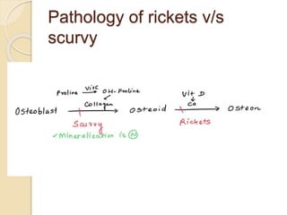
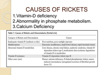
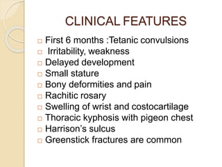
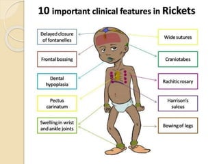
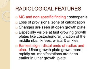
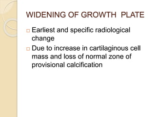
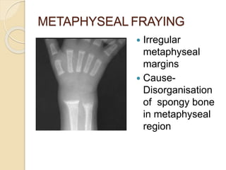
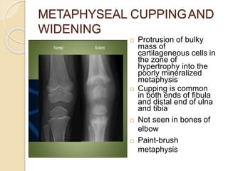
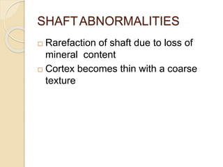
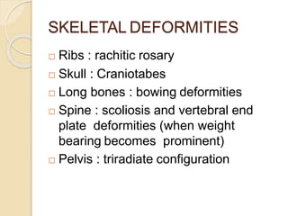
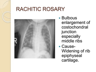
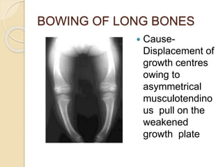
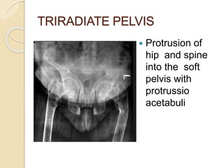
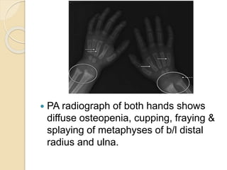
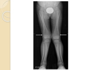
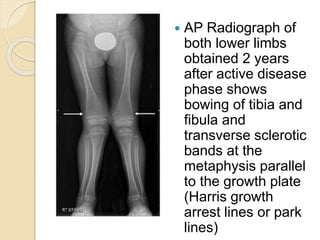
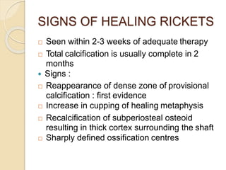
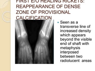
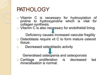
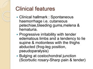
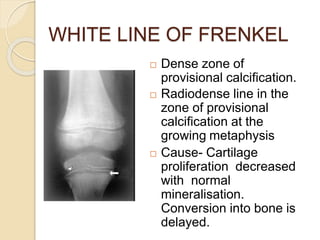
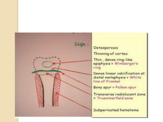
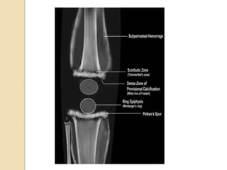
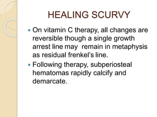
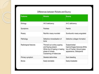
SlideShare a Scribd company logo
Imaging in Rickets & Scurvy
Presented by- Moderator-
Dr. Shivani Gupta Dr. Shalini
Agarwal
Junior Resident 1
Dept. of Radio-diagnosis
PGIMS Rohtak
PHYSIS/ GROWTH PLATE
 It is the anatomical
difference between growing
and mature bone.
 It appears radiologically as a
lucency between the
epiphysis and the
metaphysis.
 It represents the site where
longitudinal bone growth
occurs.
 It is the primary site for the
effect of metabolic and
endocrine bone disorder.
3 zones of
physis-
 Resting/ germinal zone
 Proliferating zone
 Hypertrophic
zone: zone of
maturation,
degeneration and
provisional
calcification
RICKET
S
INTRODUCTION
 It is a disease of childhood
characterized by failure of
mineralization of osteoid tissue in
developing skeleton, particularly at the
growth plate.
 Age: 6-12 months.
 At risk- Dark pigmented individuals,
infants breast-fed for prolonged
periods of time by a multiparous
mother, at higher altitudes
Pathology of rickets v/s
scurvy
PATHOLOGY
 Decrease in quantity of calcified
osteoid & increase in uncalcified
osteoid.
Osteopenia
 Cartilage cells at physis grow normally
but FAIL TO CALCIFY.
Growth plate is widened due to
overgrown & hypertrophied cartilage
CAUSES OF RICKETS
1.Vitamin-D deficiency
2.Abnormality in phosphate metabolism.
3.Calcium Deficiency
CLINICAL FEATURES
 First 6 months :Tetanic convulsions
 Irritability, weakness
 Delayed development
 Small stature
 Bony deformities and pain
 Rachitic rosary
 Swelling of wrist and costocartilage
 Thoracic kyphosis with pigeon chest
 Harrison’s sulcus
 Greenstick fractures are common
RADIOLOGICAL FEATURES
 MC and non specific finding : osteopenia
 Loss of provisional zone of calcification
 Changes are seen at open growth plate
 Especially visible at fast growing growth
plates like costochondral junction of the
middle ribs, knees, wrists & ankles.
 Earliest sign : distal ends of radius and
ulna. Ulnar growth plate grows more
rapidly so manifestations are seen
earlier in ulnar growth plate
WIDENING OF GROWTH PLATE
 Earliest and specific radiological
change
 Due to increase in cartilaginous cell
mass and loss of normal zone of
provisional calcification
METAPHYSEAL FRAYING
 Irregular
metaphyseal
margins
 Cause-
Disorganisation
of spongy bone
in metaphyseal
region
METAPHYSEAL CUPPINGAND
WIDENING
 Protrusion of bulky
mass of
cartilageneous cells in
the zone of
hypertrophy into the
poorly mineralized
metaphysis
 Cupping is common
in both ends of fibula
and distal end of ulna
and tibia
 Not seen in bones of
elbow
 Paint-brush
metaphysis
SHAFTABNORMALITIES
 Rarefaction of shaft due to loss of
mineral content
 Cortex becomes thin with a coarse
texture
SKELETAL DEFORMITIES
 Ribs : rachitic rosary
 Skull : Craniotabes
 Long bones : bowing deformities
 Spine : scoliosis and vertebral end
plate deformities (when weight
bearing becomes prominent)
 Pelvis : triradiate configuration
RACHITIC ROSARY
 Bulbous
enlargement of
costochondral
junction
especially
middle ribs
 Cause-
Widening of rib
epiphyseal
cartilage.
CRANIOTABES
 Excess osteoid
deposition in
frontal and parietal
regions with
posterior flattening
of skull due to
supine posture of
infant
 Squared
configuration of
skull
 Demineralisation
of skull
BOWING OF LONG BONES
 Cause-
Displacement of
growth centres
owing to
asymmetrical
musculotendino
us pull on the
weakened
growth plate
TRIRADIATE PELVIS
 Protrusion of
hip and spine
into the soft
pelvis with
protrussio
acetabuli
 PA radiograph of both hands shows
diffuse osteopenia, cupping, fraying &
splaying of metaphyses of b/l distal
radius and ulna.
 AP Radiograph of
both lower limbs
obtained 2 years
after active disease
phase shows
bowing of tibia and
fibula and
transverse sclerotic
bands at the
metaphysis parallel
to the growth plate
(Harris growth
arrest lines or park
lines)
SIGNS OF HEALING RICKETS
 Seen within 2-3 weeks of adequate therapy
 Total calcification is usually complete in 2
months
 Signs :
 Reappearance of dense zone of provisional
calcification : first evidence
 Increase in cupping of healing metaphysis
 Recalcification of subperiosteal osteoid
resulting in thick cortex surrounding the shaft
 Sharply defined ossification centres
FIRST E/O HEALING RICKETS:
REAPPEARANCE OF DENSE
ZONE OF PROVISIONAL
CALCIFICATION
 Seen as a
transverse line of
increased density
which appears
beyond the visible
end of shaft with
metaphysis
interposed
between two
radiolucent areas
 Complete healing and restoration of
normal structure is the rule in rickets
even if severe changes are present
during the active stage !
SCURVY
 A.k.a. Barlow’s disease or
hypovitaminosis C
Introduction
 Scurvy is a nutritional bone disorder
which occurs due to long term
deficiency of Vitamin C.
 Infantile Scurvy: due to pasteurised or
boiled milk preparations
 Age: 8-14 months
 Latent period- 4 months before
symptoms and skeletal changes
become apparent.
PATHOLOGY
 Vitamin C is necessary for hydroxylation of
proline to hydroxyproline which is vital for
collagen synthesis.
 Vitamin C is also necessary for endothelial lining
Deficiency causes increased vascular fragility
 Osteoblasts require vit C to form mature osteoid
tissue.
 Decreased osteoblastic activity
Generalised osteopenia and osteoporosis
 Cartilage proliferation is decreased but
mineralisation is normal.
Clinical features
 Clinical hallmark : Spontaneous
haemorrhage i.e. cutaneous
petechiae,bleeding gums,melena &
hematuria.
 Progressive irritability with tender
edematous limbs and a tendency to lie
supine & motionless with the thighs
abducted (frog-leg position,
pseudoparalysis)
 Bulging at costochondral junction
(Scorbutic rosary-Sharp pain & tender)
WHITE LINE OF FRENKEL
 Dense zone of
provisional calcification.
 Radiodense line in the
zone of provisional
calcification at the
growing metaphysis
 Cause- Cartilage
proliferation decreased
with normal
mineralisation.
Conversion into bone is
delayed.
TRUMMERFELD ZONE
(Scorbutic zone)
 Transverse radiolucent
band directly beneath
the zone of provisonal
calcification.
 Cause- Suppressed
osteoblastic activity
with normal
mineralisation leading
to disordered osteoid
formation.
 Trabecular bone mass is
decreased.
PELKAN’S SPUR
 Bony protuberances at
the metaphyseal
margins at right angles
to the shaft.
 Cause- Zone of
provisional calcification
extends beyond the
margins of the
metaphysis resulting in
periosteal elevation
and marginal spur
formation
WIMBERGER'S
SIGN
(Ring epiphysis)
 Epiphysis is small &
sharply marginated by
sclerotic rim with central
portion more radiolucent.
 Cause- Decreased
cartilage proliferation
and unimpaired
mineralization
(sclerosis)
 Differentiates healing
rickets and scurvy
CORNER (ANGLE) SIGN
 Irregularity of the
metaphyseal
margins.
 Cause- Infarctions
of the epiphyseal-
metaphyseal
junction
SUBPERIOSTEAL
HEMORRAGES
 Seen in ends of long
bones(femur,tibia,
humerus)
 May cause
periosteal elevation
and new bone
formation
 Cause-Increased
capillary fragility
 Coronal T2-weighted fat-suppressed MR
image of both distal femoral
metadiaphyses shows heterogeneously
increased T2 signal intensity in the
marrow (*) and around the bone (arrows)
HEALING SCURVY
 On vitamin C therapy, all changes are
reversible though a single growth
arrest line may remain in metaphysis
as residual frenkel’s line.
 Following therapy, subperiosteal
hematomas rapidly calcify and
demarcate.
Thank you !

More Related Content

What's hot

Diagnostic Imaging of Bone Tumors
Diagnostic Imaging of Bone TumorsDiagnostic Imaging of Bone Tumors
Diagnostic Imaging of Bone Tumors
Mohamed M.A. Zaitoun
 
Imaging in rickets
Imaging in ricketsImaging in rickets
Imaging in rickets
DrYashasUllasL
 
Radiology of Bone Tumours
Radiology of Bone TumoursRadiology of Bone Tumours
Radiology of Bone Tumours
Muhammad Eimaduddin
 
Tumors
TumorsTumors
Tumors
Dr Imran Jan
 
Radiological evaluation of Arthritis
Radiological evaluation of  ArthritisRadiological evaluation of  Arthritis
Radiological evaluation of Arthritis
shopnilp
 
Bone tumor radiological approach
Bone tumor radiological approachBone tumor radiological approach
Bone tumor radiological approachSitanshu Barik
 
Presentation1, radiological imaging of popliteal fossa masses.
Presentation1, radiological imaging of popliteal fossa masses.Presentation1, radiological imaging of popliteal fossa masses.
Presentation1, radiological imaging of popliteal fossa masses.
Abdellah Nazeer
 
Bone tumors
Bone tumorsBone tumors
Bone tumors
Dhruv Taneja
 
Rickets and osteomalacia
Rickets and osteomalaciaRickets and osteomalacia
Rickets and osteomalacia
tesfa tiruneh
 
Benign bone tumors
Benign bone tumorsBenign bone tumors
Benign bone tumors
macshrestha
 
Diagnostic Imaging of Paget's Disease
Diagnostic Imaging of Paget's DiseaseDiagnostic Imaging of Paget's Disease
Diagnostic Imaging of Paget's Disease
Mohamed M.A. Zaitoun
 
patterns of enhancement in hepatocellular carcinoma
patterns of enhancement in hepatocellular carcinomapatterns of enhancement in hepatocellular carcinoma
patterns of enhancement in hepatocellular carcinoma
Haseeb Manzoor
 
Bone and joint infection presentation
Bone and joint infection presentationBone and joint infection presentation
Bone and joint infection presentation
Amit Rauniyar
 
Presentation1.pptx, radiological imaging of metabolic bone diseases.
Presentation1.pptx, radiological imaging of metabolic bone diseases.Presentation1.pptx, radiological imaging of metabolic bone diseases.
Presentation1.pptx, radiological imaging of metabolic bone diseases.
Abdellah Nazeer
 
Radiology Spots PPT- 3 by Dr Chandni Wadhwani
 Radiology Spots PPT- 3 by Dr Chandni Wadhwani Radiology Spots PPT- 3 by Dr Chandni Wadhwani
Radiology Spots PPT- 3 by Dr Chandni Wadhwani
Chandni Wadhwani
 
Lytic leisons of the skull
Lytic leisons of the skullLytic leisons of the skull
Lytic leisons of the skull
Milan Silwal
 
Imaging in arthritis
Imaging in arthritisImaging in arthritis
Imaging in arthritis
Navni Garg
 
Systemic approach to bone tumor radiology
Systemic approach to bone tumor radiologySystemic approach to bone tumor radiology
Systemic approach to bone tumor radiology
Ramin Sadeghi
 
HEPATOCELLULAR CARCINOMA RADIOLOGY
HEPATOCELLULAR CARCINOMA RADIOLOGYHEPATOCELLULAR CARCINOMA RADIOLOGY
HEPATOCELLULAR CARCINOMA RADIOLOGY
RMLIMS
 
Presentation1, radiological imaging of degenerative and inflammatory disease ...
Presentation1, radiological imaging of degenerative and inflammatory disease ...Presentation1, radiological imaging of degenerative and inflammatory disease ...
Presentation1, radiological imaging of degenerative and inflammatory disease ...
Abdellah Nazeer
 

What's hot (20)

Diagnostic Imaging of Bone Tumors
Diagnostic Imaging of Bone TumorsDiagnostic Imaging of Bone Tumors
Diagnostic Imaging of Bone Tumors
 
Imaging in rickets
Imaging in ricketsImaging in rickets
Imaging in rickets
 
Radiology of Bone Tumours
Radiology of Bone TumoursRadiology of Bone Tumours
Radiology of Bone Tumours
 
Tumors
TumorsTumors
Tumors
 
Radiological evaluation of Arthritis
Radiological evaluation of  ArthritisRadiological evaluation of  Arthritis
Radiological evaluation of Arthritis
 
Bone tumor radiological approach
Bone tumor radiological approachBone tumor radiological approach
Bone tumor radiological approach
 
Presentation1, radiological imaging of popliteal fossa masses.
Presentation1, radiological imaging of popliteal fossa masses.Presentation1, radiological imaging of popliteal fossa masses.
Presentation1, radiological imaging of popliteal fossa masses.
 
Bone tumors
Bone tumorsBone tumors
Bone tumors
 
Rickets and osteomalacia
Rickets and osteomalaciaRickets and osteomalacia
Rickets and osteomalacia
 
Benign bone tumors
Benign bone tumorsBenign bone tumors
Benign bone tumors
 
Diagnostic Imaging of Paget's Disease
Diagnostic Imaging of Paget's DiseaseDiagnostic Imaging of Paget's Disease
Diagnostic Imaging of Paget's Disease
 
patterns of enhancement in hepatocellular carcinoma
patterns of enhancement in hepatocellular carcinomapatterns of enhancement in hepatocellular carcinoma
patterns of enhancement in hepatocellular carcinoma
 
Bone and joint infection presentation
Bone and joint infection presentationBone and joint infection presentation
Bone and joint infection presentation
 
Presentation1.pptx, radiological imaging of metabolic bone diseases.
Presentation1.pptx, radiological imaging of metabolic bone diseases.Presentation1.pptx, radiological imaging of metabolic bone diseases.
Presentation1.pptx, radiological imaging of metabolic bone diseases.
 
Radiology Spots PPT- 3 by Dr Chandni Wadhwani
 Radiology Spots PPT- 3 by Dr Chandni Wadhwani Radiology Spots PPT- 3 by Dr Chandni Wadhwani
Radiology Spots PPT- 3 by Dr Chandni Wadhwani
 
Lytic leisons of the skull
Lytic leisons of the skullLytic leisons of the skull
Lytic leisons of the skull
 
Imaging in arthritis
Imaging in arthritisImaging in arthritis
Imaging in arthritis
 
Systemic approach to bone tumor radiology
Systemic approach to bone tumor radiologySystemic approach to bone tumor radiology
Systemic approach to bone tumor radiology
 
HEPATOCELLULAR CARCINOMA RADIOLOGY
HEPATOCELLULAR CARCINOMA RADIOLOGYHEPATOCELLULAR CARCINOMA RADIOLOGY
HEPATOCELLULAR CARCINOMA RADIOLOGY
 
Presentation1, radiological imaging of degenerative and inflammatory disease ...
Presentation1, radiological imaging of degenerative and inflammatory disease ...Presentation1, radiological imaging of degenerative and inflammatory disease ...
Presentation1, radiological imaging of degenerative and inflammatory disease ...
 

Similar to Imaging in Rickets & Scurvy.pptx

skeletaldisordersofmetabolicorigin-151013014651-lva1-app6892.pptx
skeletaldisordersofmetabolicorigin-151013014651-lva1-app6892.pptxskeletaldisordersofmetabolicorigin-151013014651-lva1-app6892.pptx
skeletaldisordersofmetabolicorigin-151013014651-lva1-app6892.pptx
Yasiele897
 
skeletal disorders of metabolic and endocrine origin
skeletal disorders of metabolic and endocrine originskeletal disorders of metabolic and endocrine origin
skeletal disorders of metabolic and endocrine origin
yashovrattiwari1
 
metabolicendocrinedisordersaffectingbone-180222114735.pptx
metabolicendocrinedisordersaffectingbone-180222114735.pptxmetabolicendocrinedisordersaffectingbone-180222114735.pptx
metabolicendocrinedisordersaffectingbone-180222114735.pptx
Yasiele897
 
Metabolic & endocrine disorders affecting bone (Radiology)
Metabolic & endocrine disorders affecting bone (Radiology)Metabolic & endocrine disorders affecting bone (Radiology)
Metabolic & endocrine disorders affecting bone (Radiology)
Dr.Santosh Atreya
 
Fibro Osseous Lesions
Fibro Osseous LesionsFibro Osseous Lesions
Fibro Osseous Lesions
Sanchit Goyal
 
Plain Radiological findings of Primary Hyperparathyrodism.pptx
Plain Radiological findings of Primary Hyperparathyrodism.pptxPlain Radiological findings of Primary Hyperparathyrodism.pptx
Plain Radiological findings of Primary Hyperparathyrodism.pptx
Ahmed Ashour dr.
 
Plain radiological findings of primary hyperparathyrodism
Plain radiological findings of primary hyperparathyrodismPlain radiological findings of primary hyperparathyrodism
Plain radiological findings of primary hyperparathyrodism
Amr Mansour Hassan
 
OSTEONECROSIS 2003.ppt
OSTEONECROSIS 2003.pptOSTEONECROSIS 2003.ppt
OSTEONECROSIS 2003.ppt
ranjitharadhakrishna3
 
Osteopetrosis
OsteopetrosisOsteopetrosis
Osteopetrosis
Dr. Mohit Goel
 
Osteopetrosis
OsteopetrosisOsteopetrosis
Osteopetrosis
Anish Choudhary
 
Bone pathology
Bone pathologyBone pathology
Bone pathology
Dr Pooja Chaturvedi
 
Metabolic & Endocrinal Diseases affecting bone
Metabolic & Endocrinal Diseases affecting boneMetabolic & Endocrinal Diseases affecting bone
Metabolic & Endocrinal Diseases affecting bone
Dr Sahidul
 
Orthopedic Aspects Of Metabolic Bone Disease By Xiu
Orthopedic Aspects Of Metabolic Bone Disease By XiuOrthopedic Aspects Of Metabolic Bone Disease By Xiu
Orthopedic Aspects Of Metabolic Bone Disease By Xiu
Xiu Srithammasit
 
Rickets
RicketsRickets
Rickets
Pro Faather
 
tb hip.pptx
tb hip.pptxtb hip.pptx
tb hip.pptx
RAdhavan
 
Tuberculosis of Hip
Tuberculosis of Hip Tuberculosis of Hip
Tuberculosis of Hip
Dr. Arpit Joshi
 
orthopedicaspectsofmetabolicbonediseasebyxiu-091217093240-phpapp01.pptx
orthopedicaspectsofmetabolicbonediseasebyxiu-091217093240-phpapp01.pptxorthopedicaspectsofmetabolicbonediseasebyxiu-091217093240-phpapp01.pptx
orthopedicaspectsofmetabolicbonediseasebyxiu-091217093240-phpapp01.pptx
Yasiele897
 
Osteoarthritis
OsteoarthritisOsteoarthritis
Osteoarthritis
Rohit Rajeevan
 

Similar to Imaging in Rickets & Scurvy.pptx (20)

skeletaldisordersofmetabolicorigin-151013014651-lva1-app6892.pptx
skeletaldisordersofmetabolicorigin-151013014651-lva1-app6892.pptxskeletaldisordersofmetabolicorigin-151013014651-lva1-app6892.pptx
skeletaldisordersofmetabolicorigin-151013014651-lva1-app6892.pptx
 
skeletal disorders of metabolic and endocrine origin
skeletal disorders of metabolic and endocrine originskeletal disorders of metabolic and endocrine origin
skeletal disorders of metabolic and endocrine origin
 
metabolicendocrinedisordersaffectingbone-180222114735.pptx
metabolicendocrinedisordersaffectingbone-180222114735.pptxmetabolicendocrinedisordersaffectingbone-180222114735.pptx
metabolicendocrinedisordersaffectingbone-180222114735.pptx
 
Metabolic & endocrine disorders affecting bone (Radiology)
Metabolic & endocrine disorders affecting bone (Radiology)Metabolic & endocrine disorders affecting bone (Radiology)
Metabolic & endocrine disorders affecting bone (Radiology)
 
Fibro Osseous Lesions
Fibro Osseous LesionsFibro Osseous Lesions
Fibro Osseous Lesions
 
Plain Radiological findings of Primary Hyperparathyrodism.pptx
Plain Radiological findings of Primary Hyperparathyrodism.pptxPlain Radiological findings of Primary Hyperparathyrodism.pptx
Plain Radiological findings of Primary Hyperparathyrodism.pptx
 
Plain radiological findings of primary hyperparathyrodism
Plain radiological findings of primary hyperparathyrodismPlain radiological findings of primary hyperparathyrodism
Plain radiological findings of primary hyperparathyrodism
 
OSTEONECROSIS 2003.ppt
OSTEONECROSIS 2003.pptOSTEONECROSIS 2003.ppt
OSTEONECROSIS 2003.ppt
 
Osteopetrosis
OsteopetrosisOsteopetrosis
Osteopetrosis
 
Osteopetrosis
OsteopetrosisOsteopetrosis
Osteopetrosis
 
Bone pathology
Bone pathologyBone pathology
Bone pathology
 
Metabolic & Endocrinal Diseases affecting bone
Metabolic & Endocrinal Diseases affecting boneMetabolic & Endocrinal Diseases affecting bone
Metabolic & Endocrinal Diseases affecting bone
 
Orthopedic Aspects Of Metabolic Bone Disease By Xiu
Orthopedic Aspects Of Metabolic Bone Disease By XiuOrthopedic Aspects Of Metabolic Bone Disease By Xiu
Orthopedic Aspects Of Metabolic Bone Disease By Xiu
 
Rickets
RicketsRickets
Rickets
 
Bones And Muscles
Bones And MusclesBones And Muscles
Bones And Muscles
 
Bones And Muscles
Bones And MusclesBones And Muscles
Bones And Muscles
 
tb hip.pptx
tb hip.pptxtb hip.pptx
tb hip.pptx
 
Tuberculosis of Hip
Tuberculosis of Hip Tuberculosis of Hip
Tuberculosis of Hip
 
orthopedicaspectsofmetabolicbonediseasebyxiu-091217093240-phpapp01.pptx
orthopedicaspectsofmetabolicbonediseasebyxiu-091217093240-phpapp01.pptxorthopedicaspectsofmetabolicbonediseasebyxiu-091217093240-phpapp01.pptx
orthopedicaspectsofmetabolicbonediseasebyxiu-091217093240-phpapp01.pptx
 
Osteoarthritis
OsteoarthritisOsteoarthritis
Osteoarthritis
 

Recently uploaded

micro teaching on communication m.sc nursing.pdf
micro teaching on communication m.sc nursing.pdfmicro teaching on communication m.sc nursing.pdf
micro teaching on communication m.sc nursing.pdf
Anurag Sharma
 
Report Back from SGO 2024: What’s the Latest in Cervical Cancer?
Report Back from SGO 2024: What’s the Latest in Cervical Cancer?Report Back from SGO 2024: What’s the Latest in Cervical Cancer?
Report Back from SGO 2024: What’s the Latest in Cervical Cancer?
bkling
 
POST OPERATIVE OLIGURIA and its management
POST OPERATIVE OLIGURIA and its managementPOST OPERATIVE OLIGURIA and its management
POST OPERATIVE OLIGURIA and its management
touseefaziz1
 
Flu Vaccine Alert in Bangalore Karnataka
Flu Vaccine Alert in Bangalore KarnatakaFlu Vaccine Alert in Bangalore Karnataka
Flu Vaccine Alert in Bangalore Karnataka
addon Scans
 
Ophthalmology Clinical Tests for OSCE exam
Ophthalmology Clinical Tests for OSCE examOphthalmology Clinical Tests for OSCE exam
Ophthalmology Clinical Tests for OSCE exam
KafrELShiekh University
 
BRACHYTHERAPY OVERVIEW AND APPLICATORS
BRACHYTHERAPY OVERVIEW  AND  APPLICATORSBRACHYTHERAPY OVERVIEW  AND  APPLICATORS
BRACHYTHERAPY OVERVIEW AND APPLICATORS
Krishan Murari
 
MANAGEMENT OF ATRIOVENTRICULAR CONDUCTION BLOCK.pdf
MANAGEMENT OF ATRIOVENTRICULAR CONDUCTION BLOCK.pdfMANAGEMENT OF ATRIOVENTRICULAR CONDUCTION BLOCK.pdf
MANAGEMENT OF ATRIOVENTRICULAR CONDUCTION BLOCK.pdf
Jim Jacob Roy
 
Hemodialysis: Chapter 3, Dialysis Water Unit - Dr.Gawad
Hemodialysis: Chapter 3, Dialysis Water Unit - Dr.GawadHemodialysis: Chapter 3, Dialysis Water Unit - Dr.Gawad
Hemodialysis: Chapter 3, Dialysis Water Unit - Dr.Gawad
NephroTube - Dr.Gawad
 
Charaka Samhita Sutra Sthana 9 Chapter khuddakachatuspadadhyaya
Charaka Samhita Sutra Sthana 9 Chapter khuddakachatuspadadhyayaCharaka Samhita Sutra Sthana 9 Chapter khuddakachatuspadadhyaya
Charaka Samhita Sutra Sthana 9 Chapter khuddakachatuspadadhyaya
Dr KHALID B.M
 
Ocular injury ppt Upendra pal optometrist upums saifai etawah
Ocular injury  ppt  Upendra pal  optometrist upums saifai etawahOcular injury  ppt  Upendra pal  optometrist upums saifai etawah
Ocular injury ppt Upendra pal optometrist upums saifai etawah
pal078100
 
Antiulcer drugs Advance Pharmacology .pptx
Antiulcer drugs Advance Pharmacology .pptxAntiulcer drugs Advance Pharmacology .pptx
Antiulcer drugs Advance Pharmacology .pptx
Rohit chaurpagar
 
Physiology of Chemical Sensation of smell.pdf
Physiology of Chemical Sensation of smell.pdfPhysiology of Chemical Sensation of smell.pdf
Physiology of Chemical Sensation of smell.pdf
MedicoseAcademics
 
ARTIFICIAL INTELLIGENCE IN HEALTHCARE.pdf
ARTIFICIAL INTELLIGENCE IN  HEALTHCARE.pdfARTIFICIAL INTELLIGENCE IN  HEALTHCARE.pdf
ARTIFICIAL INTELLIGENCE IN HEALTHCARE.pdf
Anujkumaranit
 
Cervical & Brachial Plexus By Dr. RIG.pptx
Cervical & Brachial Plexus By Dr. RIG.pptxCervical & Brachial Plexus By Dr. RIG.pptx
Cervical & Brachial Plexus By Dr. RIG.pptx
Dr. Rabia Inam Gandapore
 
Maxilla, Mandible & Hyoid Bone & Clinical Correlations by Dr. RIG.pptx
Maxilla, Mandible & Hyoid Bone & Clinical Correlations by Dr. RIG.pptxMaxilla, Mandible & Hyoid Bone & Clinical Correlations by Dr. RIG.pptx
Maxilla, Mandible & Hyoid Bone & Clinical Correlations by Dr. RIG.pptx
Dr. Rabia Inam Gandapore
 
BENIGN PROSTATIC HYPERPLASIA.BPH. BPHpdf
BENIGN PROSTATIC HYPERPLASIA.BPH. BPHpdfBENIGN PROSTATIC HYPERPLASIA.BPH. BPHpdf
BENIGN PROSTATIC HYPERPLASIA.BPH. BPHpdf
DR SETH JOTHAM
 
Non-respiratory Functions of the Lungs.pdf
Non-respiratory Functions of the Lungs.pdfNon-respiratory Functions of the Lungs.pdf
Non-respiratory Functions of the Lungs.pdf
MedicoseAcademics
 
Pulmonary Thromboembolism - etilogy, types, medical- Surgical and nursing man...
Pulmonary Thromboembolism - etilogy, types, medical- Surgical and nursing man...Pulmonary Thromboembolism - etilogy, types, medical- Surgical and nursing man...
Pulmonary Thromboembolism - etilogy, types, medical- Surgical and nursing man...
VarunMahajani
 
Charaka Samhita Sutra sthana Chapter 15 Upakalpaniyaadhyaya
Charaka Samhita Sutra sthana Chapter 15 UpakalpaniyaadhyayaCharaka Samhita Sutra sthana Chapter 15 Upakalpaniyaadhyaya
Charaka Samhita Sutra sthana Chapter 15 Upakalpaniyaadhyaya
Dr KHALID B.M
 
Evaluation of antidepressant activity of clitoris ternatea in animals
Evaluation of antidepressant activity of clitoris ternatea in animalsEvaluation of antidepressant activity of clitoris ternatea in animals
Evaluation of antidepressant activity of clitoris ternatea in animals
Shweta
 

Recently uploaded (20)

micro teaching on communication m.sc nursing.pdf
micro teaching on communication m.sc nursing.pdfmicro teaching on communication m.sc nursing.pdf
micro teaching on communication m.sc nursing.pdf
 
Report Back from SGO 2024: What’s the Latest in Cervical Cancer?
Report Back from SGO 2024: What’s the Latest in Cervical Cancer?Report Back from SGO 2024: What’s the Latest in Cervical Cancer?
Report Back from SGO 2024: What’s the Latest in Cervical Cancer?
 
POST OPERATIVE OLIGURIA and its management
POST OPERATIVE OLIGURIA and its managementPOST OPERATIVE OLIGURIA and its management
POST OPERATIVE OLIGURIA and its management
 
Flu Vaccine Alert in Bangalore Karnataka
Flu Vaccine Alert in Bangalore KarnatakaFlu Vaccine Alert in Bangalore Karnataka
Flu Vaccine Alert in Bangalore Karnataka
 
Ophthalmology Clinical Tests for OSCE exam
Ophthalmology Clinical Tests for OSCE examOphthalmology Clinical Tests for OSCE exam
Ophthalmology Clinical Tests for OSCE exam
 
BRACHYTHERAPY OVERVIEW AND APPLICATORS
BRACHYTHERAPY OVERVIEW  AND  APPLICATORSBRACHYTHERAPY OVERVIEW  AND  APPLICATORS
BRACHYTHERAPY OVERVIEW AND APPLICATORS
 
MANAGEMENT OF ATRIOVENTRICULAR CONDUCTION BLOCK.pdf
MANAGEMENT OF ATRIOVENTRICULAR CONDUCTION BLOCK.pdfMANAGEMENT OF ATRIOVENTRICULAR CONDUCTION BLOCK.pdf
MANAGEMENT OF ATRIOVENTRICULAR CONDUCTION BLOCK.pdf
 
Hemodialysis: Chapter 3, Dialysis Water Unit - Dr.Gawad
Hemodialysis: Chapter 3, Dialysis Water Unit - Dr.GawadHemodialysis: Chapter 3, Dialysis Water Unit - Dr.Gawad
Hemodialysis: Chapter 3, Dialysis Water Unit - Dr.Gawad
 
Charaka Samhita Sutra Sthana 9 Chapter khuddakachatuspadadhyaya
Charaka Samhita Sutra Sthana 9 Chapter khuddakachatuspadadhyayaCharaka Samhita Sutra Sthana 9 Chapter khuddakachatuspadadhyaya
Charaka Samhita Sutra Sthana 9 Chapter khuddakachatuspadadhyaya
 
Ocular injury ppt Upendra pal optometrist upums saifai etawah
Ocular injury  ppt  Upendra pal  optometrist upums saifai etawahOcular injury  ppt  Upendra pal  optometrist upums saifai etawah
Ocular injury ppt Upendra pal optometrist upums saifai etawah
 
Antiulcer drugs Advance Pharmacology .pptx
Antiulcer drugs Advance Pharmacology .pptxAntiulcer drugs Advance Pharmacology .pptx
Antiulcer drugs Advance Pharmacology .pptx
 
Physiology of Chemical Sensation of smell.pdf
Physiology of Chemical Sensation of smell.pdfPhysiology of Chemical Sensation of smell.pdf
Physiology of Chemical Sensation of smell.pdf
 
ARTIFICIAL INTELLIGENCE IN HEALTHCARE.pdf
ARTIFICIAL INTELLIGENCE IN  HEALTHCARE.pdfARTIFICIAL INTELLIGENCE IN  HEALTHCARE.pdf
ARTIFICIAL INTELLIGENCE IN HEALTHCARE.pdf
 
Cervical & Brachial Plexus By Dr. RIG.pptx
Cervical & Brachial Plexus By Dr. RIG.pptxCervical & Brachial Plexus By Dr. RIG.pptx
Cervical & Brachial Plexus By Dr. RIG.pptx
 
Maxilla, Mandible & Hyoid Bone & Clinical Correlations by Dr. RIG.pptx
Maxilla, Mandible & Hyoid Bone & Clinical Correlations by Dr. RIG.pptxMaxilla, Mandible & Hyoid Bone & Clinical Correlations by Dr. RIG.pptx
Maxilla, Mandible & Hyoid Bone & Clinical Correlations by Dr. RIG.pptx
 
BENIGN PROSTATIC HYPERPLASIA.BPH. BPHpdf
BENIGN PROSTATIC HYPERPLASIA.BPH. BPHpdfBENIGN PROSTATIC HYPERPLASIA.BPH. BPHpdf
BENIGN PROSTATIC HYPERPLASIA.BPH. BPHpdf
 
Non-respiratory Functions of the Lungs.pdf
Non-respiratory Functions of the Lungs.pdfNon-respiratory Functions of the Lungs.pdf
Non-respiratory Functions of the Lungs.pdf
 
Pulmonary Thromboembolism - etilogy, types, medical- Surgical and nursing man...
Pulmonary Thromboembolism - etilogy, types, medical- Surgical and nursing man...Pulmonary Thromboembolism - etilogy, types, medical- Surgical and nursing man...
Pulmonary Thromboembolism - etilogy, types, medical- Surgical and nursing man...
 
Charaka Samhita Sutra sthana Chapter 15 Upakalpaniyaadhyaya
Charaka Samhita Sutra sthana Chapter 15 UpakalpaniyaadhyayaCharaka Samhita Sutra sthana Chapter 15 Upakalpaniyaadhyaya
Charaka Samhita Sutra sthana Chapter 15 Upakalpaniyaadhyaya
 
Evaluation of antidepressant activity of clitoris ternatea in animals
Evaluation of antidepressant activity of clitoris ternatea in animalsEvaluation of antidepressant activity of clitoris ternatea in animals
Evaluation of antidepressant activity of clitoris ternatea in animals
 

Imaging in Rickets & Scurvy.pptx

  • 1. Imaging in Rickets & Scurvy Presented by- Moderator- Dr. Shivani Gupta Dr. Shalini Agarwal Junior Resident 1 Dept. of Radio-diagnosis PGIMS Rohtak
  • 2. PHYSIS/ GROWTH PLATE  It is the anatomical difference between growing and mature bone.  It appears radiologically as a lucency between the epiphysis and the metaphysis.  It represents the site where longitudinal bone growth occurs.  It is the primary site for the effect of metabolic and endocrine bone disorder.
  • 3. 3 zones of physis-  Resting/ germinal zone  Proliferating zone  Hypertrophic zone: zone of maturation, degeneration and provisional calcification
  • 5. INTRODUCTION  It is a disease of childhood characterized by failure of mineralization of osteoid tissue in developing skeleton, particularly at the growth plate.  Age: 6-12 months.  At risk- Dark pigmented individuals, infants breast-fed for prolonged periods of time by a multiparous mother, at higher altitudes
  • 6. Pathology of rickets v/s scurvy
  • 7. PATHOLOGY  Decrease in quantity of calcified osteoid & increase in uncalcified osteoid. Osteopenia  Cartilage cells at physis grow normally but FAIL TO CALCIFY. Growth plate is widened due to overgrown & hypertrophied cartilage
  • 8. CAUSES OF RICKETS 1.Vitamin-D deficiency 2.Abnormality in phosphate metabolism. 3.Calcium Deficiency
  • 9. CLINICAL FEATURES  First 6 months :Tetanic convulsions  Irritability, weakness  Delayed development  Small stature  Bony deformities and pain  Rachitic rosary  Swelling of wrist and costocartilage  Thoracic kyphosis with pigeon chest  Harrison’s sulcus  Greenstick fractures are common
  • 10.
  • 11. RADIOLOGICAL FEATURES  MC and non specific finding : osteopenia  Loss of provisional zone of calcification  Changes are seen at open growth plate  Especially visible at fast growing growth plates like costochondral junction of the middle ribs, knees, wrists & ankles.  Earliest sign : distal ends of radius and ulna. Ulnar growth plate grows more rapidly so manifestations are seen earlier in ulnar growth plate
  • 12. WIDENING OF GROWTH PLATE  Earliest and specific radiological change  Due to increase in cartilaginous cell mass and loss of normal zone of provisional calcification
  • 13. METAPHYSEAL FRAYING  Irregular metaphyseal margins  Cause- Disorganisation of spongy bone in metaphyseal region
  • 14. METAPHYSEAL CUPPINGAND WIDENING  Protrusion of bulky mass of cartilageneous cells in the zone of hypertrophy into the poorly mineralized metaphysis  Cupping is common in both ends of fibula and distal end of ulna and tibia  Not seen in bones of elbow  Paint-brush metaphysis
  • 15. SHAFTABNORMALITIES  Rarefaction of shaft due to loss of mineral content  Cortex becomes thin with a coarse texture
  • 16. SKELETAL DEFORMITIES  Ribs : rachitic rosary  Skull : Craniotabes  Long bones : bowing deformities  Spine : scoliosis and vertebral end plate deformities (when weight bearing becomes prominent)  Pelvis : triradiate configuration
  • 17. RACHITIC ROSARY  Bulbous enlargement of costochondral junction especially middle ribs  Cause- Widening of rib epiphyseal cartilage.
  • 18. CRANIOTABES  Excess osteoid deposition in frontal and parietal regions with posterior flattening of skull due to supine posture of infant  Squared configuration of skull  Demineralisation of skull
  • 19. BOWING OF LONG BONES  Cause- Displacement of growth centres owing to asymmetrical musculotendino us pull on the weakened growth plate
  • 20. TRIRADIATE PELVIS  Protrusion of hip and spine into the soft pelvis with protrussio acetabuli
  • 21.  PA radiograph of both hands shows diffuse osteopenia, cupping, fraying & splaying of metaphyses of b/l distal radius and ulna.
  • 22.
  • 23.  AP Radiograph of both lower limbs obtained 2 years after active disease phase shows bowing of tibia and fibula and transverse sclerotic bands at the metaphysis parallel to the growth plate (Harris growth arrest lines or park lines)
  • 24. SIGNS OF HEALING RICKETS  Seen within 2-3 weeks of adequate therapy  Total calcification is usually complete in 2 months  Signs :  Reappearance of dense zone of provisional calcification : first evidence  Increase in cupping of healing metaphysis  Recalcification of subperiosteal osteoid resulting in thick cortex surrounding the shaft  Sharply defined ossification centres
  • 25. FIRST E/O HEALING RICKETS: REAPPEARANCE OF DENSE ZONE OF PROVISIONAL CALCIFICATION  Seen as a transverse line of increased density which appears beyond the visible end of shaft with metaphysis interposed between two radiolucent areas
  • 26.  Complete healing and restoration of normal structure is the rule in rickets even if severe changes are present during the active stage !
  • 27. SCURVY  A.k.a. Barlow’s disease or hypovitaminosis C
  • 28. Introduction  Scurvy is a nutritional bone disorder which occurs due to long term deficiency of Vitamin C.  Infantile Scurvy: due to pasteurised or boiled milk preparations  Age: 8-14 months  Latent period- 4 months before symptoms and skeletal changes become apparent.
  • 29. PATHOLOGY  Vitamin C is necessary for hydroxylation of proline to hydroxyproline which is vital for collagen synthesis.  Vitamin C is also necessary for endothelial lining Deficiency causes increased vascular fragility  Osteoblasts require vit C to form mature osteoid tissue.  Decreased osteoblastic activity Generalised osteopenia and osteoporosis  Cartilage proliferation is decreased but mineralisation is normal.
  • 30. Clinical features  Clinical hallmark : Spontaneous haemorrhage i.e. cutaneous petechiae,bleeding gums,melena & hematuria.  Progressive irritability with tender edematous limbs and a tendency to lie supine & motionless with the thighs abducted (frog-leg position, pseudoparalysis)  Bulging at costochondral junction (Scorbutic rosary-Sharp pain & tender)
  • 31. WHITE LINE OF FRENKEL  Dense zone of provisional calcification.  Radiodense line in the zone of provisional calcification at the growing metaphysis  Cause- Cartilage proliferation decreased with normal mineralisation. Conversion into bone is delayed.
  • 32. TRUMMERFELD ZONE (Scorbutic zone)  Transverse radiolucent band directly beneath the zone of provisonal calcification.  Cause- Suppressed osteoblastic activity with normal mineralisation leading to disordered osteoid formation.  Trabecular bone mass is decreased.
  • 33. PELKAN’S SPUR  Bony protuberances at the metaphyseal margins at right angles to the shaft.  Cause- Zone of provisional calcification extends beyond the margins of the metaphysis resulting in periosteal elevation and marginal spur formation
  • 34. WIMBERGER'S SIGN (Ring epiphysis)  Epiphysis is small & sharply marginated by sclerotic rim with central portion more radiolucent.  Cause- Decreased cartilage proliferation and unimpaired mineralization (sclerosis)  Differentiates healing rickets and scurvy
  • 35. CORNER (ANGLE) SIGN  Irregularity of the metaphyseal margins.  Cause- Infarctions of the epiphyseal- metaphyseal junction
  • 36. SUBPERIOSTEAL HEMORRAGES  Seen in ends of long bones(femur,tibia, humerus)  May cause periosteal elevation and new bone formation  Cause-Increased capillary fragility
  • 37.
  • 38.
  • 39.
  • 40.  Coronal T2-weighted fat-suppressed MR image of both distal femoral metadiaphyses shows heterogeneously increased T2 signal intensity in the marrow (*) and around the bone (arrows)
  • 41. HEALING SCURVY  On vitamin C therapy, all changes are reversible though a single growth arrest line may remain in metaphysis as residual frenkel’s line.  Following therapy, subperiosteal hematomas rapidly calcify and demarcate.
  • 42.