Downloaded 538 times


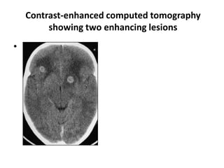


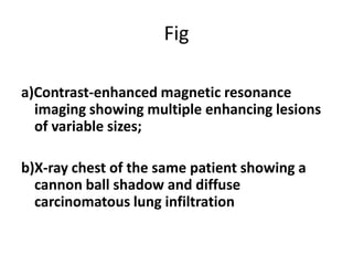


The document discusses ring enhancing lesions seen on neuroimaging. These lesions appear as hypodense masses that enhance with contrast. Common causes include metastatic lesions, primary brain tumors, pyogenic brain abscesses, tuberculomas, cysticercus granuloma, demyelinating disorders, and opportunistic infections in HIV patients such as toxoplasmosis and primary CNS lymphoma. Differential diagnosis depends on location, size, enhancement pattern and associated findings on imaging and other tests.


