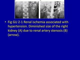
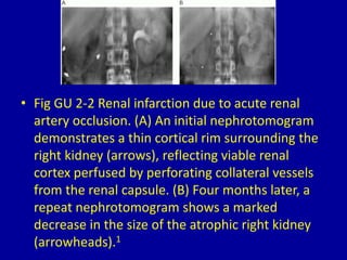
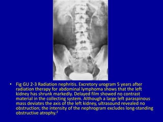
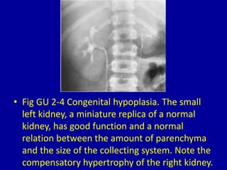
2Unilateral Small, Smooth Kidney
CLINICAL IMAGAGING
AN ATLAS OF DIFFERENTIAL DAIGNOSIS
EISENBERG
DR. Muhammad Bin Zulfiqar
PGR-FCPS III SIMS/SHL
• Fig GU 2-1 Renal ischemia associated with
hypertension. Diminished size of the right
kidney (A) due to renal artery stenosis (B)
(arrow).
• Fig GU 2-2 Renal infarction due to acute renal
artery occlusion. (A) An initial nephrotomogram
demonstrates a thin cortical rim surrounding the
right kidney (arrows), reflecting viable renal
cortex perfused by perforating collateral vessels
from the renal capsule. (B) Four months later, a
repeat nephrotomogram shows a marked
decrease in the size of the atrophic right kidney
(arrowheads).1
• Fig GU 2-3 Radiation nephritis. Excretory urogram 5 years after
radiation therapy for abdominal lymphoma shows that the left
kidney has shrunk markedly. Delayed film showed no contrast
material in the collecting system. Although a large left paraspinous
mass deviates the axis of the left kidney, ultrasound revealed no
obstruction; the intensity of the nephrogram excludes long-standing
obstructive atrophy.2
• Fig GU 2-4 Congenital hypoplasia. The small
left kidney, a miniature replica of a normal
kidney, has good function and a normal
relation between the amount of parenchyma
and the size of the collecting system. Note the
compensatory hypertrophy of the right kidney.
2 unilateral small, smooth kidney

2 unilateral small, smooth kidney

  • 1.
  • 2.
    CLINICAL IMAGAGING AN ATLASOF DIFFERENTIAL DAIGNOSIS EISENBERG DR. Muhammad Bin Zulfiqar PGR-FCPS III SIMS/SHL
  • 3.
    • Fig GU2-1 Renal ischemia associated with hypertension. Diminished size of the right kidney (A) due to renal artery stenosis (B) (arrow).
  • 4.
    • Fig GU2-2 Renal infarction due to acute renal artery occlusion. (A) An initial nephrotomogram demonstrates a thin cortical rim surrounding the right kidney (arrows), reflecting viable renal cortex perfused by perforating collateral vessels from the renal capsule. (B) Four months later, a repeat nephrotomogram shows a marked decrease in the size of the atrophic right kidney (arrowheads).1
  • 5.
    • Fig GU2-3 Radiation nephritis. Excretory urogram 5 years after radiation therapy for abdominal lymphoma shows that the left kidney has shrunk markedly. Delayed film showed no contrast material in the collecting system. Although a large left paraspinous mass deviates the axis of the left kidney, ultrasound revealed no obstruction; the intensity of the nephrogram excludes long-standing obstructive atrophy.2
  • 6.
    • Fig GU2-4 Congenital hypoplasia. The small left kidney, a miniature replica of a normal kidney, has good function and a normal relation between the amount of parenchyma and the size of the collecting system. Note the compensatory hypertrophy of the right kidney.