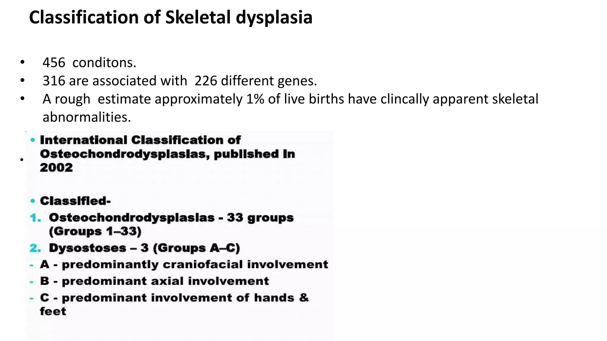
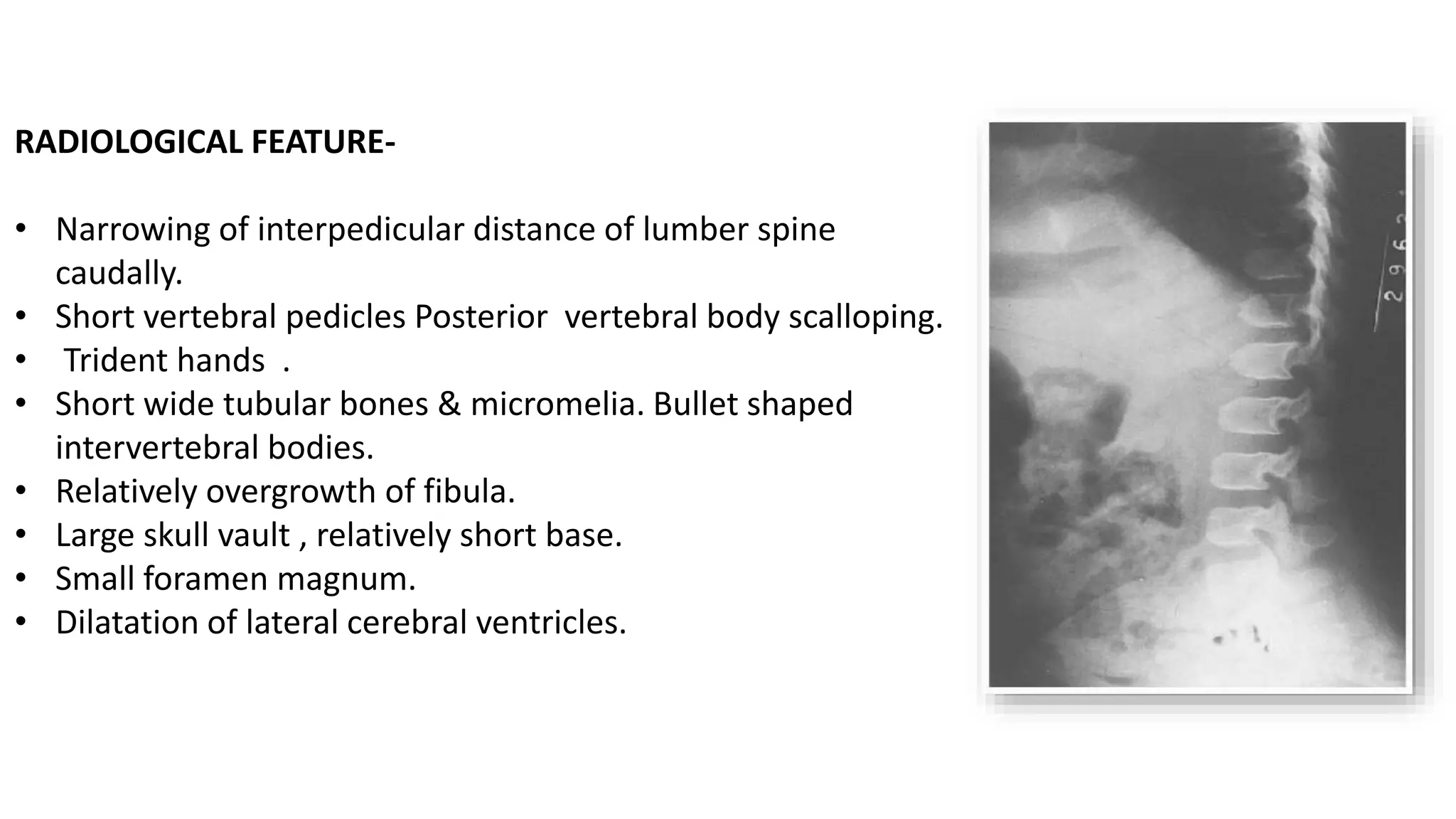
IMAGING OF SKELETAL
DYSPLASIA
MODERATOR:
PROFESSOR SUNIL KUMAR
MD
PRESENTER:
DR. NABA KUMAR BARMAN
JR II
• Osteochondrodysplasia – consists of dysplasia (abnormalities of
bone and/or cartilage growth)and osteodystrophies(abnormalities in
bone and/or cartilage texture). These are intrinsic to bone and
cartilage and as a result of gene expression , continue to evolve
throught life span of an individual.
• Dysostosis- it occurs as a result of altered blastogenesis in first 6
weeks of intra uterine life. Phenotype is fixed and previously normal
bones continues to remain so.
Classification of Skeletal dysplasia
• 456 conditons.
• 316 are associated with 226 different genes.
• A rough estimate approximately 1% of live births have clincally apparent skeletal
abnormalities.
•
• Diagnosis requires multidisciplinary approach with combined
clinical , paediatric, genetic , biochemical, radiological ,
pathological impact.
• Identification of genetic mutation allows“families ”of
conditions to be recognised.
• Clinical data used in diagnosis of skeletal dysplasia
-Stature
-Abnormal body proportion
-Abnormal LIMB SEGMENTS
-Local anomalies & deformities
-Facies
Prenatal Diagnosis
• Prenatal USG screening at between 14 and 18 weeks
gestation.
• USG findings are highly significant but are not specific.
Pregnancy termination offered on the grounds of USG findings
, radiological and histopathological evaluation to determine
the precise diagnosis and before genetic counselling is offered.
• Low dose CT for evaluation of skeletal anomalies.
• MRI – CNS, Musculoskeletal & lung volumes.
SKELETAL SURVEY
skeletal survey should be performed which should include an
• AP and lateral view of the skull,
• AP view of chest, abdomen and pelvis,
• Lateral view of the whole of the spine,
• Detailed lateral view of the cervical spine, if there is a question of atlanto-
axial subluxation,
• AP view of hands,
• AP view of knees and ankles,
• Whole of the tibia and fibula to assess bone modelling,
• Detailed views of any other areas where there is an identified abnormality.
IMPORTANT CLUES TO DYSPLASIAS
• Is the spine normal?
• Are the vertebral bodies and disc spaces of appropriate proportional height?
• Pelvis: is the acetabulum normal? Are the iliac blades normal in proportion?
• Are the epiphyses normal?
• Are the metaphyses normal?
• Are the diaphyses normal?
• Identify any skull abnormalities to include wormian bones, J-shaped sella, hypo- or
hypertelorism or sutural stenosis.
• Hands and feet: are the bones normally modelled or is there pointing of the bases of
the metacarpals? Is there polydactyly?
• Chest: is the configuration normal? Are the ribs normal?
• Is there any associated abnormality such as scoliosis?
• Is the bone density normal, osteopenic or osteosclerotic?
GROUP 1
ACHONDROPLASIA: Autosomal Dominant
CLINICAL FEATURE-
• Short stature ,disproportionate and rhizomelic
• Facial appearance : frontal bossing and depressed nasal bridge.
• The fingertip reach only to the top of the greater trochanters.
• The digit of the hand have extra space between the 3rd and the 4th digit
• so the digit seperated into three groups “trident hand”
• Kyphosis at the thoracolumbar junction
• Intelligence is normal
RADIOLOGICAL FEATURE-
• Narrowing of interpedicular distance of lumber spine
caudally.
• Short vertebral pedicles Posterior vertebral body scalloping.
• Trident hands .
• Short wide tubular bones & micromelia. Bullet shaped
intervertebral bodies.
• Relatively overgrowth of fibula.
• Large skull vault , relatively short base.
• Small foramen magnum.
• Dilatation of lateral cerebral ventricles.
Classic trident hand Champagne glass pelvis Progressively decreasing
interpedicular distance
from L1 to L5
A B C
Achondroplasia. A: Sagittal US demonstrates frontal bossing at 32 weeks’ gestation. B: Coronal
SSFSE T2w MR demonstrates short humeri but normal lung volume. C: Sagittal SSFP MR confirms
frontal bossing and documents narrowing of the foramen magnum.
Hypochondoplasia-
• Clinically the skull is never affected and patients are either normal or
mildly reduced in height.(main diff with achondroplasia)
• Radiologically lumbar lordosis with narrowing of interpedicular
distance from L1 to L5.
• distal fibula is overgrown as compared to distal tibia. Elongation of
ulnar styloid process.
Note the decreasing inter pedicular distance posterior scalloping and distal fibula
overgrowth.
PSEUDOCHONDROPLASIA
• Clinically the skull is normal.
• Radiographically .
• In spine : vertebral bodies are flat with central anterior tongues.
• Long bones : epiphyses are delayed in appearance and markedly irregular.
Markedly irregular epiphysesCentral anterior tongue
Thanatophoric dysplasia
• Rhizomelic Dwarfism
• Relative Shortening Of The
Humeri And Femora
• Femora Are Curved And In A
Classic "Telephone-receiver"
Configuration
• Ribs Are Short And Horizontal
And The Thoracic Cage Is
Hypoplastic
• Iliac Wings Are Small And
Squared
• Generalized Flattening Of The
Vertebral Bodies
• Clover Leaf Skull
Leathal neonatal skeletal dysplasia
GROUP 2 (type 2 collagen)
• Spondyloepiphyseal dysplasia congenital
• Autosomal dominant condition.
• At birth the infant is having short limbs, flat faces and widely spaced eyes.
The neck and spine are short exaggerated kyphosis and lordosis.
• Radiologically
Most prominent feature are found in spine and pelvis.
Delayed ossification complicated with premature osteoarthritis.
Anterior vertebral bodies are bulbous (pear shaped vertebra) . With age
these flatten into platyspondyly
Odontoid hypoplasia is common with cervical instability.
The proximal femoral epiphyseal ossification centres are delayed. Long bones are short and
broad. The epiphyseal ossification centers of the proximal tibia are irregular, small,misshapen
and flattened.
Delayed ossification of humeral epiphysis
mild bulbous appearance of the anterior vertebral bodies. This will flatten into platyspondyly and
persist into
adulthood. COMMENT: The platyspondyly, along with thin discs, results in extreme shortening of the
trunk.
• Spondyloepiphyseal tarda.
• It is manifested in males only and
transmitted as a x linked recessive
condition.
• Short stature is because of
platyspondyly most marked at
thoracic region.
• Radiologically there is hyperostotic
bone deposited in posterior two
third of end plate leading to heaped
up/hump vertebrae.
Hump/heaped up vertebrae
Progressive change from relatively short limbs to relatively short
trunk hence the name metatropic.(group 8)
Chondroectodermal dysplasia
• Aka ellis van Creveld syndrome
Polysyndactyly with carpal fusion(especially between capitate and hamate) in 90%
cases.Ectodermal dysplasia wit sparse hair and abnormal hypoplastic nails , complete or
incomplete absence of tooth.Cardiac defects in 50% cases.
• Belongs to short rib dysplasia group(group 9).
Chondroectodermal dysplasia showing polysyndactyly and carpal fusion
Asphyxiating thoracic dysplasia (jeune syndrome)
• Most patients die in infancy due to respiratory failure.
CLINICAL FEATURES-
• Long narrow thorax.
• Rhizomelia ,nephronophthisis.
• RADIOLOGICAL FEATURES-
• Thorax is stenotic,ribs are short
• Widened costochondral junctions.
• High clavicles(Handle bar clavicle).
• Short iliac bones, trident acetabula.
• Polydactyly.
• Premature ossification of proximal femoral epiphysis.
• Cone shaped epiphysis of phalanges.
GROUP 10
Multiple epiphyseal dysplasia
• First noted when child starts to walk with complaints of waddling gait and difficulty
running.
• It is caused by abnormal epiphyseal chondrocytes with decreased number and
arrangement leading to delayed and disordered ossification of the epiphysis.
• Bilateral and symmetric involvement of epiphyses around hip , knee and ankle.
• Irregular epiphyses leads to premature joint degeneration.
Mottled appearance of proximal
femoral epiphyses( symmetrical and
bilateral)
Fragmentation and deformity of
distal tibia and tarsals.
GROUP 11
• Metaphyseal chondrodysplasia (Pyle’s disease)
Schmid type : mild and relatively common . Metaphysis of long bones appear cupped
(resembling rickets).
Jansens type : grossly irregular mineralization seen in metaphysis of tubular bones.
Other rare types name Pena and Vaandrager (intermediate types).
Some cases are also associated with pancreatic insufficieny and neutropenia.
McKusick described a syndrome of metaphyseal lesions, sparse hairs, dwarfism in amish
families…cartilage-hair-hypoplasia.
Cupped metaphysis bilaterally. Flaring of metaphysis with typical erlynmeyer
flask deformity
GROUP18 (BENT BONE
DISEASE)
• Campomelic dysplasia
Clinically narrow thorax with
respiratory distress.
Radiologically 11 pair of ribs ,
hypoplastic scapulae, bowing of
femur and tibia with hypoplastic
fibulae and patellae.
Antenatal usg shows prenatal onset
of bowed lower limbs.
GROUP 21
• Chondrodysplasia punctata
Characterized by stippled or punctate calcification of multiple epiphyseal centres
Two forms : autosomal dominant ( conradi hunermann syndrome) and recessive form.
Pathology : epiphyseal centres undergo mucoid degeneration with hypervascularity leading to
fragmentation and followed by calcific punctate deposits over fragments.
A
GROUP 22 (neonatal osteosclerotic dysplasia)
• Aka infantile cortical hyperostosis (Caffey disese)
Patient usually presents with a triad of palpable hard bony mass over affected bone, soft
tissue swelling and hyperirritability usually before 5 months age.
Fever is almost always present with raise ESR , leucocytosis , anaemia and raised serum
alkaline phosphatase.
Most commonly involved bones are mandible, clavicle, and ribs.
Most common long bone involved is ulna.
Periosteal new bone formation with cortical thickening. Abnormality limited to diaphysis.
GROUP 23(increased bone density )
1.Pyknodysostosis
Transmitted as an autosomal recessive trait and is characterized by increased bone
density , skeletal fragility and dwarfism.
Short limbs with propensity to transverse pathologic fracture.
Receding jaw and anomalous dentition.
Hands and feet are stubby with spoon shaped nails and clubbing.
Madelung’s deformity is also seen.
Hypoplasia of lateral ends of clavicle and terminal tuft of finger and toes.
Spool shaped vertebra with large anterior and posterior defects.
Unlike osteopetrosis, the medullary canal is maintained in patients with pyknodysostosis;
therefore, anemia and splenomegaly are not associated with this dysplasia.
Spool shaped
vertebra Terminal tuft hypoplasiaHypoplastic Mandible
2.Osteopoikilosis
Characterised by small round to ovoid (1-10mm) radio opacities in epiphysis and
metaphysis.
Associated with cutaneous abnormalities like scleroderma like reaction , keloid
formation, dermatofibrosis.(bushke ollendorf syndrome).
It has a predilection for carpal and tarsal bones as well as long tubular bones.
3.Melorheostosis (leri’s disease)
• In greek it means limb flow bone. There is hyperostotic appearance likened to
flowing wax from a lighted candle.
• Usually monomelic and predilection towards lower limb bones.
• Murray and mc Credie suggested that the lesions follow sclerotome and
myotome that are supplied individual spinal sensory nerve. Thus disease of
sensory nerve results in segmental distribution.
• 4. Osteopetrosis (Albers Schonberg disease , Marble / Chalk bone disease)
Occurs due to lack of resorption of primitive osteoccohndrous tissue the
persistence of which leads to non formation of normal mature adult bone with
medullary canal containing marrow tissue.
Types
Benign heterogenous autosomal dominant
Severe malignant autosomal recessive
Intermediate recessive
Recessive with tubular acidosis(carbonic anhydrase II deficiency syndrome)
• Clinical features includes anaemia, hepatosplenomegaly,
lymphadenopathy , thrombocytopenia and failure to thrive.
• Radiologically
--- homogenous increased density of the involved bone without
trabeculations or differentaition into cortex and medulla. Increased
propensity to transverse fractures with normal healing.
--- erlynmeyer flask deformity (in long bones).
--- bone within bone or endobone appeareance.
--- rugger jersey spine.
--- sandwich vertebra.
The most frequent causes of death are massive hemorrhage and
recurrent infection. Leukemia and sarcoma are known sequelae
A B C
C D
GROUP 24 (increased bone density with diaphyseal involvement)
• Diaphyseal dysplasia (aka camurati-englemann disease)
It has a predilection towards long bones and starts in mid diaphysis extending proximally
and distally sparing metaphysis and epiphysis.
Symmetric in distribution with fusiform widening of diaphyseal portion of long bone.
Muscle weakness and tenderness over affected bone is seen (improvement of bone pain
with administration of corticosteroids).Carpal and tarsal bones are spared.
• Symmetrical widening and sclerosis of diaphysis sparing epi and metaphysis.
A B
GROUP 25
Osteogenesis imperfecta
Caused due to quantitative and qualitative defects in synthesis of type 1 collagen
2/4 major clinical criteria are required for clinical diagnosis:
>Osteoporosis with abnormal fragility of skeleton.
>Premature otosclerosis.
>Blue sclera
>Dentiogenesis imperfecta
Sillence offered a new classification of osteogenesis imperfecta.
It occurs in about 1/20,000 births
The most serious involvement is in the skeleton, but changes in ligaments, skin, sclera, the
inner ear, and dentition are also noted. The disease is thought to have an autosomal
dominant transmission
Clinical findings I II III IV V
Incidence 1:30,000,AD
Most common
1:30,000
AR
Rare
SPORADIC
Unknown (rare)
AD
Unknown
(rare)
Severity Mild Lethal Severe Mild/moderately
severe
Moderate
Death Old age Stillborn By 30
years
Old age Old age
Sclerae Blue Blue Blue then
grey
White White
Hearing
impairment
Frequent --------- Rare Rare Rare
Teeth IA normal IB DI --------- DI IVA normal IVB DI Normal
Stature Normal --------- Short Normal/mildly
short
Normal/mildly
short
Osteogenesis Imperfecta Clinical (Based on the Sillence Classification)and Radiological Findings
I II III IV V
Radiological
findings
Fractures at
birth
<10% Multiple Frequent Rare Rare
Osseous
fragility
Moderate/
mild
Severe Moderate/
severe
Moderate/
mild
Moderate
Deformity Mild ------- Severe Variable Moderate/se
vere
The cardinal roentgen features of osteogenesis imperfecta are a diffuse
decrease in bone density, pencil-thin cortices and multiple fractures
Fairbank subdivided osteogenesis imperfecta into three groups : thin and
gracile bones; short, thick bones; and cystic bones.
THIN AND GRACILE
BONES.
SHORT AND THICK
BONES.
CYSTIC BONES
A
B
C
Diffuse osteoporosis with
pencil thin cortex
WORMIAN BONES.
BICONCAVE LENS
VERTEBRA
Images shows a child born with osteogenesis imperfecta type 2 with
multiple fractures and bowing. Wormian bones are seen in skull.
GROUP 27 (mucopolysaccharidosis)
McKusick
classified these entities into six distinct syndromes:
MPS-I (MPS-I-H): Hurler’s syndrome.
MPS-I-S: Scheie’s syndrome.
MPS-I-H-S: Hurler-Scheie syndrome.
MPS-II: Hunter’s syndrome.
MPS-III (subtypes A, B, C, and D): Sanfilippo’s syndrome.
MPS-IV: (subtypes A and B) Morquio’s syndrome.
MPS-VI: Maroteaux-Lamy syndrome.
MPS-VII: Sly’s syndrom
Among 7 of them relatively common are type 1(hurler’s disease)
and type 4 (morquio’s disease)
HURLER’S DISEASE
Hurler’s syndrome is a rare autosomal recessive disorder of mucopolysaccharide
metabolism that leads to excessive lipoid accumulation in the central nervous
system and other viscera. It occurs in approximately 1/100,000 births. The
excessive mucopolysaccharides excreted in the urine are dermatan sulfate and
heparin sulfate.
Hurler’s syndrome represents the classical prototype of mucopolysaccharide
disorder
• Radiological Features included
1. Characteristic j shaped enlarged sella
2. Lower thoracic and upper lumbar vertebra are small and beaked inferiorly
3. Ribs are overly wide showing paddle or spatulated appeareance.
A. J-Shaped Sella Turcica. Note the characteristic
enlarged, J-shaped sella.
B. Thoracolumbar Kyphosis. Note that the lower
thoracic and upper lumbar vertebral bodies are small
and are
beaked inferiorly, resulting in a thoracolumbar
kyphosis
C. Paddle Ribs. Observe that the ribs are overly
wide,
producing a paddled or spatulated appearance.
Note the characteristic varus deformity of the
humerus (arrows).
D. Hands. Observe that the metacarpals and
phalanges are short and wide.
Clinical Features
• Patients often appear normal at birth, with the manifestation of skeletal changes
becoming apparent only upon weight bearing.
• During early childhood individuals develop marked dwarfism, dorsal kyphoscoliosis,
weakness, and muscular hypotonia. Adult height rarely exceeds 4 ft. Sternum-
pectus carinatum
• The neck is short, and the head appears sunken into the chest. The nose is short,
with a depressed bridge, and the eyes are wide-set. The maxillae are wide, with
deformed, poorly spaced teeth Genu valgum and flexion contractures of the
extremities are common.
• Atlantoaxial dislocations occur owing to odontoid hypoplasia and may result in
paraplegia and respiratory paralysis. Mental capacity is generally normal, but
deafness often develops. Cardiomegaly - aortic valvular involvement. Most patients
survive iupto the 3rd and 4th decades.
MORQUIO’S SYNDROME
1.Sternum is horizontal and protuberant.
2.Platyspondyly with central beaking of vertebrae.Intervertebral disc height are
maintained.
3.Odontoid agenesis and joint laxity is common
A
GROUP 32 (cleidocranial dysplasia)
• Autosomal dominant disorder caused by
mutation in chromosome 6
, characterized by faulty ossification of the
intramembranous and enchondral bones.
• Skull and clavicular anomalies as well as mid
line defects are classic features of this
dysplasia.
• Delayed ossification of calavaria with
wormian bone formation.
• Widening of principal sutures giving hot cross
bun appearance.
• Underdeveloped facial bones.
• Clavicle absent bilaterally in 10%cases.
• Pubic diastases.
Hypoplastic middle part of mid
clavicle with pseudoarthrosis
Wormian bones with wide
open major sutures
Pelvic diastases
GROUP29 (DISORGANISED DEVELOPMENT
OF SKELETAL COMPONENTS GROUP
• MULTIPLE CARTILAGINOUS EXOSTOSES
• ENCHONDROMATOSIS (OLLIER’S DISEASE)
• FIBROUS DYSPLASIA
• NEUROFIBROMATOSIS
MULTIPLE CARTILAGINOUS EXOSTOSES
• CLINICAL FEATURES-
• Multiple bony prominences.
• Ulnare deviation of wrist.
• Limitation of joint movements.
• RADIOLOGICAL FEATURES-
• Ulnare deviation of wrist, skull vault spared.
• Multiple flate /protuberant,polypoid /sessile exostoses.
• Reverse Madelung deformity.
• Vertebral bodies rarely involved.
• Iliac crest and scapulae may be involved.
ENCHONDROMATOSIS (OLLIER’S DISEASE)
CLINICAL FEATURES-
• Asymmetrical limb shortening.
• Expansion of affected bones.
• Occasional pathological fractures.
RADIOLOGICAL FEATURES-
• Typically assymetrical.
• Shortening of affected long bones.
• Rounded /streaky radiolucencies particularly in metaphysis.
• Expnsion of bones with cortical thinning.
• Areas of calcifications within lesions.
• Joint deformity Reverse madelung deformity.
FIBROUS DYSPLASIA
CLINICAL FEATURES-
• Pain and deformity of involved bones. Monostotic or polystotic.
RADIOLOGICAL FEATURES-
• Asymmetrical thickening of skull vault with sclerosis of the base;
multiple roounded opacities.
• Obliteration of paranasal sinuses.
• Marked facial defomity(LEONTIASIS OSSEA).
• Ground glass or rdiolucent areas of trabecular alteration in the long
bones with patchy sclerosis and expansion with cortical thining and
end osteal scalloping.
• Pathological fracture and deformities due to bone softening
SHEPHERD’S CROOK femoral necks.
• Localised or asymmetrical overgrowth.
B
A
NEUROFIBROMATOSIS
CLINICAL FEATURES-
• Focal gigantism,Macrocrania, Axillary freckling.
• Multiple café au lait macules.
• Molluscum fibrosum and anteromedial bowing of tibia.
RADIOLOGICAL FEATURES-
• Aplasia/hypoplasia of the sphenoid wings.
• Dumb-bell neurofibromas/lateral meningocoele.
• Hypoplasia of posterosuperior wall.(Pulsatile exophthalmos)
• Mesodermal dysplasia ,ribbon ribs & rib notching.
• Angular kyphoscoliosis.
• Posterior scalloping of vertbral bodies(dural ectasia).
• Pseudoarthrosis of tibia fibula or clavicle.
• Fibrous cortical defects & Intraosseous cysts.
OTHER EPIPHYSEAL DYSPLASIA
• MARFANS SYNDROME.
Autosomal dominant entity consisting of long slender tubular bones with
ocular abnormalities and cardiovascular abnormalities.
The extremities are markedly elongated with sparse soft tissue owing to
muscular hypoplasia and scarcity of soft tissue(arachnodactyly).
Ectopia lentis is most common ocular abnormality.
Atrial septal defect is the most common congenital cardiac lesion.
Aka floppy valve syndrome.
Steinbergs thumb sign positive.
Increased meta carpal index(8.4-10.4)
Arachnodactyly (spider like fingers)
HOLT ORAM SYNDROME
• Autosomal dominant transmission with cardiac and arm and hand
anomalies.
• Most common cardiac defect is atrial septal defect.
• Radial ray deficiency is also seen. Anomalies of thumb are the most
common skeletal finding.(triphalangeal thumb).
• Extra carpals and carpal fusion , absent thumb may also be seen.
Absent 1st digit Radius deficiency
A B
MCQ
Q 1 Features of Thanatophoric dysplasia except
a) Pear shaped vertebral bodies.
b) Platyspondyly and autosomal dominant.
c) Telephone reciever femora and Rhizomelia.
d) Trident acetabula with irregular metaphysis
Essentials of Skeletal Radiology,3rd Edition,Skeletal Dysplasia,chapter -8
© 2005 Lippincott Williams & Wilkins,Lindsay J. RoweTerry R. Yochum
Q 2 Which is not true for multiple epiphyseal
dysplasia
a) Early osteoarthritis.
b) Late osteoarthritis.
c) Joint stiffness ± limp.
d) Autosomal dominant.
Essentials of Skeletal Radiology,3rd Edition,Skeletal Dysplasia,chapter -8
© 2005 Lippincott Williams & Wilkins,Lindsay J. RoweTerry R. Yochum
ANS -A
Essentials of Skeletal Radiology,3rd Edition,Skeletal Dysplasia,chapter -8
© 2005 Lippincott Williams & Wilkins,Lindsay J. RoweTerry R. Yochum
ANS -B
Q 3 Drippping candle wax appearance characteristic
feature of
a) Melorheostosis
b) Osteopetrosis
c) Pyknodysotosis
d) Osteosclerosis
Essentials of Skeletal Radiology,3rd Edition,Skeletal Dysplasia,chapter -8
© 2005 Lippincott Williams & Wilkins,Lindsay J. RoweTerry R. Yochum
ANS-A
Q4- Which one is true
a)Thanatophoric dysplasia-Autosomal recessive and
Telephone receiver femora.
b)Jeune syndrome-Autosomal dominant and handle
bar clavicle.
c)Achondroplasia-Chevron deformity and Coronal
cleft vertebrae.
d)Apert Syndrome-Autosomal dominant and
Coronal craniosynostosis.
Essentials of Skeletal Radiology,3rd Edition,Skeletal Dysplasia,chapter -8
© 2005 Lippincott Williams & Wilkins,Lindsay J. RoweTerry R. Yochum
ANS-D
Q-5 Periosteal cloaking present in
a)Mucolipidosis typeII
b)Multiple cartilaginous exostosis.
c)Enchondromatosis.
d)Morquio syndrome.
Essentials of Skeletal Radiology,3rd Edition,Skeletal Dysplasia,chapter -8
© 2005 Lippincott Williams & Wilkins,Lindsay J. RoweTerry R. Yochum
ANS-D
Q-6 All are features of Cleidocranial dysplasia
except
a)Frontal bossing and small scapulae.
b) Multiple wormian bones.
c) Supernumerary teeth.
d)Mitten/sock defomity.
Essentials of Skeletal Radiology,3rd Edition,Skeletal Dysplasia,chapter -8
© 2005 Lippincott Williams & Wilkins,Lindsay J. RoweTerry R. Yochum
ANS-D
Q-7 Campomelic dysplasia also known as
a) Bent bone disease.
b)Staight bone disease.
c)Dumb-bell bone disease.
d)Marble bone disease.
Essentials of Skeletal Radiology,3rd Edition,Skeletal Dysplasia,chapter -8
© 2005 Lippincott Williams & Wilkins,Lindsay J. RoweTerry R. Yochum
ANS-A
Q-8 All are false combination except
a)Achondroplasia- Champgne glass appearance
pelvis and bullet shaped vertebrae.
b)Osteopetrosis-Bone within bone appearance and
decreased bone density.
c)Mucopolysaccharidosis- Microcephaly and
hooked shaped vertebrae .
d)Infantile cortical hyperostosis- Periosteal new
bone and cortical thining.
Essentials of Skeletal Radiology,3rd Edition,Skeletal Dysplasia,chapter -8
© 2005 Lippincott Williams & Wilkins,Lindsay J. RoweTerry R. Yochum
ANS-A
Q-9 In Metatropic dysplasia
a) Progressive change from short limb to short
trunk hence name, ‘metatropic’.
b) Progressive change from short trunk to long limb
hence name, ‘metatropic’.
c) Progressive change from long limb to short trunk
hence name, ‘metatropic’.
d) Progressive change from long limb to long trunk
hence name, ‘metatropic’.
Essentials of Skeletal Radiology,3rd Edition,Skeletal Dysplasia,chapter -8
© 2005 Lippincott Williams & Wilkins,Lindsay J. RoweTerry R. Yochum
ANS-A
Q-10 Craniosynostosis syndromes are
a)Pfeiffer Syndrome
b)Apert Syndrome
c)Both of the above.
d)None of the above.
ANS -A
THANK YOU

skeletal dysplasia ppt

  • 1.
    IMAGING OF SKELETAL DYSPLASIA MODERATOR: PROFESSORSUNIL KUMAR MD PRESENTER: DR. NABA KUMAR BARMAN JR II
  • 2.
    • Osteochondrodysplasia –consists of dysplasia (abnormalities of bone and/or cartilage growth)and osteodystrophies(abnormalities in bone and/or cartilage texture). These are intrinsic to bone and cartilage and as a result of gene expression , continue to evolve throught life span of an individual. • Dysostosis- it occurs as a result of altered blastogenesis in first 6 weeks of intra uterine life. Phenotype is fixed and previously normal bones continues to remain so.
  • 3.
    Classification of Skeletaldysplasia • 456 conditons. • 316 are associated with 226 different genes. • A rough estimate approximately 1% of live births have clincally apparent skeletal abnormalities. •
  • 4.
    • Diagnosis requiresmultidisciplinary approach with combined clinical , paediatric, genetic , biochemical, radiological , pathological impact. • Identification of genetic mutation allows“families ”of conditions to be recognised. • Clinical data used in diagnosis of skeletal dysplasia -Stature -Abnormal body proportion -Abnormal LIMB SEGMENTS -Local anomalies & deformities -Facies
  • 5.
    Prenatal Diagnosis • PrenatalUSG screening at between 14 and 18 weeks gestation. • USG findings are highly significant but are not specific. Pregnancy termination offered on the grounds of USG findings , radiological and histopathological evaluation to determine the precise diagnosis and before genetic counselling is offered. • Low dose CT for evaluation of skeletal anomalies. • MRI – CNS, Musculoskeletal & lung volumes.
  • 6.
    SKELETAL SURVEY skeletal surveyshould be performed which should include an • AP and lateral view of the skull, • AP view of chest, abdomen and pelvis, • Lateral view of the whole of the spine, • Detailed lateral view of the cervical spine, if there is a question of atlanto- axial subluxation, • AP view of hands, • AP view of knees and ankles, • Whole of the tibia and fibula to assess bone modelling, • Detailed views of any other areas where there is an identified abnormality.
  • 7.
    IMPORTANT CLUES TODYSPLASIAS • Is the spine normal? • Are the vertebral bodies and disc spaces of appropriate proportional height? • Pelvis: is the acetabulum normal? Are the iliac blades normal in proportion? • Are the epiphyses normal? • Are the metaphyses normal? • Are the diaphyses normal? • Identify any skull abnormalities to include wormian bones, J-shaped sella, hypo- or hypertelorism or sutural stenosis. • Hands and feet: are the bones normally modelled or is there pointing of the bases of the metacarpals? Is there polydactyly? • Chest: is the configuration normal? Are the ribs normal? • Is there any associated abnormality such as scoliosis? • Is the bone density normal, osteopenic or osteosclerotic?
  • 8.
    GROUP 1 ACHONDROPLASIA: AutosomalDominant CLINICAL FEATURE- • Short stature ,disproportionate and rhizomelic • Facial appearance : frontal bossing and depressed nasal bridge. • The fingertip reach only to the top of the greater trochanters. • The digit of the hand have extra space between the 3rd and the 4th digit • so the digit seperated into three groups “trident hand” • Kyphosis at the thoracolumbar junction • Intelligence is normal
  • 9.
    RADIOLOGICAL FEATURE- • Narrowingof interpedicular distance of lumber spine caudally. • Short vertebral pedicles Posterior vertebral body scalloping. • Trident hands . • Short wide tubular bones & micromelia. Bullet shaped intervertebral bodies. • Relatively overgrowth of fibula. • Large skull vault , relatively short base. • Small foramen magnum. • Dilatation of lateral cerebral ventricles.
  • 10.
    Classic trident handChampagne glass pelvis Progressively decreasing interpedicular distance from L1 to L5
  • 11.
  • 12.
    Achondroplasia. A: SagittalUS demonstrates frontal bossing at 32 weeks’ gestation. B: Coronal SSFSE T2w MR demonstrates short humeri but normal lung volume. C: Sagittal SSFP MR confirms frontal bossing and documents narrowing of the foramen magnum.
  • 13.
    Hypochondoplasia- • Clinically theskull is never affected and patients are either normal or mildly reduced in height.(main diff with achondroplasia) • Radiologically lumbar lordosis with narrowing of interpedicular distance from L1 to L5. • distal fibula is overgrown as compared to distal tibia. Elongation of ulnar styloid process.
  • 14.
    Note the decreasinginter pedicular distance posterior scalloping and distal fibula overgrowth.
  • 15.
    PSEUDOCHONDROPLASIA • Clinically theskull is normal. • Radiographically . • In spine : vertebral bodies are flat with central anterior tongues. • Long bones : epiphyses are delayed in appearance and markedly irregular. Markedly irregular epiphysesCentral anterior tongue
  • 16.
    Thanatophoric dysplasia • RhizomelicDwarfism • Relative Shortening Of The Humeri And Femora • Femora Are Curved And In A Classic "Telephone-receiver" Configuration • Ribs Are Short And Horizontal And The Thoracic Cage Is Hypoplastic • Iliac Wings Are Small And Squared • Generalized Flattening Of The Vertebral Bodies • Clover Leaf Skull Leathal neonatal skeletal dysplasia
  • 17.
    GROUP 2 (type2 collagen) • Spondyloepiphyseal dysplasia congenital • Autosomal dominant condition. • At birth the infant is having short limbs, flat faces and widely spaced eyes. The neck and spine are short exaggerated kyphosis and lordosis. • Radiologically Most prominent feature are found in spine and pelvis. Delayed ossification complicated with premature osteoarthritis. Anterior vertebral bodies are bulbous (pear shaped vertebra) . With age these flatten into platyspondyly Odontoid hypoplasia is common with cervical instability.
  • 18.
    The proximal femoralepiphyseal ossification centres are delayed. Long bones are short and broad. The epiphyseal ossification centers of the proximal tibia are irregular, small,misshapen and flattened.
  • 19.
    Delayed ossification ofhumeral epiphysis mild bulbous appearance of the anterior vertebral bodies. This will flatten into platyspondyly and persist into adulthood. COMMENT: The platyspondyly, along with thin discs, results in extreme shortening of the trunk.
  • 20.
    • Spondyloepiphyseal tarda. •It is manifested in males only and transmitted as a x linked recessive condition. • Short stature is because of platyspondyly most marked at thoracic region. • Radiologically there is hyperostotic bone deposited in posterior two third of end plate leading to heaped up/hump vertebrae. Hump/heaped up vertebrae
  • 21.
    Progressive change fromrelatively short limbs to relatively short trunk hence the name metatropic.(group 8)
  • 22.
    Chondroectodermal dysplasia • Akaellis van Creveld syndrome Polysyndactyly with carpal fusion(especially between capitate and hamate) in 90% cases.Ectodermal dysplasia wit sparse hair and abnormal hypoplastic nails , complete or incomplete absence of tooth.Cardiac defects in 50% cases. • Belongs to short rib dysplasia group(group 9). Chondroectodermal dysplasia showing polysyndactyly and carpal fusion
  • 23.
    Asphyxiating thoracic dysplasia(jeune syndrome) • Most patients die in infancy due to respiratory failure. CLINICAL FEATURES- • Long narrow thorax. • Rhizomelia ,nephronophthisis. • RADIOLOGICAL FEATURES- • Thorax is stenotic,ribs are short • Widened costochondral junctions. • High clavicles(Handle bar clavicle). • Short iliac bones, trident acetabula. • Polydactyly. • Premature ossification of proximal femoral epiphysis. • Cone shaped epiphysis of phalanges.
  • 24.
    GROUP 10 Multiple epiphysealdysplasia • First noted when child starts to walk with complaints of waddling gait and difficulty running. • It is caused by abnormal epiphyseal chondrocytes with decreased number and arrangement leading to delayed and disordered ossification of the epiphysis. • Bilateral and symmetric involvement of epiphyses around hip , knee and ankle. • Irregular epiphyses leads to premature joint degeneration.
  • 25.
    Mottled appearance ofproximal femoral epiphyses( symmetrical and bilateral) Fragmentation and deformity of distal tibia and tarsals.
  • 26.
    GROUP 11 • Metaphysealchondrodysplasia (Pyle’s disease) Schmid type : mild and relatively common . Metaphysis of long bones appear cupped (resembling rickets). Jansens type : grossly irregular mineralization seen in metaphysis of tubular bones. Other rare types name Pena and Vaandrager (intermediate types). Some cases are also associated with pancreatic insufficieny and neutropenia. McKusick described a syndrome of metaphyseal lesions, sparse hairs, dwarfism in amish families…cartilage-hair-hypoplasia.
  • 27.
    Cupped metaphysis bilaterally.Flaring of metaphysis with typical erlynmeyer flask deformity
  • 28.
    GROUP18 (BENT BONE DISEASE) •Campomelic dysplasia Clinically narrow thorax with respiratory distress. Radiologically 11 pair of ribs , hypoplastic scapulae, bowing of femur and tibia with hypoplastic fibulae and patellae. Antenatal usg shows prenatal onset of bowed lower limbs.
  • 29.
    GROUP 21 • Chondrodysplasiapunctata Characterized by stippled or punctate calcification of multiple epiphyseal centres Two forms : autosomal dominant ( conradi hunermann syndrome) and recessive form. Pathology : epiphyseal centres undergo mucoid degeneration with hypervascularity leading to fragmentation and followed by calcific punctate deposits over fragments. A
  • 30.
    GROUP 22 (neonatalosteosclerotic dysplasia) • Aka infantile cortical hyperostosis (Caffey disese) Patient usually presents with a triad of palpable hard bony mass over affected bone, soft tissue swelling and hyperirritability usually before 5 months age. Fever is almost always present with raise ESR , leucocytosis , anaemia and raised serum alkaline phosphatase. Most commonly involved bones are mandible, clavicle, and ribs. Most common long bone involved is ulna. Periosteal new bone formation with cortical thickening. Abnormality limited to diaphysis.
  • 32.
    GROUP 23(increased bonedensity ) 1.Pyknodysostosis Transmitted as an autosomal recessive trait and is characterized by increased bone density , skeletal fragility and dwarfism. Short limbs with propensity to transverse pathologic fracture. Receding jaw and anomalous dentition. Hands and feet are stubby with spoon shaped nails and clubbing. Madelung’s deformity is also seen. Hypoplasia of lateral ends of clavicle and terminal tuft of finger and toes. Spool shaped vertebra with large anterior and posterior defects. Unlike osteopetrosis, the medullary canal is maintained in patients with pyknodysostosis; therefore, anemia and splenomegaly are not associated with this dysplasia.
  • 33.
    Spool shaped vertebra Terminaltuft hypoplasiaHypoplastic Mandible
  • 34.
    2.Osteopoikilosis Characterised by smallround to ovoid (1-10mm) radio opacities in epiphysis and metaphysis. Associated with cutaneous abnormalities like scleroderma like reaction , keloid formation, dermatofibrosis.(bushke ollendorf syndrome). It has a predilection for carpal and tarsal bones as well as long tubular bones.
  • 35.
    3.Melorheostosis (leri’s disease) •In greek it means limb flow bone. There is hyperostotic appearance likened to flowing wax from a lighted candle. • Usually monomelic and predilection towards lower limb bones. • Murray and mc Credie suggested that the lesions follow sclerotome and myotome that are supplied individual spinal sensory nerve. Thus disease of sensory nerve results in segmental distribution.
  • 36.
    • 4. Osteopetrosis(Albers Schonberg disease , Marble / Chalk bone disease) Occurs due to lack of resorption of primitive osteoccohndrous tissue the persistence of which leads to non formation of normal mature adult bone with medullary canal containing marrow tissue. Types Benign heterogenous autosomal dominant Severe malignant autosomal recessive Intermediate recessive Recessive with tubular acidosis(carbonic anhydrase II deficiency syndrome)
  • 37.
    • Clinical featuresincludes anaemia, hepatosplenomegaly, lymphadenopathy , thrombocytopenia and failure to thrive. • Radiologically --- homogenous increased density of the involved bone without trabeculations or differentaition into cortex and medulla. Increased propensity to transverse fractures with normal healing. --- erlynmeyer flask deformity (in long bones). --- bone within bone or endobone appeareance. --- rugger jersey spine. --- sandwich vertebra. The most frequent causes of death are massive hemorrhage and recurrent infection. Leukemia and sarcoma are known sequelae
  • 38.
  • 40.
  • 41.
    GROUP 24 (increasedbone density with diaphyseal involvement) • Diaphyseal dysplasia (aka camurati-englemann disease) It has a predilection towards long bones and starts in mid diaphysis extending proximally and distally sparing metaphysis and epiphysis. Symmetric in distribution with fusiform widening of diaphyseal portion of long bone. Muscle weakness and tenderness over affected bone is seen (improvement of bone pain with administration of corticosteroids).Carpal and tarsal bones are spared. • Symmetrical widening and sclerosis of diaphysis sparing epi and metaphysis. A B
  • 42.
    GROUP 25 Osteogenesis imperfecta Causeddue to quantitative and qualitative defects in synthesis of type 1 collagen 2/4 major clinical criteria are required for clinical diagnosis: >Osteoporosis with abnormal fragility of skeleton. >Premature otosclerosis. >Blue sclera >Dentiogenesis imperfecta Sillence offered a new classification of osteogenesis imperfecta. It occurs in about 1/20,000 births The most serious involvement is in the skeleton, but changes in ligaments, skin, sclera, the inner ear, and dentition are also noted. The disease is thought to have an autosomal dominant transmission
  • 43.
    Clinical findings III III IV V Incidence 1:30,000,AD Most common 1:30,000 AR Rare SPORADIC Unknown (rare) AD Unknown (rare) Severity Mild Lethal Severe Mild/moderately severe Moderate Death Old age Stillborn By 30 years Old age Old age Sclerae Blue Blue Blue then grey White White Hearing impairment Frequent --------- Rare Rare Rare Teeth IA normal IB DI --------- DI IVA normal IVB DI Normal Stature Normal --------- Short Normal/mildly short Normal/mildly short Osteogenesis Imperfecta Clinical (Based on the Sillence Classification)and Radiological Findings
  • 44.
    I II IIIIV V Radiological findings Fractures at birth <10% Multiple Frequent Rare Rare Osseous fragility Moderate/ mild Severe Moderate/ severe Moderate/ mild Moderate Deformity Mild ------- Severe Variable Moderate/se vere The cardinal roentgen features of osteogenesis imperfecta are a diffuse decrease in bone density, pencil-thin cortices and multiple fractures Fairbank subdivided osteogenesis imperfecta into three groups : thin and gracile bones; short, thick bones; and cystic bones.
  • 45.
    THIN AND GRACILE BONES. SHORTAND THICK BONES. CYSTIC BONES A B C
  • 46.
    Diffuse osteoporosis with pencilthin cortex WORMIAN BONES. BICONCAVE LENS VERTEBRA
  • 47.
    Images shows achild born with osteogenesis imperfecta type 2 with multiple fractures and bowing. Wormian bones are seen in skull.
  • 48.
    GROUP 27 (mucopolysaccharidosis) McKusick classifiedthese entities into six distinct syndromes: MPS-I (MPS-I-H): Hurler’s syndrome. MPS-I-S: Scheie’s syndrome. MPS-I-H-S: Hurler-Scheie syndrome. MPS-II: Hunter’s syndrome. MPS-III (subtypes A, B, C, and D): Sanfilippo’s syndrome. MPS-IV: (subtypes A and B) Morquio’s syndrome. MPS-VI: Maroteaux-Lamy syndrome. MPS-VII: Sly’s syndrom Among 7 of them relatively common are type 1(hurler’s disease) and type 4 (morquio’s disease)
  • 49.
    HURLER’S DISEASE Hurler’s syndromeis a rare autosomal recessive disorder of mucopolysaccharide metabolism that leads to excessive lipoid accumulation in the central nervous system and other viscera. It occurs in approximately 1/100,000 births. The excessive mucopolysaccharides excreted in the urine are dermatan sulfate and heparin sulfate. Hurler’s syndrome represents the classical prototype of mucopolysaccharide disorder • Radiological Features included 1. Characteristic j shaped enlarged sella 2. Lower thoracic and upper lumbar vertebra are small and beaked inferiorly 3. Ribs are overly wide showing paddle or spatulated appeareance.
  • 50.
    A. J-Shaped SellaTurcica. Note the characteristic enlarged, J-shaped sella. B. Thoracolumbar Kyphosis. Note that the lower thoracic and upper lumbar vertebral bodies are small and are beaked inferiorly, resulting in a thoracolumbar kyphosis C. Paddle Ribs. Observe that the ribs are overly wide, producing a paddled or spatulated appearance. Note the characteristic varus deformity of the humerus (arrows). D. Hands. Observe that the metacarpals and phalanges are short and wide.
  • 51.
    Clinical Features • Patientsoften appear normal at birth, with the manifestation of skeletal changes becoming apparent only upon weight bearing. • During early childhood individuals develop marked dwarfism, dorsal kyphoscoliosis, weakness, and muscular hypotonia. Adult height rarely exceeds 4 ft. Sternum- pectus carinatum • The neck is short, and the head appears sunken into the chest. The nose is short, with a depressed bridge, and the eyes are wide-set. The maxillae are wide, with deformed, poorly spaced teeth Genu valgum and flexion contractures of the extremities are common. • Atlantoaxial dislocations occur owing to odontoid hypoplasia and may result in paraplegia and respiratory paralysis. Mental capacity is generally normal, but deafness often develops. Cardiomegaly - aortic valvular involvement. Most patients survive iupto the 3rd and 4th decades. MORQUIO’S SYNDROME
  • 52.
    1.Sternum is horizontaland protuberant. 2.Platyspondyly with central beaking of vertebrae.Intervertebral disc height are maintained. 3.Odontoid agenesis and joint laxity is common A
  • 53.
    GROUP 32 (cleidocranialdysplasia) • Autosomal dominant disorder caused by mutation in chromosome 6 , characterized by faulty ossification of the intramembranous and enchondral bones. • Skull and clavicular anomalies as well as mid line defects are classic features of this dysplasia. • Delayed ossification of calavaria with wormian bone formation. • Widening of principal sutures giving hot cross bun appearance. • Underdeveloped facial bones. • Clavicle absent bilaterally in 10%cases. • Pubic diastases.
  • 54.
    Hypoplastic middle partof mid clavicle with pseudoarthrosis Wormian bones with wide open major sutures Pelvic diastases
  • 55.
    GROUP29 (DISORGANISED DEVELOPMENT OFSKELETAL COMPONENTS GROUP • MULTIPLE CARTILAGINOUS EXOSTOSES • ENCHONDROMATOSIS (OLLIER’S DISEASE) • FIBROUS DYSPLASIA • NEUROFIBROMATOSIS
  • 56.
    MULTIPLE CARTILAGINOUS EXOSTOSES •CLINICAL FEATURES- • Multiple bony prominences. • Ulnare deviation of wrist. • Limitation of joint movements. • RADIOLOGICAL FEATURES- • Ulnare deviation of wrist, skull vault spared. • Multiple flate /protuberant,polypoid /sessile exostoses. • Reverse Madelung deformity. • Vertebral bodies rarely involved. • Iliac crest and scapulae may be involved.
  • 57.
    ENCHONDROMATOSIS (OLLIER’S DISEASE) CLINICALFEATURES- • Asymmetrical limb shortening. • Expansion of affected bones. • Occasional pathological fractures. RADIOLOGICAL FEATURES- • Typically assymetrical. • Shortening of affected long bones. • Rounded /streaky radiolucencies particularly in metaphysis. • Expnsion of bones with cortical thinning. • Areas of calcifications within lesions. • Joint deformity Reverse madelung deformity.
  • 58.
    FIBROUS DYSPLASIA CLINICAL FEATURES- •Pain and deformity of involved bones. Monostotic or polystotic. RADIOLOGICAL FEATURES- • Asymmetrical thickening of skull vault with sclerosis of the base; multiple roounded opacities. • Obliteration of paranasal sinuses. • Marked facial defomity(LEONTIASIS OSSEA). • Ground glass or rdiolucent areas of trabecular alteration in the long bones with patchy sclerosis and expansion with cortical thining and end osteal scalloping. • Pathological fracture and deformities due to bone softening SHEPHERD’S CROOK femoral necks. • Localised or asymmetrical overgrowth.
  • 59.
  • 60.
    NEUROFIBROMATOSIS CLINICAL FEATURES- • Focalgigantism,Macrocrania, Axillary freckling. • Multiple café au lait macules. • Molluscum fibrosum and anteromedial bowing of tibia. RADIOLOGICAL FEATURES- • Aplasia/hypoplasia of the sphenoid wings. • Dumb-bell neurofibromas/lateral meningocoele. • Hypoplasia of posterosuperior wall.(Pulsatile exophthalmos) • Mesodermal dysplasia ,ribbon ribs & rib notching. • Angular kyphoscoliosis. • Posterior scalloping of vertbral bodies(dural ectasia). • Pseudoarthrosis of tibia fibula or clavicle. • Fibrous cortical defects & Intraosseous cysts.
  • 61.
    OTHER EPIPHYSEAL DYSPLASIA •MARFANS SYNDROME. Autosomal dominant entity consisting of long slender tubular bones with ocular abnormalities and cardiovascular abnormalities. The extremities are markedly elongated with sparse soft tissue owing to muscular hypoplasia and scarcity of soft tissue(arachnodactyly). Ectopia lentis is most common ocular abnormality. Atrial septal defect is the most common congenital cardiac lesion. Aka floppy valve syndrome. Steinbergs thumb sign positive. Increased meta carpal index(8.4-10.4)
  • 62.
  • 63.
    HOLT ORAM SYNDROME •Autosomal dominant transmission with cardiac and arm and hand anomalies. • Most common cardiac defect is atrial septal defect. • Radial ray deficiency is also seen. Anomalies of thumb are the most common skeletal finding.(triphalangeal thumb). • Extra carpals and carpal fusion , absent thumb may also be seen.
  • 64.
    Absent 1st digitRadius deficiency A B
  • 65.
    MCQ Q 1 Featuresof Thanatophoric dysplasia except a) Pear shaped vertebral bodies. b) Platyspondyly and autosomal dominant. c) Telephone reciever femora and Rhizomelia. d) Trident acetabula with irregular metaphysis Essentials of Skeletal Radiology,3rd Edition,Skeletal Dysplasia,chapter -8 © 2005 Lippincott Williams & Wilkins,Lindsay J. RoweTerry R. Yochum
  • 66.
    Q 2 Whichis not true for multiple epiphyseal dysplasia a) Early osteoarthritis. b) Late osteoarthritis. c) Joint stiffness ± limp. d) Autosomal dominant. Essentials of Skeletal Radiology,3rd Edition,Skeletal Dysplasia,chapter -8 © 2005 Lippincott Williams & Wilkins,Lindsay J. RoweTerry R. Yochum ANS -A
  • 67.
    Essentials of SkeletalRadiology,3rd Edition,Skeletal Dysplasia,chapter -8 © 2005 Lippincott Williams & Wilkins,Lindsay J. RoweTerry R. Yochum ANS -B Q 3 Drippping candle wax appearance characteristic feature of a) Melorheostosis b) Osteopetrosis c) Pyknodysotosis d) Osteosclerosis
  • 68.
    Essentials of SkeletalRadiology,3rd Edition,Skeletal Dysplasia,chapter -8 © 2005 Lippincott Williams & Wilkins,Lindsay J. RoweTerry R. Yochum ANS-A Q4- Which one is true a)Thanatophoric dysplasia-Autosomal recessive and Telephone receiver femora. b)Jeune syndrome-Autosomal dominant and handle bar clavicle. c)Achondroplasia-Chevron deformity and Coronal cleft vertebrae. d)Apert Syndrome-Autosomal dominant and Coronal craniosynostosis.
  • 69.
    Essentials of SkeletalRadiology,3rd Edition,Skeletal Dysplasia,chapter -8 © 2005 Lippincott Williams & Wilkins,Lindsay J. RoweTerry R. Yochum ANS-D Q-5 Periosteal cloaking present in a)Mucolipidosis typeII b)Multiple cartilaginous exostosis. c)Enchondromatosis. d)Morquio syndrome.
  • 70.
    Essentials of SkeletalRadiology,3rd Edition,Skeletal Dysplasia,chapter -8 © 2005 Lippincott Williams & Wilkins,Lindsay J. RoweTerry R. Yochum ANS-D Q-6 All are features of Cleidocranial dysplasia except a)Frontal bossing and small scapulae. b) Multiple wormian bones. c) Supernumerary teeth. d)Mitten/sock defomity.
  • 71.
    Essentials of SkeletalRadiology,3rd Edition,Skeletal Dysplasia,chapter -8 © 2005 Lippincott Williams & Wilkins,Lindsay J. RoweTerry R. Yochum ANS-D Q-7 Campomelic dysplasia also known as a) Bent bone disease. b)Staight bone disease. c)Dumb-bell bone disease. d)Marble bone disease.
  • 72.
    Essentials of SkeletalRadiology,3rd Edition,Skeletal Dysplasia,chapter -8 © 2005 Lippincott Williams & Wilkins,Lindsay J. RoweTerry R. Yochum ANS-A Q-8 All are false combination except a)Achondroplasia- Champgne glass appearance pelvis and bullet shaped vertebrae. b)Osteopetrosis-Bone within bone appearance and decreased bone density. c)Mucopolysaccharidosis- Microcephaly and hooked shaped vertebrae . d)Infantile cortical hyperostosis- Periosteal new bone and cortical thining.
  • 73.
    Essentials of SkeletalRadiology,3rd Edition,Skeletal Dysplasia,chapter -8 © 2005 Lippincott Williams & Wilkins,Lindsay J. RoweTerry R. Yochum ANS-A Q-9 In Metatropic dysplasia a) Progressive change from short limb to short trunk hence name, ‘metatropic’. b) Progressive change from short trunk to long limb hence name, ‘metatropic’. c) Progressive change from long limb to short trunk hence name, ‘metatropic’. d) Progressive change from long limb to long trunk hence name, ‘metatropic’.
  • 74.
    Essentials of SkeletalRadiology,3rd Edition,Skeletal Dysplasia,chapter -8 © 2005 Lippincott Williams & Wilkins,Lindsay J. RoweTerry R. Yochum ANS-A Q-10 Craniosynostosis syndromes are a)Pfeiffer Syndrome b)Apert Syndrome c)Both of the above. d)None of the above.
  • 75.
  • 76.

Editor's Notes

  • #3  abnormal ossification of specific bones – individually or in combination.
  • #10 BULLET VERTEBRAE LAT IMAGE
  • #12 A.Observe the splayed and cupped metaphyses as well as the shortening of the leg. B-SHORT HUMERUS C-SHORT THICK TUBULAR BONE
  • #31 Epiphyseal sparing MANDIBULAR INVOLVEMENT. Note the bilateral mandibular hyperostosis, with soft tissue swelling. COMMENT: The most commonly involved bones are the mandible, clavicle, and ribs. Note involvement of the ulna, with sparing of the radius and humerus. COMMENT: The ulna is the most frequently involved long bone in infantile cortical hyperostosis.
  • #33 Spool-shaped vertebrae and persistence of anterior infantile notching are frequently present. Primary spondylolysis of C2 has been reported. The facial appearance is characteristic, with a beaked nose, prognathic jaw, small face, and a prominent forehead. Failure of closure of the cranial sutures produces an enlarged head. Dentition is anomalous, and the palate is high and arched. Lowered intelligence has been noted in ~ 10% of cases
  • #34 TERMINAL TUFT HYPOPLASIA. Hand. Note the acroosteolysis, which is a consistent finding in pyknodysostosis. There is also a generalized increase in bone density
  • #35 METAPHYSEAL AND EPIPHYSEAL RADIOPACITIES. Knee and Pelvis. Note the small roundish densities that are found surrounding the acetabula and proximal femora.
  • #39 HOMOGENEOUS INCREASED DENSITY. AP Thorax. Note the homogeneous increased density without trabeculation or differentiation between cortical and medullary regions throughout the ribs
  • #41 CURVED LINE PARALLELING THE ILIAC CREST. A. Pelvis. Note the single curved line paralleling each iliac crest, with a bone within a bone appearance (arrows). A similar appearance is visualized in the femoral head and supra-acetabular region bilaterally. B. Pathologic Fracture of the Femur. Note the thick dense bands paralleling the iliac crests and the pathologic subcapital fracture of the femur. Thickening of the pubic rami is secondary to previous fractures (arrows). Of incidental note is a musculotendinous exostosis at the lesser trochanter (arrowhead).
  • #45  The thin and gracile type is most frequent and these are generally osteogenesis imperfecta tarda patients. Patients with the congenita form often have the thick bone radiographic appearance. This description, however, is misleading because although the boneis wider than normal owing to fracture deformity, the cortices are paper thin and the overall density is diminished. The cystic type is rare and is characterized by osteopenic, flared metaphyses. This flared, cystic appearance may extend into diaphyses
  • #46 Intraosseous hemorrhage is believed to be the mechanism leading to the cystic appearance of bone in osteogenesis imperfecta.
  • #51 Note also the osteoporosis.
  • #53 Pectus Carinatum. The sternum is horizontally oriented and protuberant (arrows). C. Lateral Lumbar Spine. Note the characteristic central beaking in the lumbar spine. Also note that the disc heights are norma
  • #54 CLEIDOCRANIAL DYSPLASIA. Shoulders. Extreme hypermobility of the shoulders is secondary to agenesis or hypoplasia of the clavicles. This patient can approximate his shoulders under the chin