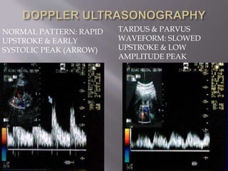
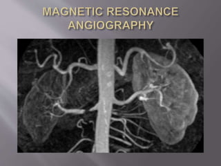
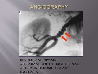
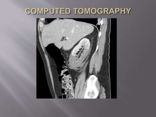
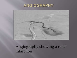
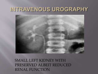
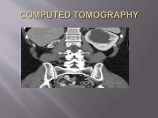
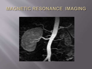
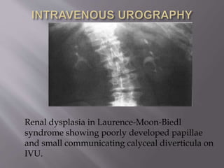
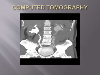
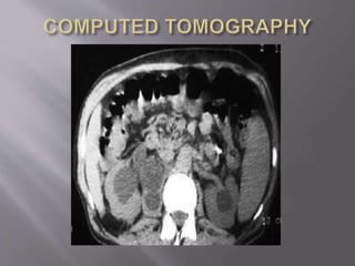
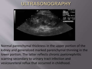
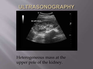
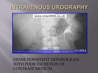
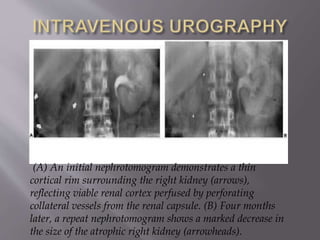
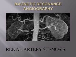
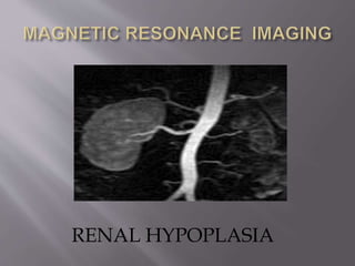
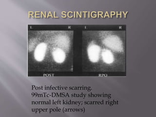
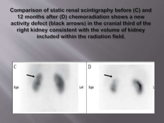
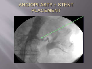
BY
DR.MAIMUNA A. HALLIRU
RADIOLOGY DEPARTMENT
AMINU KANO TEACHING HOSPITAL
02-09-2013
 INTRODUCTION
 CAUSES OF UNILATERAL SMALL KIDNEY
 ROLE OF IMAGING IN ESTABLISHING A
DIAGNOSIS
 SUMMARY/CONCLUSION
 DEFINITION OF UNILATERAL
SMALL KIDNEY
 PRE-RENAL/VASCULAR
 INTRA-RENAL/PARENCHYMAL
 POST-RENAL/COLLECTING SYSTEM
 RENAL ARTERY STENOSIS
NORMAL PATTERN: RAPID
UPSTROKE & EARLY
SYSTOLIC PEAK (ARROW)
TARDUS & PARVUS
WAVEFORM: SLOWED
UPSTROKE & LOW
AMPLITUDE PEAK
Delayed nephrogram on CT
CT- arterial phase :
Differential perfusion
DENSE PERSISTENT NEPHROGRAM
WITH POOR EXCRETION OF
CONTRAST MEDIUM
BILATERAL RENAL
ARTERY STENOSIS
BEADED, ANEURYSMAL
APPEARANCE OF THE RIGHT RENAL
ARTERY IN FIBROMUSCULAR
DYSPLASIA
RENAL INFARCTION
(A) An initial nephrotomogram demonstrates a thin
cortical rim surrounding the right kidney (arrows),
reflecting viable renal cortex perfused by perforating
collateral vessels from the renal capsule. (B) Four months
later, a repeat nephrotomogram shows a marked decrease in
the size of the atrophic right kidney (arrowheads).
C+ portal venous phase CT demonstrates a wedge of
poorly / non-enhancing renal parenchyma at the upper pole
CT demonstrates a non-enhancing
thrombus extending into the renal vein
Heterogeneous mass at the
upper pole of the kidney.
Angiography showing a renal
infarction
RADIATION
NEPHROPATHY
 CONGENITAL HYPOPLASIA
SMALL LEFT KIDNEY WITH
PRESERVED ALBEIT REDUCED
RENAL FUNCTION
RENAL DYSPLASIA
Renal dysplasia in Laurence-Moon-Biedl
syndrome showing poorly developed papillae
and small communicating calyceal diverticula on
IVU.
 POST-INFECTIVE ATROPHY
Post infective scarring.
99mTc-DMSA study showing
normal left kidney; scarred right
upper pole (arrows)
 REFLUX NEPHROPATHY
Demonstrates bilateral diffuse calyceal
clubbing (arrows) and deformity
accompanied by thinning of the
adjacent renal parenchyma
(arrowheads)
Normal parenchymal thickness in the upper portion of the
kidney and generalized marked parenchymal thinning in the
lower portion. The latter reflects chronic pyelonephritic
scarring secondary to urinary tract infection and
vesicoureteral reflux that occurred in childhood.
 POST-OBSTRUCTIVE
ATROPHY
 CONVENTIONAL RADIOGRAPHY
ULTRASONOGRAPHY:
B-MODE & DOPPLER
Normal parenchymal thickness in the upper portion of the
kidney and generalized marked parenchymal thinning in the
lower portion. The latter reflects chronic pyelonephritic
scarring secondary to urinary tract infection and
vesicoureteral reflux that occurred in childhood.
Heterogeneous mass at the
upper pole of the kidney.
NORMAL PATTERN: RAPID
UPSTROKE & EARLY
SYSTOLIC PEAK (ARROW)
TARDUS & PARVUS
WAVEFORM: SLOWED
UPSTROKE & LOW
AMPLITUDE PEAK
 INTRAVENOUS UROGRAPHY
DENSE PERSISTENT NEPHROGRAM
WITH POOR EXCRETION OF
CONTRAST MEDIUM
(A) An initial nephrotomogram demonstrates a thin
cortical rim surrounding the right kidney (arrows),
reflecting viable renal cortex perfused by perforating
collateral vessels from the renal capsule. (B) Four months
later, a repeat nephrotomogram shows a marked decrease in
the size of the atrophic right kidney (arrowheads).
SMALL LEFT KIDNEY WITH
PRESERVED ALBEIT REDUCED
RENAL FUNCTION
 COMPUTED
TOMOGRAPHY/CTA
NARROWED SEGMENT OF LEFT MAIN
RENAL ARTERY
COMPUTED TOMOGRAPHY
CORTICAL DEFECTS ON CECT IN RENAL
INFARCTION
RENAL SCARRING IN REFLUX
NEPHROPATHY
 MAGNETIC RESONANCE
IMAGING/ MRA
RENAL ARTERY STENOSIS
RENAL HYPOPLASIA
RENAL SCINTIGRAPHY
Post infective scarring.
99mTc-DMSA study showing
normal left kidney; scarred right
upper pole (arrows)
 DIGITAL SUBTRACTED
ANGIOGRAPHY
BILATERAL RENAL
ARTERY STENOSIS
Angiography showing a renal
infarction
 CONCLUSION/SUMMARY

Unilateral small kidney