This document discusses the different types of hypersensitivity reactions (allergies) as classified by Gell and Coombs in 1968. It describes the five main types of hypersensitivity: Type I mediated by IgE antibodies; Type II involving antibody-dependent cytotoxicity; Type III caused by immune complex deposition; Type IV mediated by T-cells; and Type V which stimulates cells rather than destroying them. Each type is characterized by its antigen, antibodies or cells involved, time course, and examples of diseases associated with that type of hypersensitivity reaction.
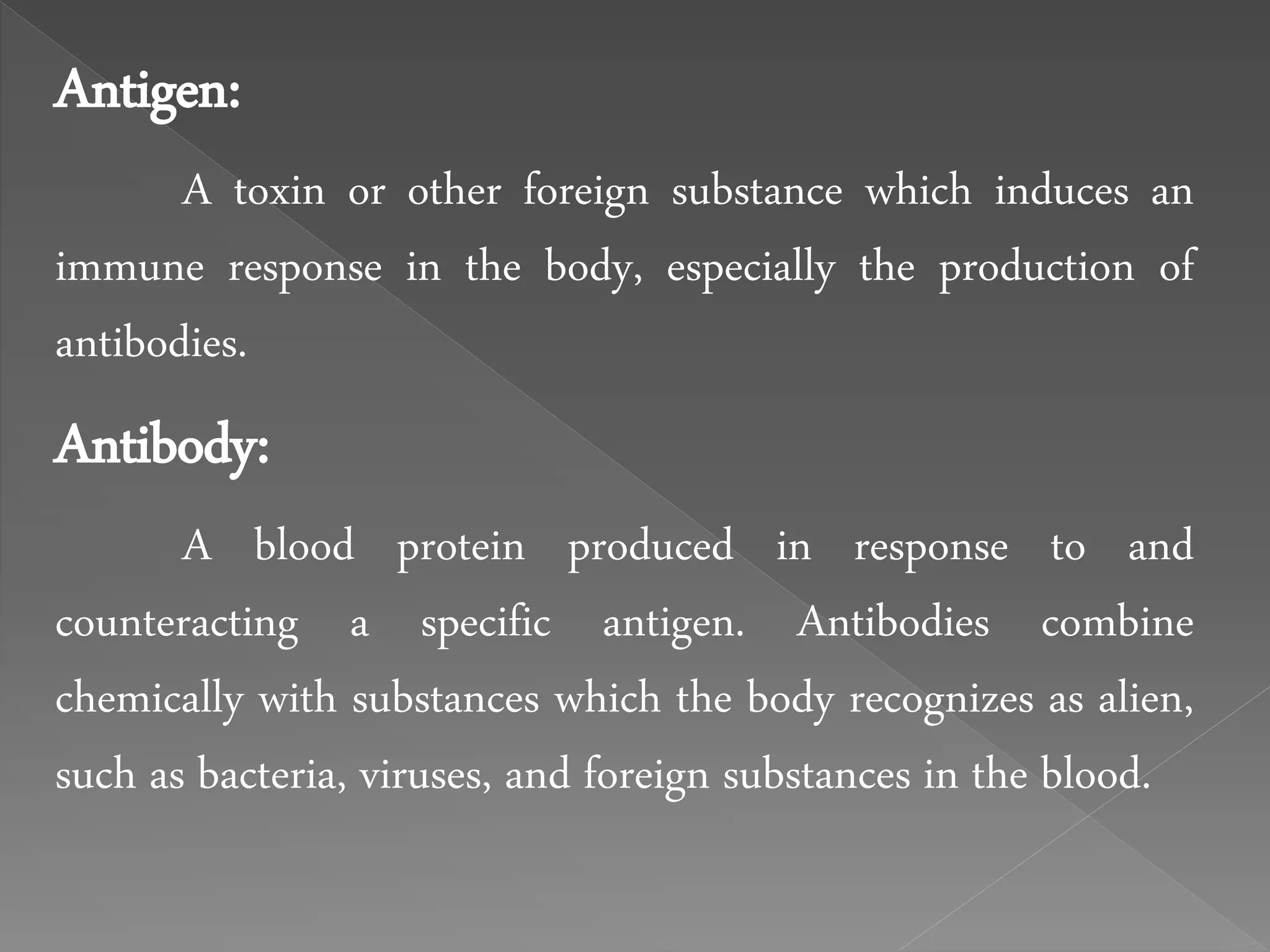
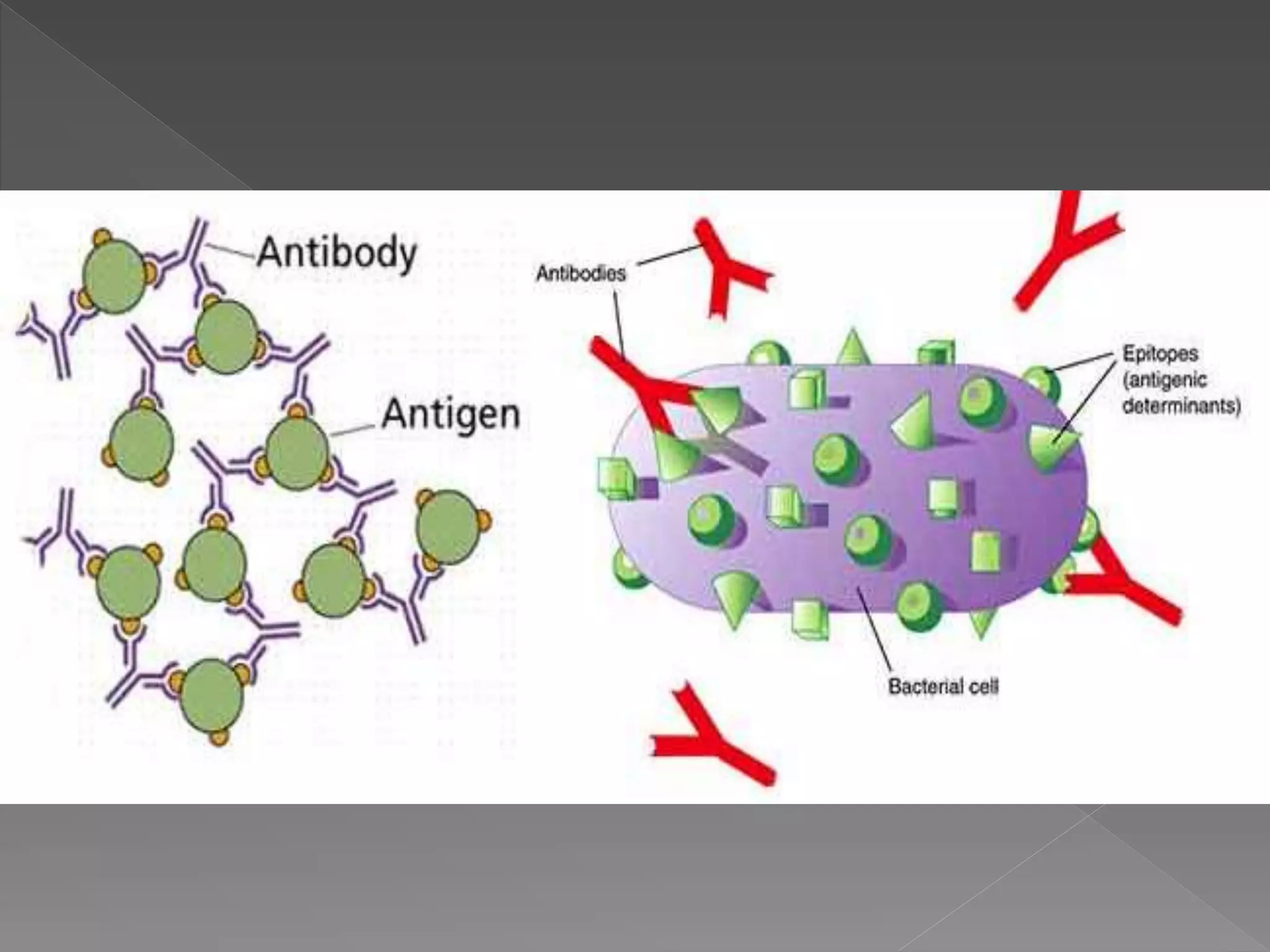
Antigen:
A toxin orother foreign substance which induces an
immune response in the body, especially the production of
antibodies.
Antibody:
A blood protein produced in response to and
counteracting a specific antigen. Antibodies combine
chemically with substances which the body recognizes as alien,
such as bacteria, viruses, and foreign substances in the blood.
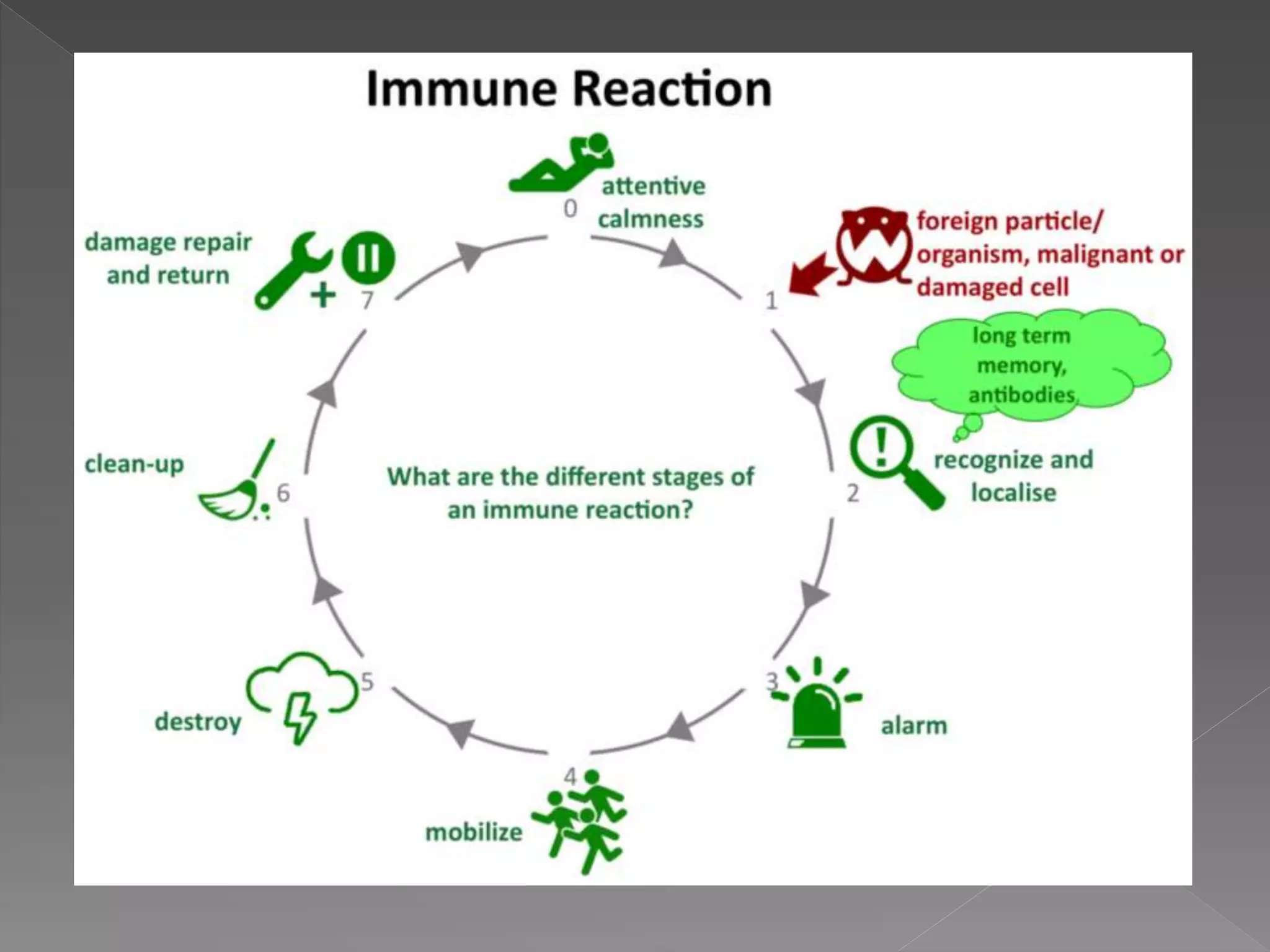
5.
A bodily responseto an antigen that occurs when
lymphocytes identify the antigenic molecule as foreign and
induce the formation of antibodies and lymphocytes capable
of reacting with it and rendering it harmless. called a immune
reaction.
7.
The inductionstate of excessive immune response with resulting
damage to normal tissues of body when exposed to an apparently
(directly) innocuous (harmless) antigens.
In this stage the individual more sensitive to antigen exposure.
8.
The antigenicsubstance which induce allergic reaction is
called allergen.
As the individual that are expose to normal antigen are
said to be immunized, individuals expose to the allergens
are said to be sensitized.
9.
When theimmune reaction manifest in a short duration of time,
within minutes, the hypersensitivity is called immediate type.
Most of the hypersensitivity to drugs like penicillin belong to this
category.
Salient feature:
Reaction appear and disappears rapidly.
Involves interaction of antigen and antibody.
Handle by B cells by the production of antibodies.
It produces urticaria,wheal and granulocyte accumulation.
10.
When theimmune reaction manifest slowly after 24 hours to 72
hours, the hypersensitivity is called delayed hypersensitivity.
Salient feature:
Appears slowly and lasts longer.
It produces erythema, induration and lymphocyte infiltration.
It involves reaction between antigens and T-cells.
desensitization cannot be easily done by drugs.
It is suppressed by corticosteroids.
11.
In 1968,P.G.H.Gelland R.R.A.Coombs proposed a classification of
immunopathological hypersensitivity reactions into four distinct
categories .
Type I (IgE-mediated/Anaphylactic Hypersensitivity)
Type II (Antibody Dependent Cytotoxic Hypersensitivity)
Type III (Immune Complex Mediated Hypersensitivity)
Type IV (Cell-Mediated Delayed Hypersensitivity)
Type V (Stimulatory Hypersensitivity).
12.
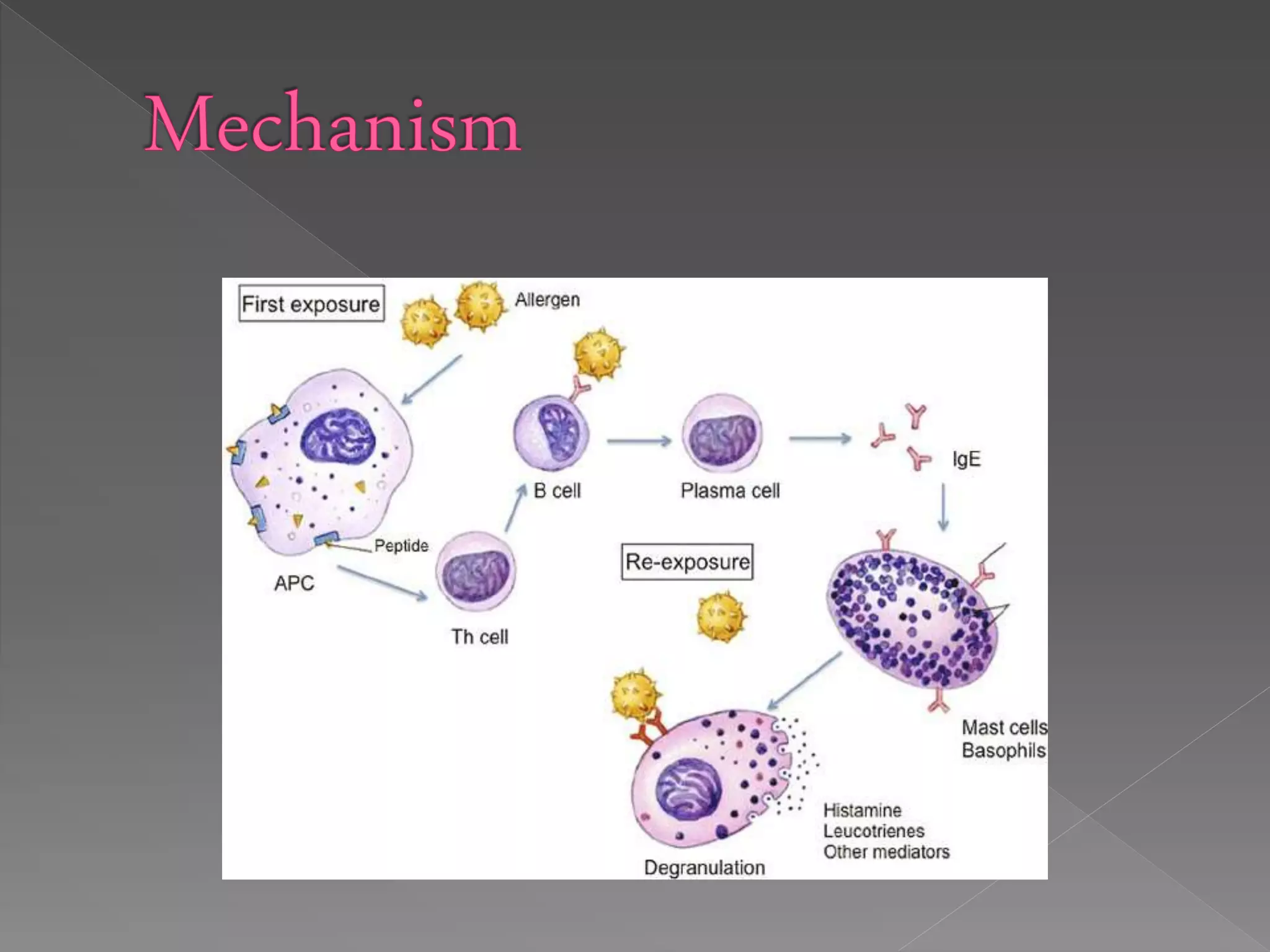
It isalso known as immediate or anaphylactic hypersensitivity.
The reaction may involve skin (urticaria and eczema), eyes
(conjunctivitis),nasopharynx (rhinorrhea, rhinitis),bronchopulmonary
tissues (asthma) and gastrointestinal tract (gastroenteritis).
The reaction may cause from minor inconvenience to death.
The reaction takes 15-30 minutes from the time of exposure to the
antigen. Sometimes the reaction may have a delayed onset (10-12
hours).
13.
Immediate hypersensitivityis mediated by IgE.
The primary cellular component in this hypersensitivity is mast cell or basophil. The
reaction is amplified and/or modified by platelets, neutrophils and eosinophils.
The mechanism of reaction involves preferential production of IgE, in response to
certain antigens, allergens .
IgE has very high affinity for its receptor on mast cells and basophils.
A subsequent exposure to the same allergen cross links the cell-bound IgE and triggers
the release of various pharmacologically active Substances.
Cross-linking of IgE Fc-receptor is important in mast cell triggering.
Mast cell degranulation is preceded by increased Ca++ influx, which is a crucial process;
ionophores which increase cytoplasmic Ca++ also promote degranulation.
Anaphylaxis isa serious, life threatening allergic reaction. The
most common anaphylactic reactions are to foods, insect
stings, medications and latex.
This reaction typically affects more than one part of the body at
the same time.
Anaphylaxis requires immediate medical treatment, including a
prompt injection of epinephrine.
18.
Pollen, dander,dust mites, certain
foods, or chemical/physical
irritants.
Hives, welts, scaling or other
signs of skin irritation, Itching of
the eyes, nose or skin, Redness of
the eyes, A runny nose, Sinus
pain and/or swelling, Sneezing.
19.
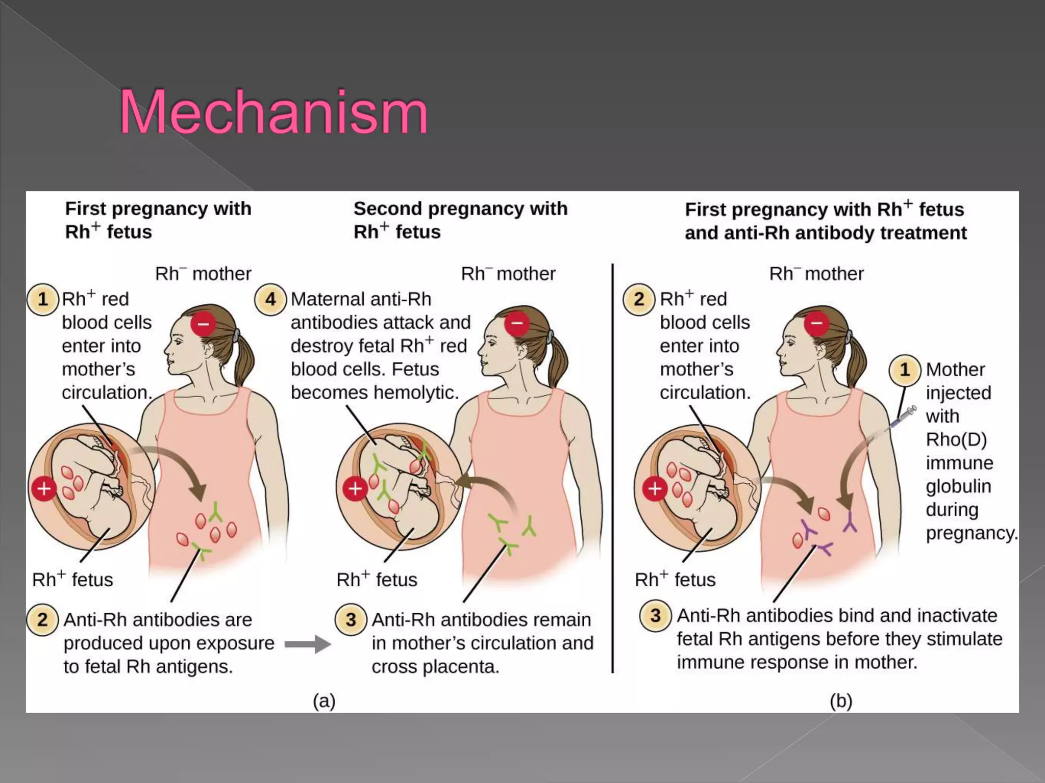
It isprimarily mediated by antibodies of IgM or IgG class and
complement.
Phagocytes and K cells may also play a role.
The reaction time is minutes to hours.
The antigens are normally endogenous, although exogenous chemicals
(haptens) which can attach to cell membranes can also lead to type II
hypersensitivity.
It ismediated by soluble immune complexes. They are mostly of IgG
class, although IgM may also be involved.
The reaction may be general (e.g., serum sickness) or may involve
individual organs including skin (e.g., systemic lupus erythematosus,
Arthus reaction), kidneys (e.g., lupus nephritis), lungs (e.g.,
aspergillosis), blood vessels (e.g., polyarteritis), joints (e.g., rheumatoid
arthritis) or other organs.
This reaction may be the pathogenic mechanism of diseases caused by
many microorganisms.
The reaction may take 3-10 hours after exposure to the antigen.
23.
The antigenis soluble and not
attached to the organ involved.
Primary components are soluble
immune complexes and
complement (C3a, 4a and 5a).
The damage is caused by platelets
and neutrophils.
The lesion contains primarily
neutrophils and deposits of
immune complexes and
complement.
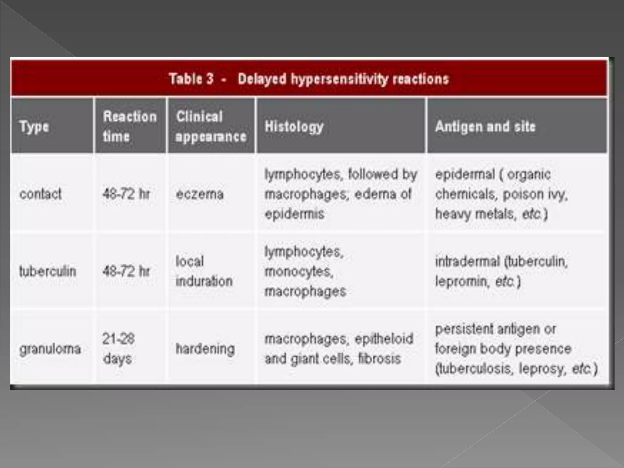
The classicalexample of this hypersensitivity is tuberculin (Montoux)
reaction which peaks 48 hours after the injection of antigen.
Delayed hypersensitivity include T lymphocytes and monocytes and/or
macrophages.
Cytotoxic T cells (Tc) cause direct damage whereas helper T (TH1) cells
secrete cytokines which activate cytotoxic T cells and recruit and activate
monocytes and macrophages, which cause the bulk of the damage .
26.
The delayedhypersensitivity lesions mainly contain monocytes and a
few T cells.
Major lymphokines involved in delayed hypersensitivity reaction
include monocyte chemotactic factor, interleukin-2,interferons, TNF,
etc.
Type IV hypersensitivity can be classified into three categories
depending on the time of onset and clinical and histological
presentation.
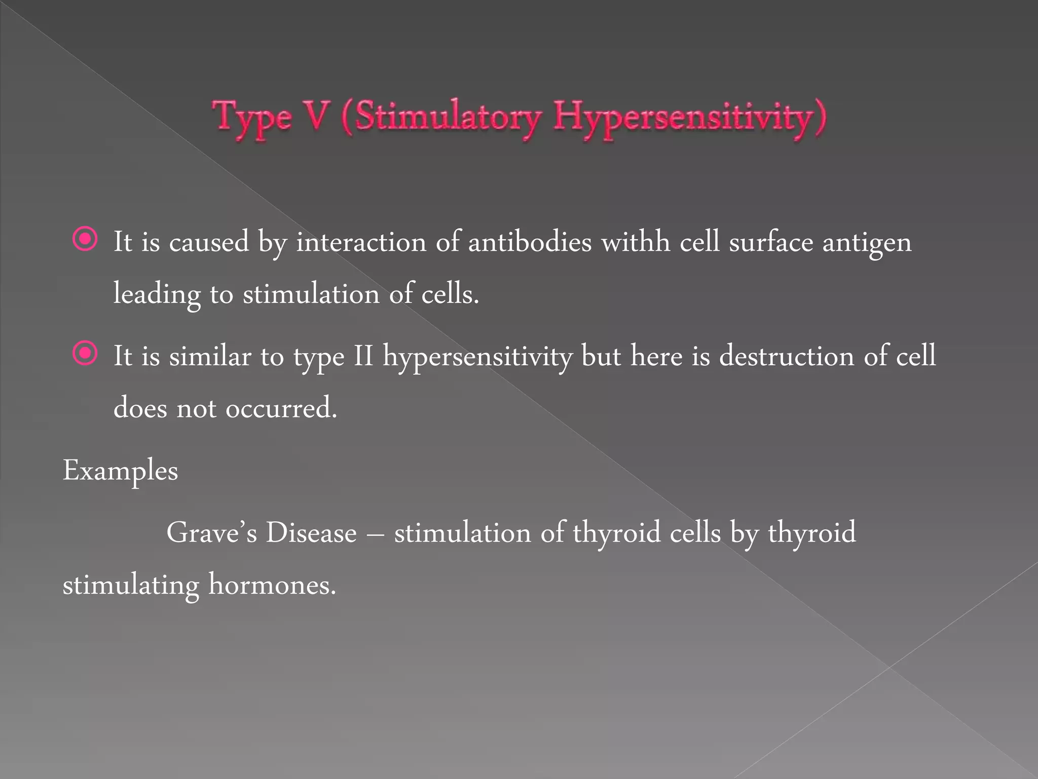
It iscaused by interaction of antibodies withh cell surface antigen
leading to stimulation of cells.
It is similar to type II hypersensitivity but here is destruction of cell
does not occurred.
Examples
Grave’s Disease – stimulation of thyroid cells by thyroid
stimulating hormones.