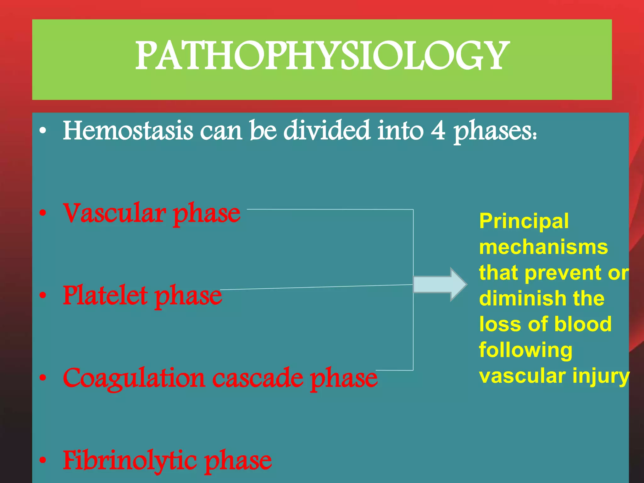
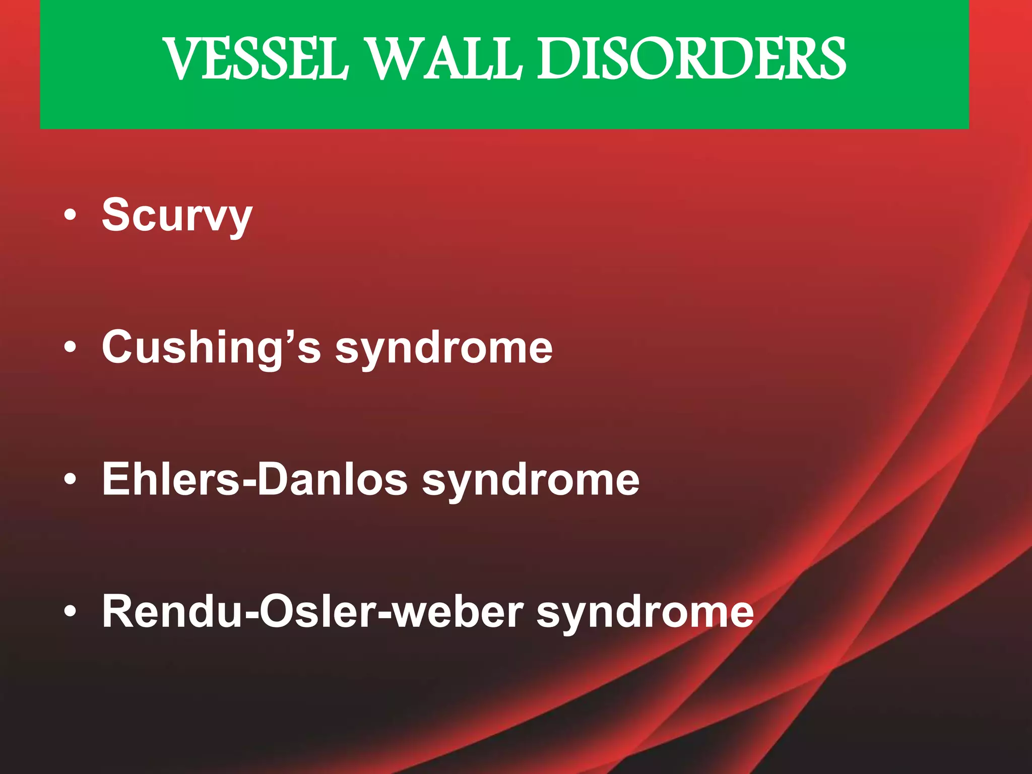
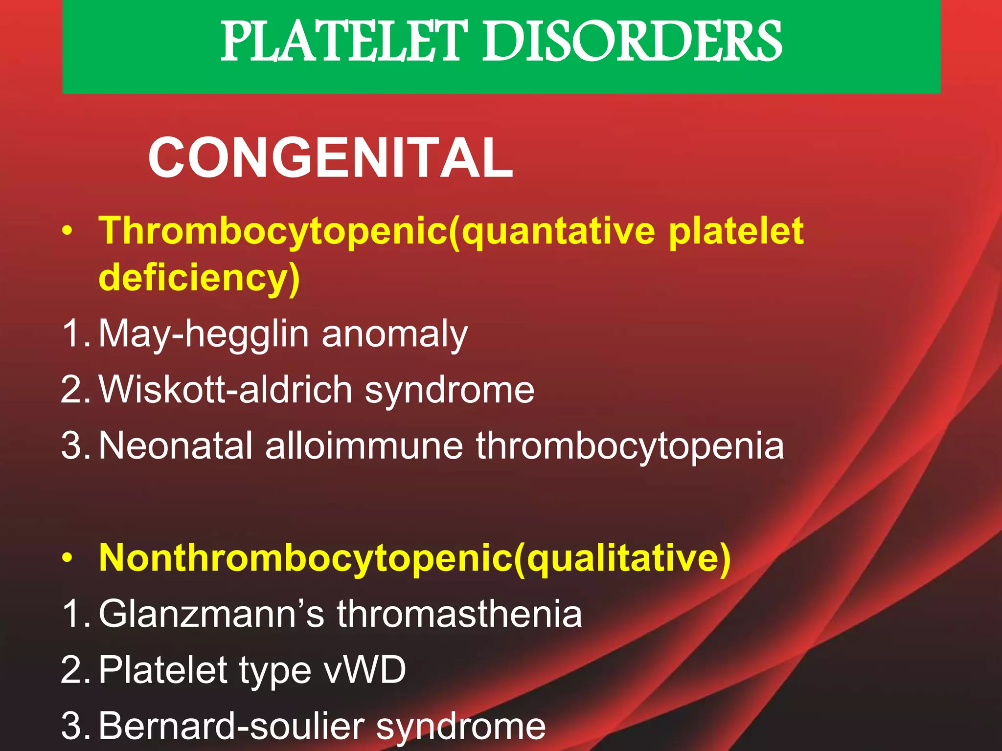
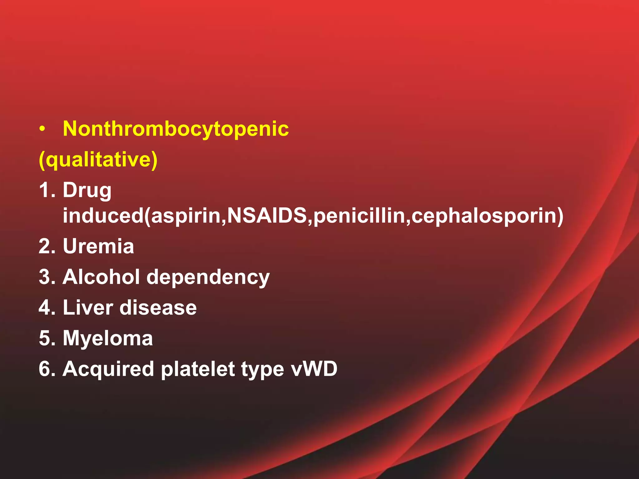
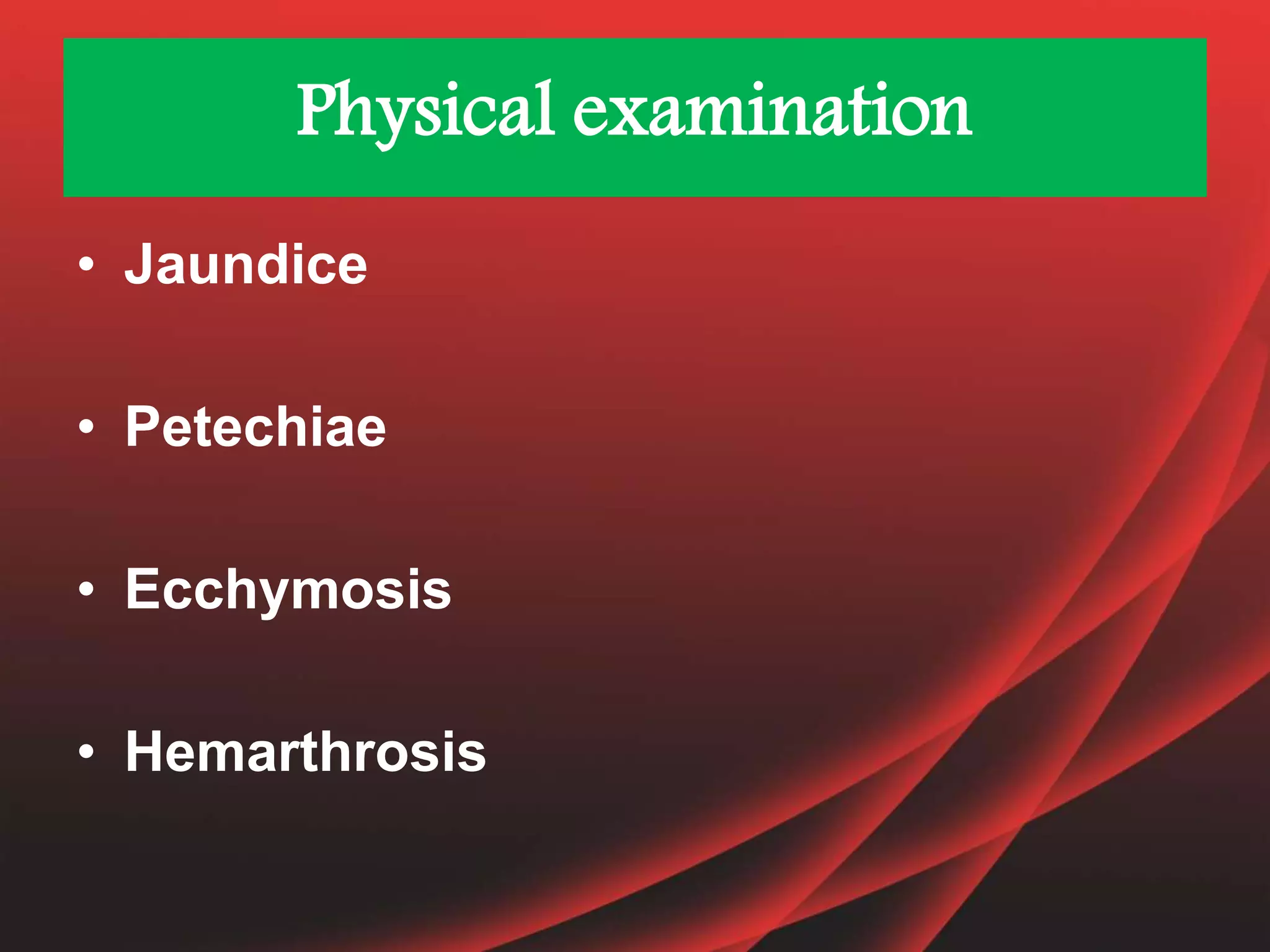
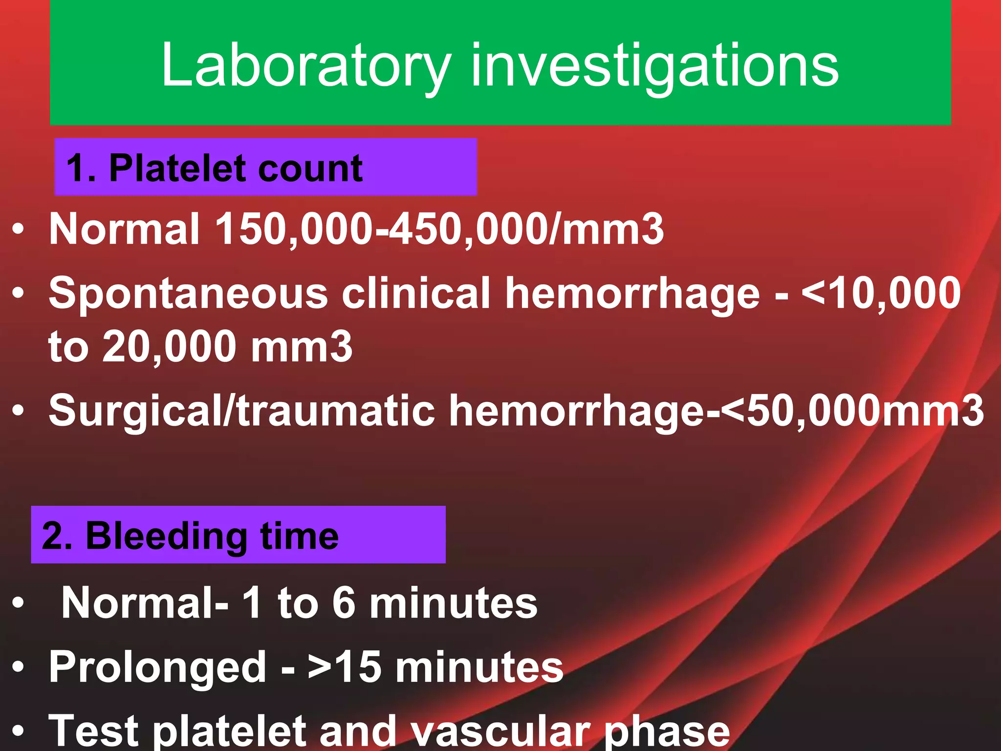
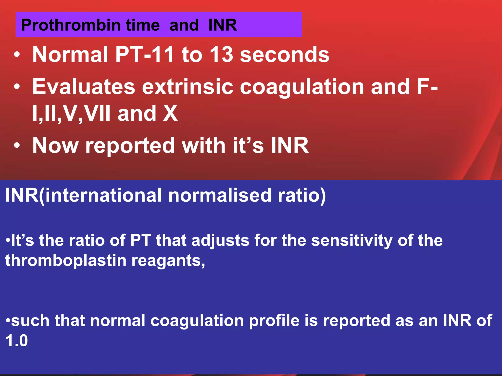
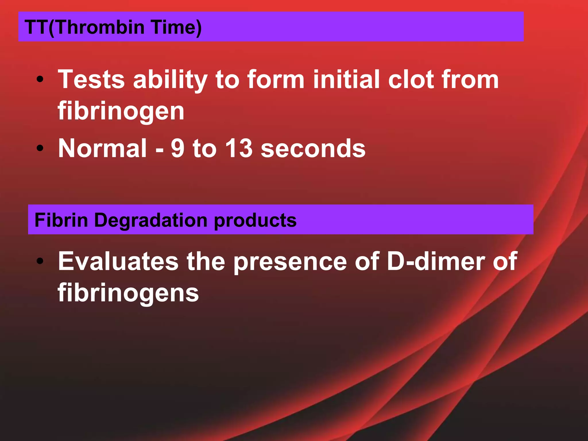
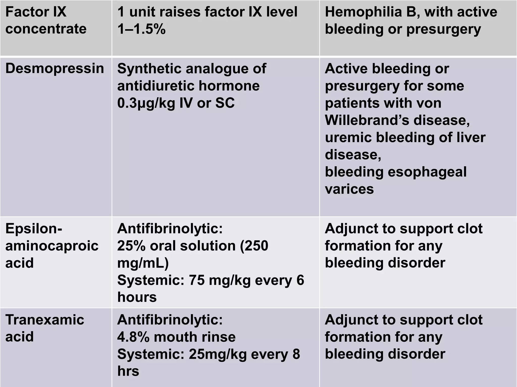
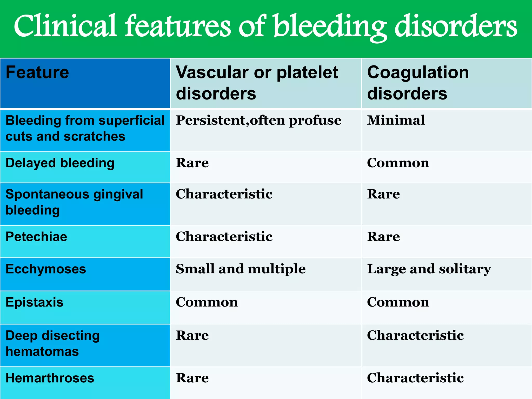
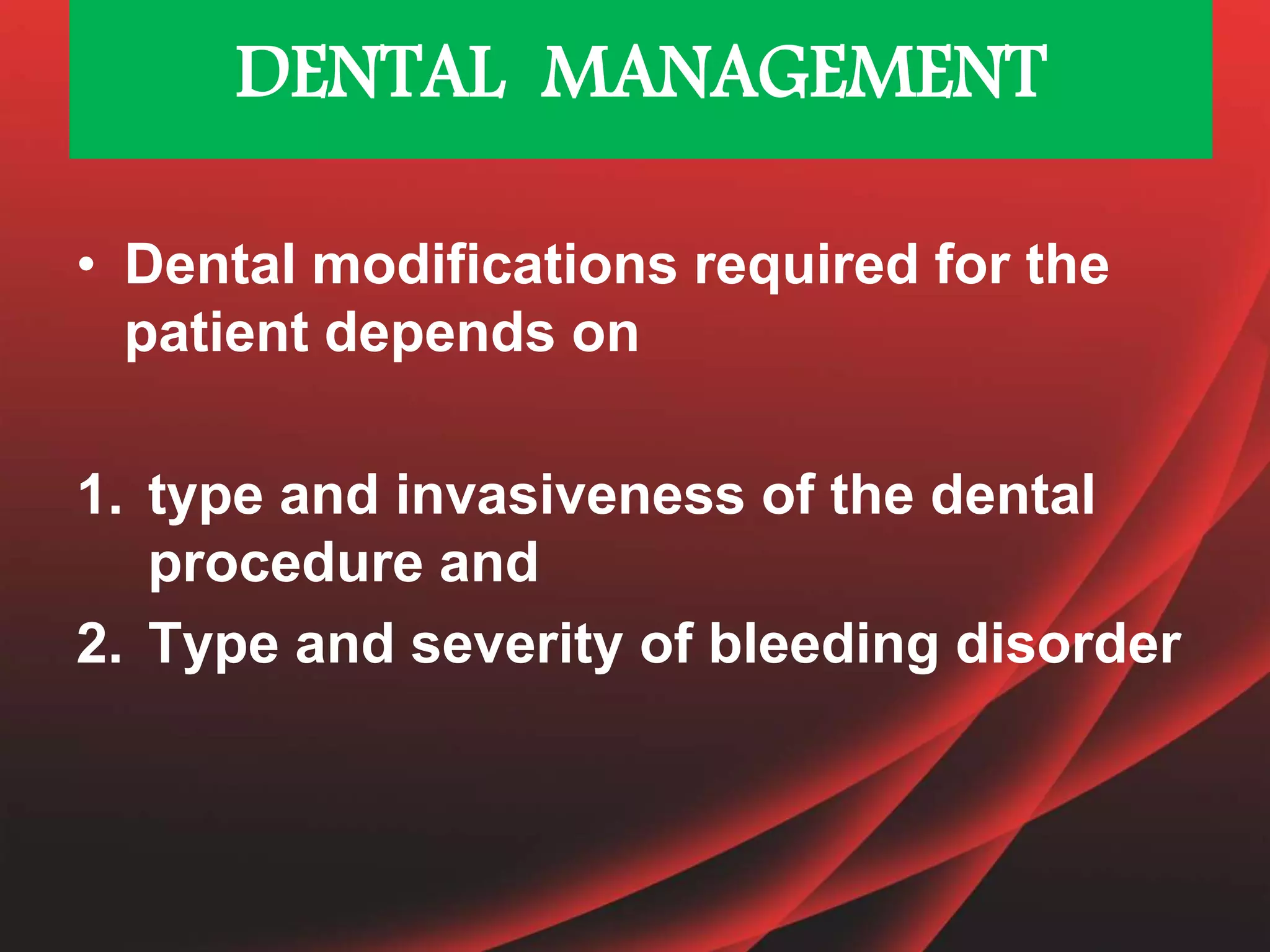
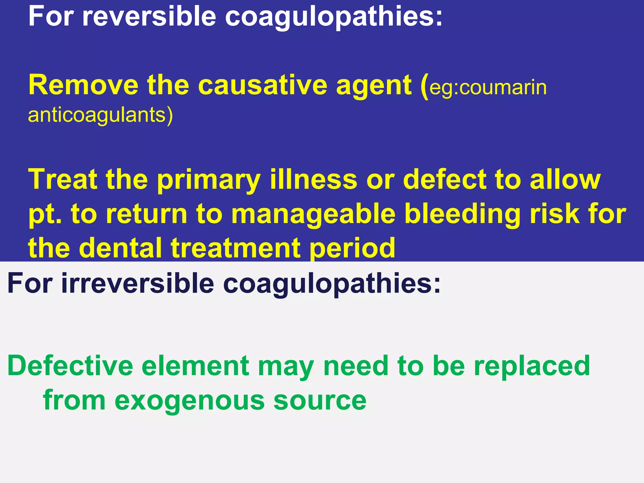
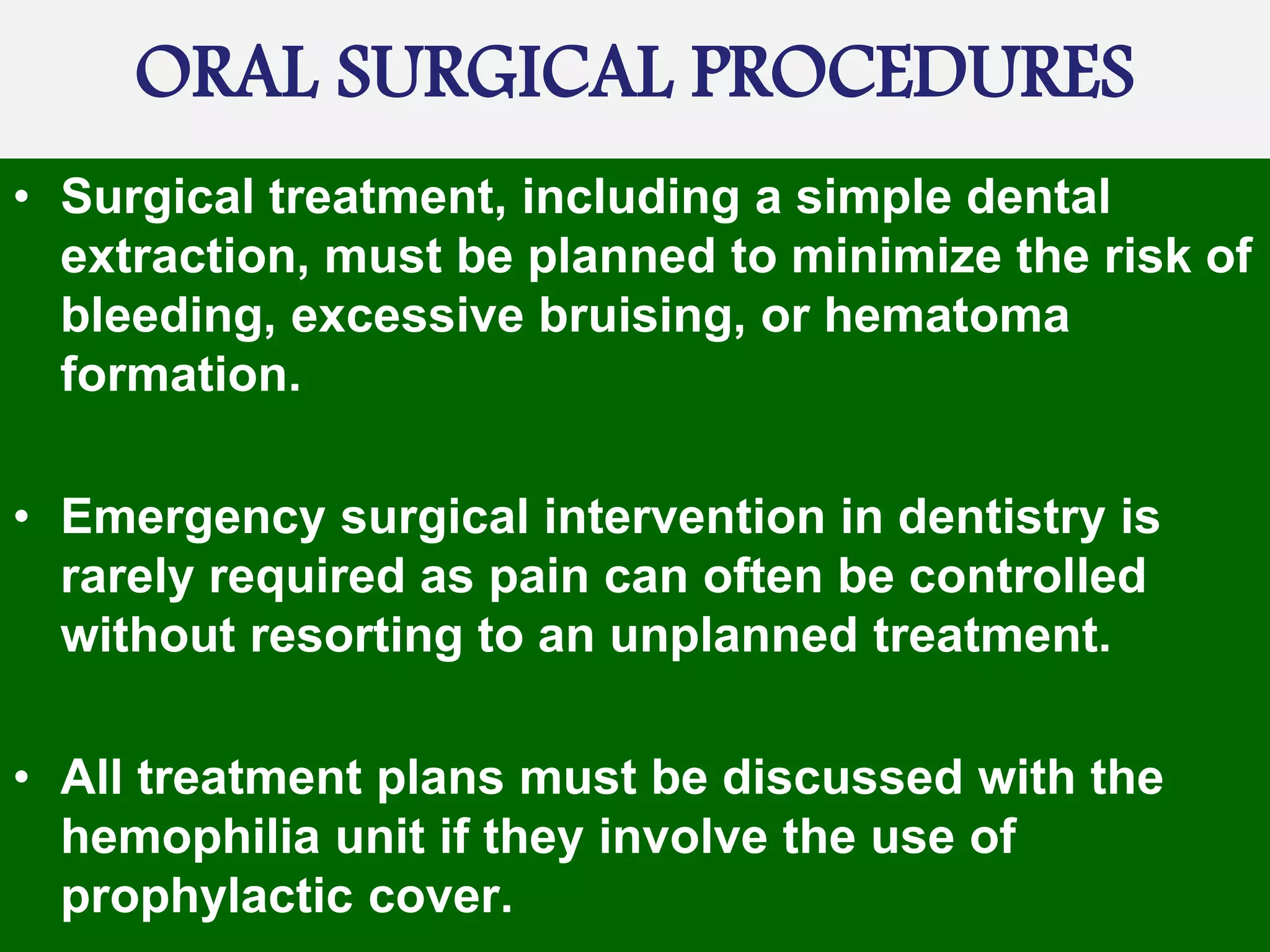
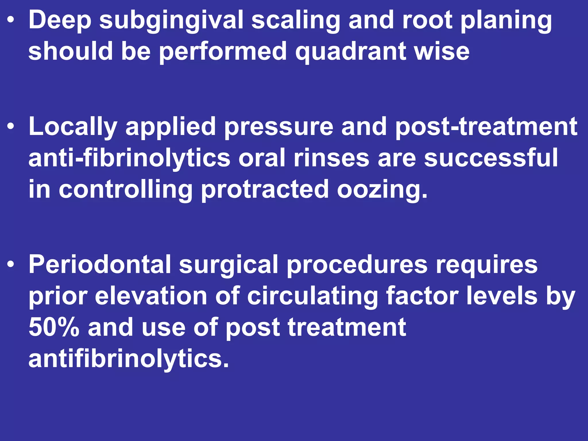
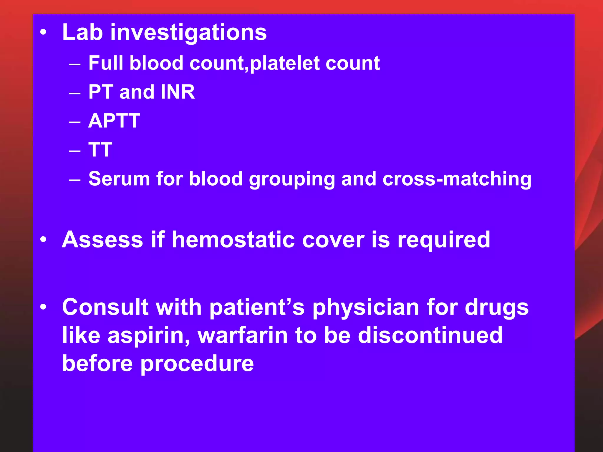
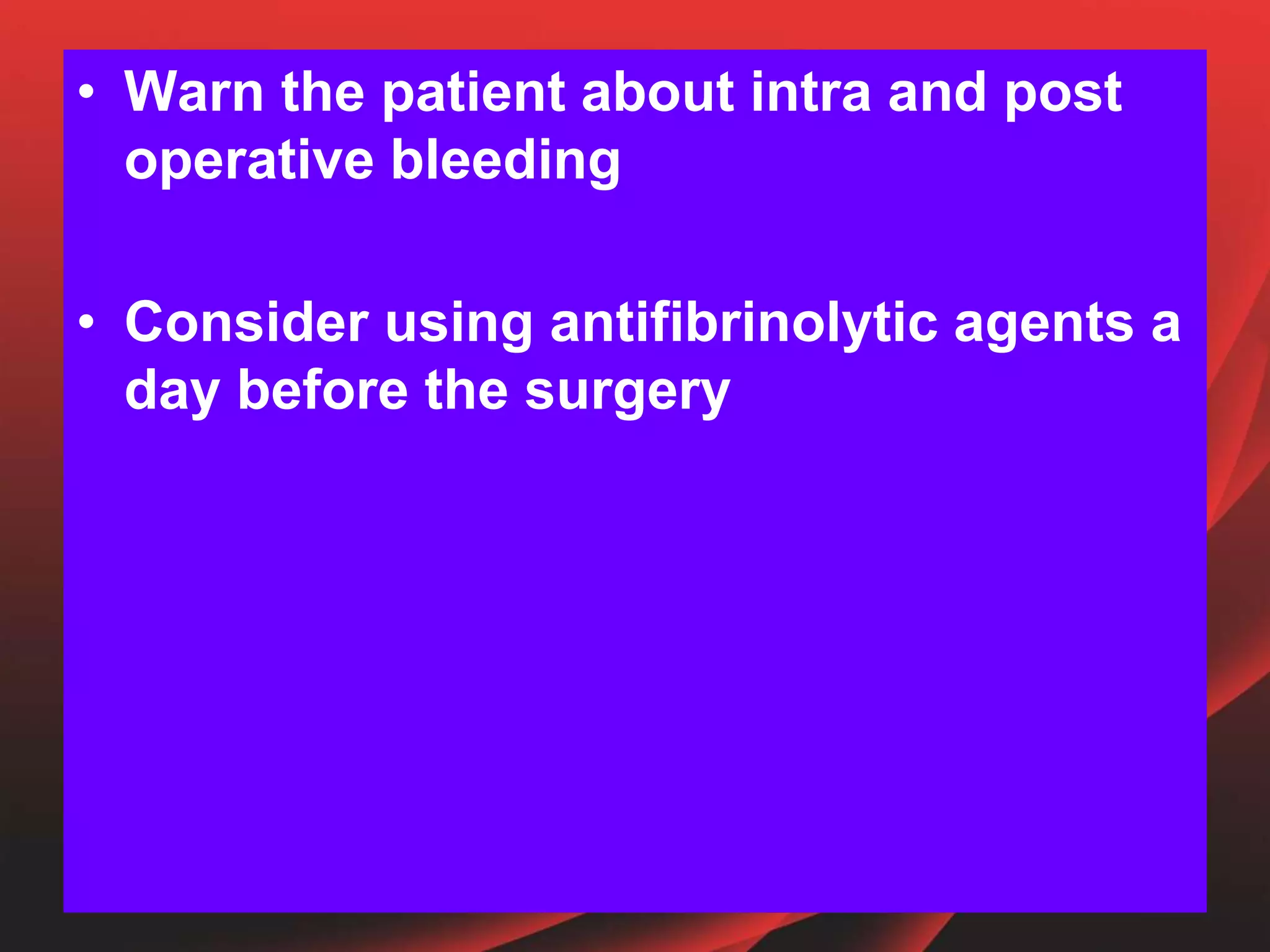
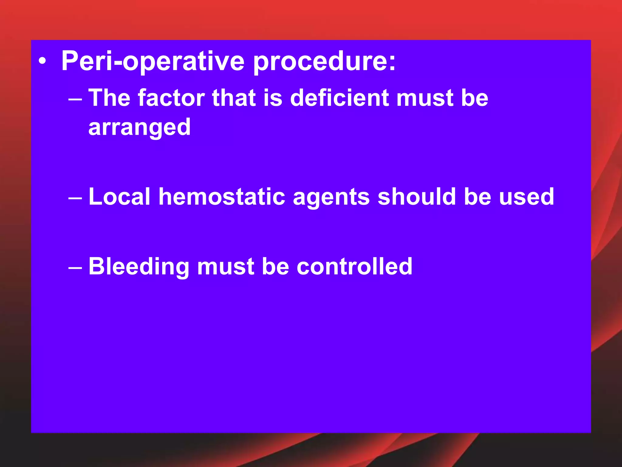
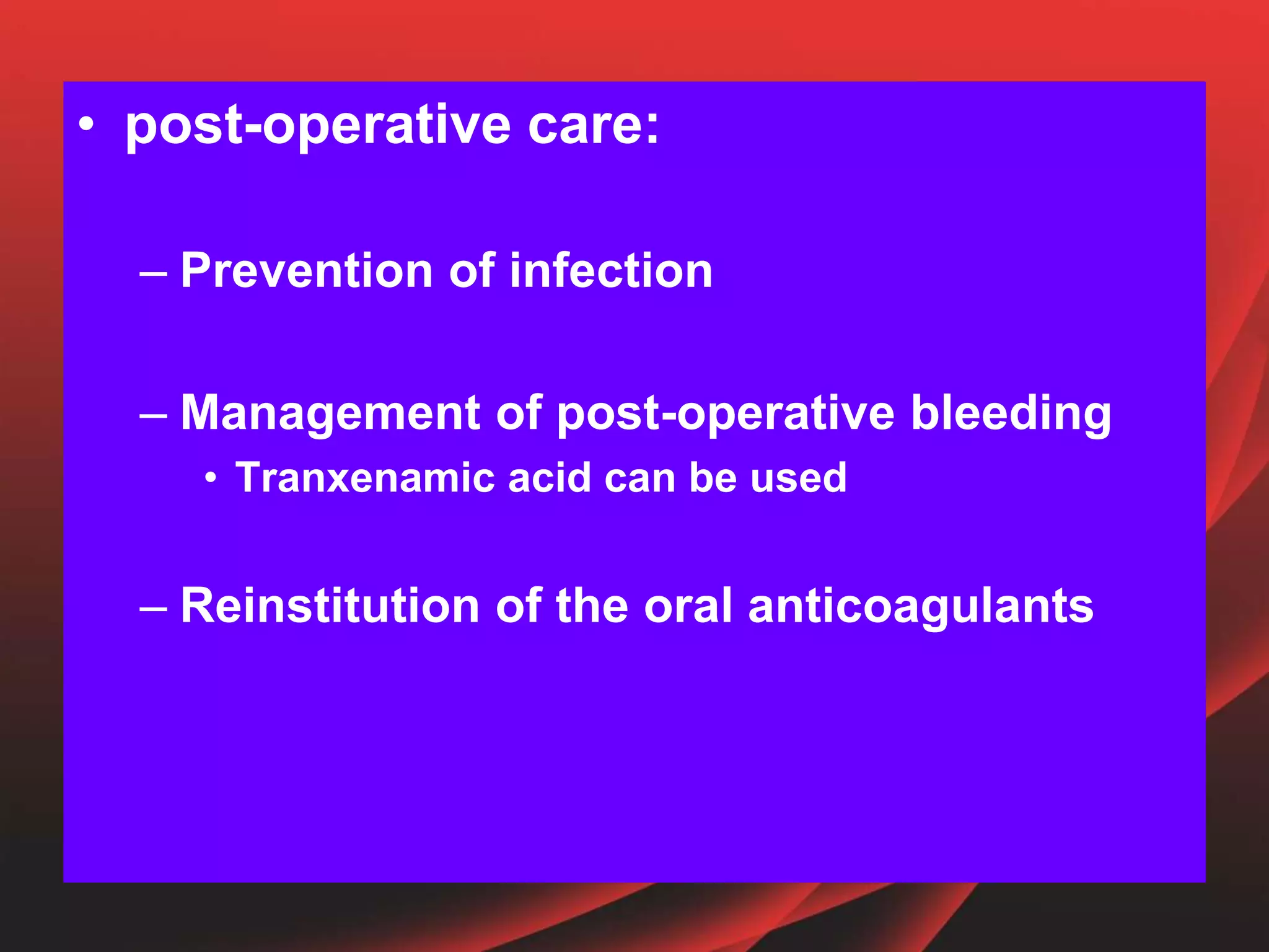
BLEEDING AND
CLOTTING
DISORDERS
RESOURCE FACULTY
DR.JYOTSNA RIMAL
Additional professor & HOD
DR.ICHHA KUMAR MAHARJAN
Associate professor
PRESENTER:
KASHMIRA POKHREL
483
BDS-2011
CONTENTS
• PATHOPHYSIOLOGY
• BLEEDING DISORDERS
• COAGULATION DISORDERS
• LABORATORY INVESTGATIONS
• ORAL MANIFESTATIONS
• DENTAL CONSIDERATION
• DENTAL MANAGEMENT
• CONCLUSION
PATHOPHYSIOLOGY
• Hemostasis can be divided into 4 phases:
• Vascular phase
• Platelet phase
• Coagulation cascade phase
• Fibrinolytic phase
Principal
mechanisms
that prevent or
diminish the
loss of blood
following
vascular injury
VASCULAR PHASE
Tissue injury
Vasoconstriction of the microvascular
bed
Serotonin, histamine,PG’s
PLATELET PHASE
Circulating platelets exposed to
vascular injury
Normal vWF, endothelial cells,
collagen, basement membrane, elasti
microfibrils and other cellular debris
Physical and chemical changes
Aggregation of
platelet
Primary platelet plug (adheres to basement membrane)
Increase in size of plug
Activates F-X
Conversion of
prothrombin to thrombin
Platelet intermixed with other cellular
components(RBC,WBC) which further contracts to
reduce bleeding and seal vascular bed
COAGULATION CASCADE
FIBRINOLYTIC PHASE
• VESSEL WALL DISORDERS
• PLATELET DISORDERS
• COAGULATION DISORDERS
CLASSIFICATION
• Scurvy
• Cushing’s syndrome
• Ehlers-Danlos syndrome
• Rendu-Osler-weber syndrome
VESSEL WALL DISORDERS
• Thrombocytopenic(quantative platelet
deficiency)
1.May-hegglin anomaly
2.Wiskott-aldrich syndrome
3.Neonatal alloimmune thrombocytopenia
• Nonthrombocytopenic(qualitative)
1.Glanzmann’s thromasthenia
2.Platelet type vWD
3.Bernard-soulier syndrome
CONGENITAL
PLATELET DISORDERS
ACQUIRED
• Thrombocytopenic(quantitative)
1. Autoimmune or idiopathic
thrombocytopenia purpura
2. Thrombotic thrombocytopenia
purpura
3. Cytotoxic chemotherapy
4. Drug-induced (eg, quinine,
quinidine, gold salts,
trimethoprim/ sulfamethoxazole,
rifampin)
5. .Leukemia
6.Aplastic anemia
7.Myelodysplasia
8.Systemic lupus
erythematosus
9.Associated with
infection: HIV,
mononucleosis, malaria
10.Disseminated
intravascular
coagulation
• Nonthrombocytopenic
(qualitative)
1. Drug
induced(aspirin,NSAIDS,penicillin,cephalosporin)
2. Uremia
3. Alcohol dependency
4. Liver disease
5. Myeloma
6. Acquired platelet type vWD
• CONGENITAL COAGULOPATHIES
1. Hemophilia A&B
2. Factors deficiency
3. von Willebrand’s disease
• ANTICOAGULANT -RELATED COAGULOPATHIES
1. Heparin
2. Coumarin
• DISEASE-RELATED COAGULOPATHIES
1. Liver disease
2. Vitamin K deficiency
3. Disseminated intravascular coagulation
4. Fibrinolytic disorders
COAGULATION DISORDERS
• History taking
• Physical examination
• Laboratory Investigations
• Observation
IDENTIFICATION OF PATIENT
WITH BLEEDING DISORDER
HISTORY TAKING
H/O frequent
• epistaxis,
• spontaneous gingival and mucosal bleeding,
• prolonged bleeding from superficial cuts
• Excessive menstrual flow
• Easy bruising
• hematuria
CONTD….
• Family history
• Past H/O bleeding after surgical procedures
• Identification of medicine(heparin, aspirin,
NSAIDS,coumarin, cytotoxic chemotherapy)
• Active medical conditions (hepatitis,cirrhosis,renal
disease, hematological malignancy,
thrombocytopenia)
• H/O heavy alcohol intake
Physical examination
• Jaundice
• Petechiae
• Ecchymosis
• Hemarthrosis
Laboratory investigations
• Normal 150,000-450,000/mm3
• Spontaneous clinical hemorrhage - <10,000
to 20,000 mm3
• Surgical/traumatic hemorrhage-<50,000mm3
• Normal- 1 to 6 minutes
• Prolonged - >15 minutes
• Test platelet and vascular phase
1. Platelet count
2. Bleeding time
Prothrombin time and INR
• Normal PT-11 to 13 seconds
• Evaluates extrinsic coagulation and F-
I,II,V,VII and X
• Now reported with it’s INR
INR(international normalised ratio)
•It’s the ratio of PT that adjusts for the sensitivity of the
thromboplastin reagants,
•such that normal coagulation profile is reported as an INR of
1.0
aPTT(activated partial thromboplastin time)
• Activator – rare earth
• Measures effectiveness of the intrinsic
pathway
• Considered normal if the control aPTT
& test aPTT are within 10 secs of each
other.
• Control aPTT = 15-35secs.
• It is altered in hemophilias A & B. and
with the use of heparin.
• Tests ability to form initial clot from
fibrinogen
• Normal - 9 to 13 seconds
• Evaluates the presence of D-dimer of
fibrinogens
TT(Thrombin Time)
Fibrin Degradation products
• Normal-60 to 150%
• Torniquet test to assess Rumpel-leede
phenomenon
Factor assays
Tests of capillary Fragility
Bleeding Disorder Platelet
Count
PT/ INR aPTT BT
Thrombocytopenia
Leukemia
F VIII, IX, XI deficiency
Heparin anticoagulation
F II, V, X deficiency
Vitamin K deficiency
Intestinal malabsorption
F VII deficiency
Coumarin anticoagulation
Liver disease
von Willebrand’s disease
DIC
Severe liver disease
F XIII deficiency
Vascular wall defect
↓
N
N
N
N, ↓
↓
N
N
N
N
↑
↑
N
↑
N
N
N
↑
↑
N
N, ↑
↑
N
N
↑
N
N
N
↑
↑
N
↑
Principal Agents for Systemic Management
Agent Description Indications
Platelets 1 unit = 50 mL; may raise
count by 6,000 Platelet
count
• < 10,000 in non bleeding
individuals
• < 50,000 presurgical level
• < 50,000 in actively
bleeding individuals
Fresh frozen
plasma
1 unit = 150–250 mL
Contains factors II, VII, IX,
X, XI, XII, XIII and heat-
labile V and VII
• Undiagnosed bleeding
disorder with active bleeding
• Severe liver disease
Cryo-
precipitate
1 unit = 10–15 mL • Hemophilia A
• von Willebrand’s disease,
• when factor concentrates
and DDAVP are unavailable
• Fibrinogen deficiency
Factor VIII
concentrate
1 unit raises factor VIII
level 2%
• Hemophilia A with active
bleeding
• Presurgery
Factor IX
concentrate
1 unit raises factor IX level
1–1.5%
Hemophilia B, with active
bleeding or presurgery
Desmopressin Synthetic analogue of
antidiuretic hormone
0.3μg/kg IV or SC
Active bleeding or
presurgery for some
patients with von
Willebrand’s disease,
uremic bleeding of liver
disease,
bleeding esophageal
varices
Epsilon-
aminocaproic
acid
Antifibrinolytic:
25% oral solution (250
mg/mL)
Systemic: 75 mg/kg every 6
hours
Adjunct to support clot
formation for any
bleeding disorder
Tranexamic
acid
Antifibrinolytic:
4.8% mouth rinse
Systemic: 25mg/kg every 8
hrs
Adjunct to support clot
formation for any
bleeding disorder
Local hemostatic agents
• ABSORBANT GELATIN SPONGE
(GELFOAM)
 Dental size - 20x20x7mm3
• OXIDISED CELLULOSE
(SURGICEL)
• TOPICAL THROMBIN
• Obtained from bovine plasma
• Applied as dry powder or freshly prepared
solution
• TRANXENAMIC ACID
• 500mg tablets
• 1000mg/10ml injection
• Epsilon Amino caproic Acid
• 50mg/kg
• Oral rinse 250mg/ml
• Fibrin sealants/Fibrin glue
• Cryoprecipitate
• 10,000units thrombin powder
• 10ml saline
• 10ml Calcium Chloride
• Application of local pressure
• suture
Clinical features of bleeding disorders
Feature Vascular or platelet
disorders
Coagulation
disorders
Bleeding from superficial
cuts and scratches
Persistent,often profuse Minimal
Delayed bleeding Rare Common
Spontaneous gingival
bleeding
Characteristic Rare
Petechiae Characteristic Rare
Ecchymoses Small and multiple Large and solitary
Epistaxis Common Common
Deep disecting
hematomas
Rare Characteristic
Hemarthroses Rare Characteristic
Oral manifestations
• Petechiae
• Ecchymoses
• Spontaneous gingival bleeding
Contd….
• Brown colored teeth due to
depositsof hemosiderin as a
result of continous long term
bleeding.
• Hemarthrosis (rarely)
DENTAL MANAGEMENT
• Dental modifications required for the
patient depends on
1. type and invasiveness of the dental
procedure and
2. Type and severity of bleeding disorder
• For reversible coagulopathies:
Remove the causative agent (eg:coumarin
anticoagulants)
Treat the primary illness or defect to allow
pt. to return to manageable bleeding risk for
the dental treatment period
For irreversible coagulopathies:
Defective element may need to be replaced
from exogenous source
Consultation with hematologist
This may involve treatment
either in specialized hospital
facilities or local general
dentist’s office
PLATELET DISORDERS
• Platelet level >50,000mm3 required prior to
surgical procedures.
• Avoidance of aspirin therapy recommended
1 week prior to extensive oral surgical
procedures
• Aspirin is rarely witheld in case of minor
oral surgical procedures such as extraction
where local hemostatic agents can be use
• When extensive surgery in emergency is
indicated DDAVP can be used
• DDAVP decreases the aspirin induced
prolongation of BT and prevents post
operative oozing
Considerations in
HEMOPHILIA A and B
and
vWD
ORAL SURGICAL PROCEDURES
• Surgical treatment, including a simple dental
extraction, must be planned to minimize the risk of
bleeding, excessive bruising, or hematoma
formation.
• Emergency surgical intervention in dentistry is
rarely required as pain can often be controlled
without resorting to an unplanned treatment.
• All treatment plans must be discussed with the
hemophilia unit if they involve the use of
prophylactic cover.
PREVENTIVE AND
PERIODONTAL THERAPY
• Periodontal probing and supragingival
scaling can be done routinely
• Severly inflamed and swollen tissues are
best treated initially with chlorhexidine oral
rinses and gross debridement with hand
instruments to allow gingival shrinkage
• Deep subgingival scaling and root planing
should be performed quadrant wise
• Locally applied pressure and post-treatment
anti-fibrinolytics oral rinses are successful
in controlling protracted oozing.
• Periodontal surgical procedures requires
prior elevation of circulating factor levels by
50% and use of post treatment
antifibrinolytics.
RESTORATIVE AND
PROSTHODONTIC THERAPY
• Rubber dam isolation advised
 to minimize the risk of lacerating soft
tissue and
avoid creating ecchymoses and hematomas
with high speed evacuators or saliva
ejectors
• Removable prosthodontic appliances can be
fabricated without complications
Denture trauma should be minimized
ENDODONTIC THERAPY
• Instrumentation should not extend beyond
apex
• Filling beyond the apical seal also should be
avoided
• Application of epinephrine intrapulpally to
apical area provides hemostasis
PEDIATRIC DENTAL THERAPY
• Administration of factor concentrate before
extraction
• Pulpotomies to be performed without
excessive pulpal bleeding
• Topical fluoride application
• Pit and fissure sealant
ORTHODONTIC THERAPY
• Care must be taken to avoid mucosal
laceration by orthodontic bands, brackets
and wires.
• Fixed orthodontic appliance prefered over
removable functional appliance
• Use of extraoral force and
• shorter treatment duration
PAIN CONTROL
• Selection of pain control method based on
patient’s pain threshold and invasiveness of
the procedure
• Hypnosis, IV diazepam, nitrous
oxide/oxygen analgesia can be used
• Anesthetic with vasoconstrictor should be
used when possible
• Hemostatic cover(20-30%) required for:
 inferior alveolar ,posterior superior
alveolar,infraorbital, lingual and long buccal
nerve block
As these injections place anesthetic solutions
in highly vascularised loose connective
tissue with no distinct boundaries where
formation of dissecting hematoma is
possible
• Hemostatic cover not required for:
Intrapulpal, periodontal ligament, gingival
papillary anesthesia
In mild disease-buccal, labial and palatal
infiltration for maxillary teeth can be
attempted slow injection and local
pressure for 3-4 minutes
PATIENTS ON
ANTICOAGULANTS
• Higher INR result in high bleeding risk
• Non surgical dental treatment can be
successfully accomplished without
alteration of anti coagulant regimen
• For surgical procedures, physician consult
is advised
• Thromboembolic complication is small and
hemorrhagic risk is high coumarin therapy
can be discontinued 2 days prior to surgery
with prompt reinstitution post operatively.
• Moderate thromboembolic and hemorrhagic
risks-coumarin therapy can be maintained
within therapeutic range and local measures
used to control postoperative oozing
• High thromboembolic and hemorrhagic risk-
requires hospitalization
Managed with combination unfractioned
heparin-coumarin method
Coumarin is withheld 24 hrs prior to surgery
Heparin therapy instituted on admission is
stopped 6-8 hours preoperatively
Coumarin reinstituted on the night of the
procedure
 heparin reinstituted 6-8 hrs after surgery
when adequate clot has formed
• Use of aditional hemostatic agents
recommended
CONCLUSION
• Pre-operative assessment:
– Proper history
• Medical history
• Family history
• Drug intake history
• General physical examination
• Oral examination
• Lab investigations
– Full blood count,platelet count
– PT and INR
– APTT
– TT
– Serum for blood grouping and cross-matching
• Assess if hemostatic cover is required
• Consult with patient’s physician for drugs
like aspirin, warfarin to be discontinued
before procedure
• Warn the patient about intra and post
operative bleeding
• Consider using antifibrinolytic agents a
day before the surgery
• Peri-operative procedure:
– The factor that is deficient must be
arranged
– Local hemostatic agents should be used
– Bleeding must be controlled
• post-operative care:
– Prevention of infection
– Management of post-operative bleeding
• Tranxenamic acid can be used
– Reinstitution of the oral anticoagulants
Which of the following phase
does not prevent bleeding?
a) Vascular phase
b) Platelet phase
c) Coagulation phase
d) Fibrinolytic phase
Hemostatic cover is required in patients
with bleeding disorder in following
anesthetic techniques:
a)Inferior alveolar nerve block
b)Buccal infiltration
c)Lingual nerve block
d)a and c both
e)All of the above
When extensive surgery is
indicated aspirin should be
avoided prior to
a) 2 days
b) 24 hours
c) 7 days
d) Not required
Patients on anti-coagulant therapy
with high thromboembolic and
hemorrhagic risk is managed by
a) coumarin therapy can be discontinued 2
days prior to surgery
b) combination unfractioned heparin-
coumarin method
c) Aspirin therapy
d) None
References
• Burket’s Oral Medicine - 1Oth&11th Edition
• Textbook of oral medicine -2nd Edition ByAnil
Ghom
• Davidson’s principles and practice of medicine-
20th Edition
• Medical problems in dentistry-6th Edition-
crispian scully
Oral consideration and laboratory investigations of bleeding and clotting disorder

Oral consideration and laboratory investigations of bleeding and clotting disorder

  • 1.
    BLEEDING AND CLOTTING DISORDERS RESOURCE FACULTY DR.JYOTSNARIMAL Additional professor & HOD DR.ICHHA KUMAR MAHARJAN Associate professor PRESENTER: KASHMIRA POKHREL 483 BDS-2011
  • 2.
    CONTENTS • PATHOPHYSIOLOGY • BLEEDINGDISORDERS • COAGULATION DISORDERS • LABORATORY INVESTGATIONS • ORAL MANIFESTATIONS • DENTAL CONSIDERATION • DENTAL MANAGEMENT • CONCLUSION
  • 3.
    PATHOPHYSIOLOGY • Hemostasis canbe divided into 4 phases: • Vascular phase • Platelet phase • Coagulation cascade phase • Fibrinolytic phase Principal mechanisms that prevent or diminish the loss of blood following vascular injury
  • 4.
    VASCULAR PHASE Tissue injury Vasoconstrictionof the microvascular bed Serotonin, histamine,PG’s
  • 5.
    PLATELET PHASE Circulating plateletsexposed to vascular injury Normal vWF, endothelial cells, collagen, basement membrane, elasti microfibrils and other cellular debris Physical and chemical changes Aggregation of platelet Primary platelet plug (adheres to basement membrane) Increase in size of plug Activates F-X Conversion of prothrombin to thrombin Platelet intermixed with other cellular components(RBC,WBC) which further contracts to reduce bleeding and seal vascular bed
  • 6.
  • 8.
  • 9.
    • VESSEL WALLDISORDERS • PLATELET DISORDERS • COAGULATION DISORDERS CLASSIFICATION
  • 10.
    • Scurvy • Cushing’ssyndrome • Ehlers-Danlos syndrome • Rendu-Osler-weber syndrome VESSEL WALL DISORDERS
  • 11.
    • Thrombocytopenic(quantative platelet deficiency) 1.May-hegglinanomaly 2.Wiskott-aldrich syndrome 3.Neonatal alloimmune thrombocytopenia • Nonthrombocytopenic(qualitative) 1.Glanzmann’s thromasthenia 2.Platelet type vWD 3.Bernard-soulier syndrome CONGENITAL PLATELET DISORDERS
  • 12.
    ACQUIRED • Thrombocytopenic(quantitative) 1. Autoimmuneor idiopathic thrombocytopenia purpura 2. Thrombotic thrombocytopenia purpura 3. Cytotoxic chemotherapy 4. Drug-induced (eg, quinine, quinidine, gold salts, trimethoprim/ sulfamethoxazole, rifampin) 5. .Leukemia 6.Aplastic anemia 7.Myelodysplasia 8.Systemic lupus erythematosus 9.Associated with infection: HIV, mononucleosis, malaria 10.Disseminated intravascular coagulation
  • 13.
    • Nonthrombocytopenic (qualitative) 1. Drug induced(aspirin,NSAIDS,penicillin,cephalosporin) 2.Uremia 3. Alcohol dependency 4. Liver disease 5. Myeloma 6. Acquired platelet type vWD
  • 14.
    • CONGENITAL COAGULOPATHIES 1.Hemophilia A&B 2. Factors deficiency 3. von Willebrand’s disease • ANTICOAGULANT -RELATED COAGULOPATHIES 1. Heparin 2. Coumarin • DISEASE-RELATED COAGULOPATHIES 1. Liver disease 2. Vitamin K deficiency 3. Disseminated intravascular coagulation 4. Fibrinolytic disorders COAGULATION DISORDERS
  • 15.
    • History taking •Physical examination • Laboratory Investigations • Observation IDENTIFICATION OF PATIENT WITH BLEEDING DISORDER
  • 16.
    HISTORY TAKING H/O frequent •epistaxis, • spontaneous gingival and mucosal bleeding, • prolonged bleeding from superficial cuts • Excessive menstrual flow • Easy bruising • hematuria
  • 17.
    CONTD…. • Family history •Past H/O bleeding after surgical procedures • Identification of medicine(heparin, aspirin, NSAIDS,coumarin, cytotoxic chemotherapy) • Active medical conditions (hepatitis,cirrhosis,renal disease, hematological malignancy, thrombocytopenia) • H/O heavy alcohol intake
  • 18.
    Physical examination • Jaundice •Petechiae • Ecchymosis • Hemarthrosis
  • 19.
    Laboratory investigations • Normal150,000-450,000/mm3 • Spontaneous clinical hemorrhage - <10,000 to 20,000 mm3 • Surgical/traumatic hemorrhage-<50,000mm3 • Normal- 1 to 6 minutes • Prolonged - >15 minutes • Test platelet and vascular phase 1. Platelet count 2. Bleeding time
  • 20.
    Prothrombin time andINR • Normal PT-11 to 13 seconds • Evaluates extrinsic coagulation and F- I,II,V,VII and X • Now reported with it’s INR INR(international normalised ratio) •It’s the ratio of PT that adjusts for the sensitivity of the thromboplastin reagants, •such that normal coagulation profile is reported as an INR of 1.0
  • 21.
    aPTT(activated partial thromboplastintime) • Activator – rare earth • Measures effectiveness of the intrinsic pathway • Considered normal if the control aPTT & test aPTT are within 10 secs of each other. • Control aPTT = 15-35secs. • It is altered in hemophilias A & B. and with the use of heparin.
  • 22.
    • Tests abilityto form initial clot from fibrinogen • Normal - 9 to 13 seconds • Evaluates the presence of D-dimer of fibrinogens TT(Thrombin Time) Fibrin Degradation products
  • 23.
    • Normal-60 to150% • Torniquet test to assess Rumpel-leede phenomenon Factor assays Tests of capillary Fragility
  • 24.
    Bleeding Disorder Platelet Count PT/INR aPTT BT Thrombocytopenia Leukemia F VIII, IX, XI deficiency Heparin anticoagulation F II, V, X deficiency Vitamin K deficiency Intestinal malabsorption F VII deficiency Coumarin anticoagulation Liver disease von Willebrand’s disease DIC Severe liver disease F XIII deficiency Vascular wall defect ↓ N N N N, ↓ ↓ N N N N ↑ ↑ N ↑ N N N ↑ ↑ N N, ↑ ↑ N N ↑ N N N ↑ ↑ N ↑
  • 25.
    Principal Agents forSystemic Management Agent Description Indications Platelets 1 unit = 50 mL; may raise count by 6,000 Platelet count • < 10,000 in non bleeding individuals • < 50,000 presurgical level • < 50,000 in actively bleeding individuals Fresh frozen plasma 1 unit = 150–250 mL Contains factors II, VII, IX, X, XI, XII, XIII and heat- labile V and VII • Undiagnosed bleeding disorder with active bleeding • Severe liver disease Cryo- precipitate 1 unit = 10–15 mL • Hemophilia A • von Willebrand’s disease, • when factor concentrates and DDAVP are unavailable • Fibrinogen deficiency Factor VIII concentrate 1 unit raises factor VIII level 2% • Hemophilia A with active bleeding • Presurgery
  • 26.
    Factor IX concentrate 1 unitraises factor IX level 1–1.5% Hemophilia B, with active bleeding or presurgery Desmopressin Synthetic analogue of antidiuretic hormone 0.3μg/kg IV or SC Active bleeding or presurgery for some patients with von Willebrand’s disease, uremic bleeding of liver disease, bleeding esophageal varices Epsilon- aminocaproic acid Antifibrinolytic: 25% oral solution (250 mg/mL) Systemic: 75 mg/kg every 6 hours Adjunct to support clot formation for any bleeding disorder Tranexamic acid Antifibrinolytic: 4.8% mouth rinse Systemic: 25mg/kg every 8 hrs Adjunct to support clot formation for any bleeding disorder
  • 27.
    Local hemostatic agents •ABSORBANT GELATIN SPONGE (GELFOAM)  Dental size - 20x20x7mm3 • OXIDISED CELLULOSE (SURGICEL)
  • 28.
    • TOPICAL THROMBIN •Obtained from bovine plasma • Applied as dry powder or freshly prepared solution • TRANXENAMIC ACID • 500mg tablets • 1000mg/10ml injection
  • 29.
    • Epsilon Aminocaproic Acid • 50mg/kg • Oral rinse 250mg/ml • Fibrin sealants/Fibrin glue • Cryoprecipitate • 10,000units thrombin powder • 10ml saline • 10ml Calcium Chloride
  • 30.
    • Application oflocal pressure • suture
  • 31.
    Clinical features ofbleeding disorders Feature Vascular or platelet disorders Coagulation disorders Bleeding from superficial cuts and scratches Persistent,often profuse Minimal Delayed bleeding Rare Common Spontaneous gingival bleeding Characteristic Rare Petechiae Characteristic Rare Ecchymoses Small and multiple Large and solitary Epistaxis Common Common Deep disecting hematomas Rare Characteristic Hemarthroses Rare Characteristic
  • 32.
    Oral manifestations • Petechiae •Ecchymoses • Spontaneous gingival bleeding
  • 33.
    Contd…. • Brown coloredteeth due to depositsof hemosiderin as a result of continous long term bleeding. • Hemarthrosis (rarely)
  • 34.
    DENTAL MANAGEMENT • Dentalmodifications required for the patient depends on 1. type and invasiveness of the dental procedure and 2. Type and severity of bleeding disorder
  • 35.
    • For reversiblecoagulopathies: Remove the causative agent (eg:coumarin anticoagulants) Treat the primary illness or defect to allow pt. to return to manageable bleeding risk for the dental treatment period For irreversible coagulopathies: Defective element may need to be replaced from exogenous source
  • 36.
    Consultation with hematologist Thismay involve treatment either in specialized hospital facilities or local general dentist’s office
  • 37.
    PLATELET DISORDERS • Plateletlevel >50,000mm3 required prior to surgical procedures. • Avoidance of aspirin therapy recommended 1 week prior to extensive oral surgical procedures • Aspirin is rarely witheld in case of minor oral surgical procedures such as extraction where local hemostatic agents can be use
  • 38.
    • When extensivesurgery in emergency is indicated DDAVP can be used • DDAVP decreases the aspirin induced prolongation of BT and prevents post operative oozing
  • 39.
  • 40.
    ORAL SURGICAL PROCEDURES •Surgical treatment, including a simple dental extraction, must be planned to minimize the risk of bleeding, excessive bruising, or hematoma formation. • Emergency surgical intervention in dentistry is rarely required as pain can often be controlled without resorting to an unplanned treatment. • All treatment plans must be discussed with the hemophilia unit if they involve the use of prophylactic cover.
  • 41.
    PREVENTIVE AND PERIODONTAL THERAPY •Periodontal probing and supragingival scaling can be done routinely • Severly inflamed and swollen tissues are best treated initially with chlorhexidine oral rinses and gross debridement with hand instruments to allow gingival shrinkage
  • 42.
    • Deep subgingivalscaling and root planing should be performed quadrant wise • Locally applied pressure and post-treatment anti-fibrinolytics oral rinses are successful in controlling protracted oozing. • Periodontal surgical procedures requires prior elevation of circulating factor levels by 50% and use of post treatment antifibrinolytics.
  • 43.
    RESTORATIVE AND PROSTHODONTIC THERAPY •Rubber dam isolation advised  to minimize the risk of lacerating soft tissue and avoid creating ecchymoses and hematomas with high speed evacuators or saliva ejectors • Removable prosthodontic appliances can be fabricated without complications Denture trauma should be minimized
  • 44.
    ENDODONTIC THERAPY • Instrumentationshould not extend beyond apex • Filling beyond the apical seal also should be avoided • Application of epinephrine intrapulpally to apical area provides hemostasis
  • 45.
    PEDIATRIC DENTAL THERAPY •Administration of factor concentrate before extraction • Pulpotomies to be performed without excessive pulpal bleeding • Topical fluoride application • Pit and fissure sealant
  • 46.
    ORTHODONTIC THERAPY • Caremust be taken to avoid mucosal laceration by orthodontic bands, brackets and wires. • Fixed orthodontic appliance prefered over removable functional appliance • Use of extraoral force and • shorter treatment duration
  • 47.
    PAIN CONTROL • Selectionof pain control method based on patient’s pain threshold and invasiveness of the procedure • Hypnosis, IV diazepam, nitrous oxide/oxygen analgesia can be used • Anesthetic with vasoconstrictor should be used when possible
  • 48.
    • Hemostatic cover(20-30%)required for:  inferior alveolar ,posterior superior alveolar,infraorbital, lingual and long buccal nerve block As these injections place anesthetic solutions in highly vascularised loose connective tissue with no distinct boundaries where formation of dissecting hematoma is possible
  • 49.
    • Hemostatic covernot required for: Intrapulpal, periodontal ligament, gingival papillary anesthesia In mild disease-buccal, labial and palatal infiltration for maxillary teeth can be attempted slow injection and local pressure for 3-4 minutes
  • 50.
    PATIENTS ON ANTICOAGULANTS • HigherINR result in high bleeding risk • Non surgical dental treatment can be successfully accomplished without alteration of anti coagulant regimen
  • 51.
    • For surgicalprocedures, physician consult is advised • Thromboembolic complication is small and hemorrhagic risk is high coumarin therapy can be discontinued 2 days prior to surgery with prompt reinstitution post operatively. • Moderate thromboembolic and hemorrhagic risks-coumarin therapy can be maintained within therapeutic range and local measures used to control postoperative oozing
  • 52.
    • High thromboembolicand hemorrhagic risk- requires hospitalization Managed with combination unfractioned heparin-coumarin method Coumarin is withheld 24 hrs prior to surgery Heparin therapy instituted on admission is stopped 6-8 hours preoperatively Coumarin reinstituted on the night of the procedure  heparin reinstituted 6-8 hrs after surgery when adequate clot has formed
  • 53.
    • Use ofaditional hemostatic agents recommended
  • 54.
    CONCLUSION • Pre-operative assessment: –Proper history • Medical history • Family history • Drug intake history • General physical examination • Oral examination
  • 55.
    • Lab investigations –Full blood count,platelet count – PT and INR – APTT – TT – Serum for blood grouping and cross-matching • Assess if hemostatic cover is required • Consult with patient’s physician for drugs like aspirin, warfarin to be discontinued before procedure
  • 56.
    • Warn thepatient about intra and post operative bleeding • Consider using antifibrinolytic agents a day before the surgery
  • 57.
    • Peri-operative procedure: –The factor that is deficient must be arranged – Local hemostatic agents should be used – Bleeding must be controlled
  • 58.
    • post-operative care: –Prevention of infection – Management of post-operative bleeding • Tranxenamic acid can be used – Reinstitution of the oral anticoagulants
  • 60.
    Which of thefollowing phase does not prevent bleeding? a) Vascular phase b) Platelet phase c) Coagulation phase d) Fibrinolytic phase
  • 61.
    Hemostatic cover isrequired in patients with bleeding disorder in following anesthetic techniques: a)Inferior alveolar nerve block b)Buccal infiltration c)Lingual nerve block d)a and c both e)All of the above
  • 62.
    When extensive surgeryis indicated aspirin should be avoided prior to a) 2 days b) 24 hours c) 7 days d) Not required
  • 63.
    Patients on anti-coagulanttherapy with high thromboembolic and hemorrhagic risk is managed by a) coumarin therapy can be discontinued 2 days prior to surgery b) combination unfractioned heparin- coumarin method c) Aspirin therapy d) None
  • 64.
    References • Burket’s OralMedicine - 1Oth&11th Edition • Textbook of oral medicine -2nd Edition ByAnil Ghom • Davidson’s principles and practice of medicine- 20th Edition • Medical problems in dentistry-6th Edition- crispian scully