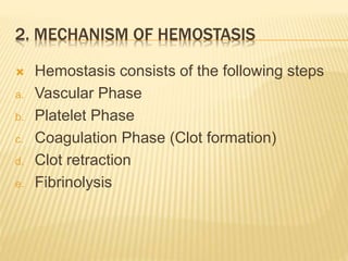
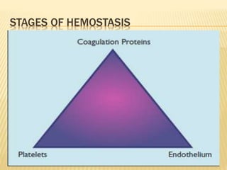
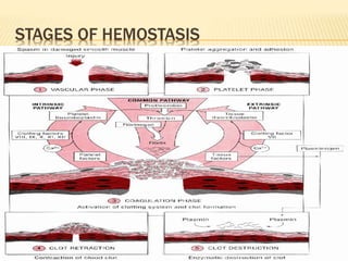
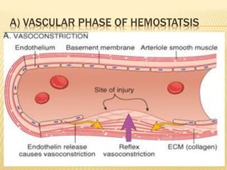
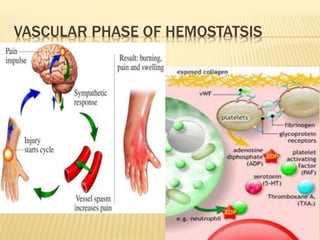
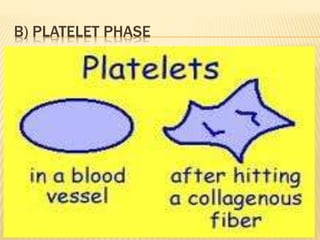
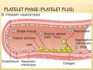
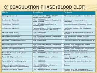
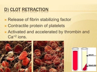
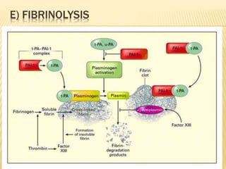
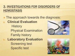
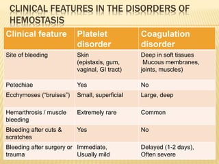
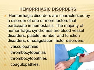
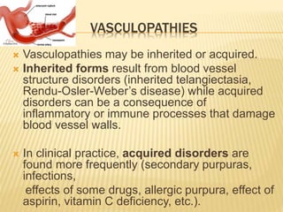
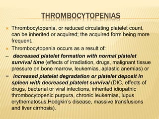
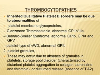
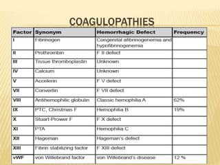
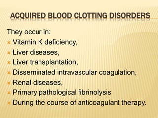
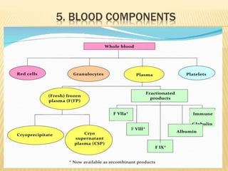
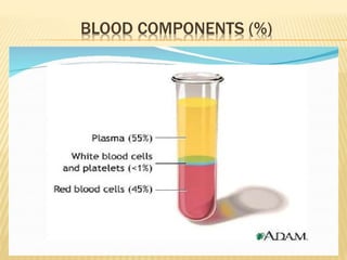
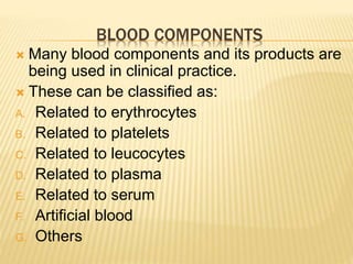
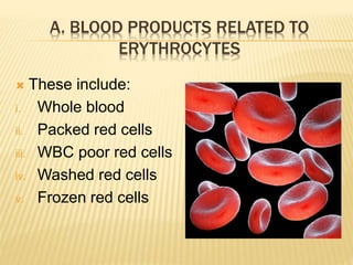
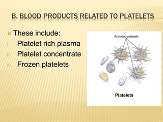
HEMOSTASIS 
& 
BLOOD TRANSFUSION 
By 
Dr. Abdul Qadeer Memon 
MBBS; FCPS; FICS 
Assistant Professor in General Surgery 
King Faisal University College of Medicine 
Kingdom of Saudi Arabia
OBJECTIVES 
1. Definition of hemostatsis 
2. Mechanism of hemostasis 
3. Investigations for disorders of hemostasis 
4. Major disorders of hemostasis 
5. Blood components 
6. Indications for component therapy 
7. Complications of blood transfusion
1. DEFINITION OF HEMOSTATSIS 
 Heme = Blood + Stasis = To halt 
(Stop) 
 It is the process of forming clots in the wall 
of damaged blood vessels & preventing 
blood loss while maintaining blood in a 
fluid state within the vascular system. 
 Spontaneous arrest of bleeding by 
physiological process.
MAINTAINS BLOOD FLOW 
PREVENTS BLOOD 
LOSS
2. MECHANISM OF HEMOSTASIS 
 Hemostasis consists of the following steps 
a. Vascular Phase 
b. Platelet Phase 
c. Coagulation Phase (Clot formation) 
d. Clot retraction 
e. Fibrinolysis
STAGES OF HEMOSTASIS
A) VASCULAR PHASE OF HEMOSTATSIS
VASCULAR PHASE OF HEMOSTATSIS
B) PLATELET PHASE
PLATELET PHASE (PLATELET PLUG)
C) COAGULATION PHASE (BLOOD CLOT)
D) CLOT RETRACTION 
 Release of fibrin stabilizing factor 
 Contractile protein of platelets 
 Activated and accelerated by thrombin and 
Ca+2 ions.
E) FIBRINOLYSIS
?
3. INVESTIGATIONS FOR DISORDERS OF 
HEMOSTASIS 
 The approach towards the diagnosis: 
a. Clinical Evaluation 
History 
Physical Examination 
Family history 
b. Laboratory Evaluation 
Screening test 
Specific test
CLINICAL FEATURES IN THE DISORDERS OF 
HEMOSTASIS 
Clinical feature Platelet 
disorder 
Coagulation 
disorder 
Site of bleeding Skin 
(epistaxis, gum, 
vaginal, GI tract) 
Deep in soft tissues 
Mucous membranes, 
joints, muscles) 
Petechiae Yes No 
Ecchymoses (“bruises”) Small, superficial Large, deep 
Hemarthrosis / muscle 
bleeding 
Extremely rare Common 
Bleeding after cuts & 
scratches 
Yes No 
Bleeding after surgery or 
trauma 
Immediate, 
Usually mild 
Delayed (1-2 days), 
Often severe
PLATELET COAGULATION 
PETECHIAE, PURPURA HEMATOMA, JOINT BL.
TESTS FOR PRIMARY HEMOSTASIS I.E PLATELET-MEDIATED 
COAGULATION 
 Bleeding time Platelet & vascular phases 
 PFA – 100 system Platelet function 
 Platelet count Quantification of platelets 
 Blood smear Quantitative & 
morphological 
abnormalities of platelets , 
Detection of underlying 
haemotological disorder
TESTS FOR SECONDARY HEMOSTASIS I.E 
PLASMA PROTEIN-MEDIATED COAGULATION 
 Clotting time Crude test of coagulation 
phase 
 Prothrombin time Extrensic & common 
pathway 
 Activated partial 
thromboplastin time Intrinsic & common pathway
OTHER TESTS FOR HEMOSTASIS 
 Thrombin time 
 INR = International Normalized Ratio
PLATELET COUNT 
NORMAL 150,000 - 400,000 
Cells/mm3 
< 100,000 Thrombocytopenia 
50,000 - 100,000 Mild Thrombocytopenia 
< 50,000 Severe Thrombocytopenia
BLEEDING TIME (BT) 
The duration of bleeding after controlled, 
standardized puncture of the earlobe or forearm 
Provides assessment of platelet count and 
function 
Normal value 
2-8 minutes
CLOTTING (COAGULATION) TIME (CT) 
 The time required for blood to clot in a glass 
tube 
Normal value 
5-15 Min
PROTHROMBIN TIME (PT) 
 The rate at which prothrombin is converted to 
thrombin in citrated blood with added calcium; 
 Measures Effectiveness of the Extrinsic 
Pathway 
Measures the activity of V, VII, X, II , I 
Normal value 
11-16 Sec
ACTIVATED PARTIAL THROMBOPLASTIN 
TIME (APTT) 
 Period required for clot formation in re-calcified 
blood plasma after contact activation 
& addition of platelet substitutes. 
It measures effectiveness of 
Intrinsic pathway & 
common pathway 
Normal value 
25-40 Sec
THROMBIN TIME (TT) 
 Time for Thrombin To Convert Fibrinogen into 
Fibrin 
 A Measure of Fibrinolytic Pathway 
Normal value 
9-13 sec
INR (INTERNATIONAL NORMALOIZED RATIO) 
 It is a laboratory measurement of how long it takes 
blood to form a clot. It is used to determine the 
effects of oral anticoagulants (e.g. Warfarin / 
Coudamin) on the clotting system. 
 All PT results are standardized by this calculation: 
 INR= ( Patient PT / Control PT) 
ISI 
 ISI= International sensitivity index, Given by the manufacturer for each 
particular thromboplastin reagent and instrument combination
SIGNIFICANCE OF INR
SPECIFIC TESTS 
 Tests for specific Platelet Functions 
1. Platelet aggregation test 
2. Flow cytometry 
3. Test for platelet secretion 
4. Clot retraction test 
5. Platelet procoagulant activity 
 Test for Coagulation Phase 
1. Quantitative estimation of Fibrinogen 
2. Coagulation factor assays 
3. F XIII Qualitative assay 
 Latex agglutination test for Fibrinolysis
?
4. MAJOR DISORDERS OF HEMOSTASIS 
INHERITED 
 Vascular 
 Platelet 
 Coagulation 
 Fibrinolytic 
ACQUIRED 
 Vascular 
 Platelet 
 Coagulation
HEMORRHAGIC DISORDERS 
 Hemorrhagic disorders are characterized by 
a disorder of one or more factors that 
participate in hemostasis. The majority of 
hemorrhagic syndromes are blood vessel 
disorders, platelet number and function 
disorders, or coagulation factor disorders: 
a. vasculopathies 
b. thrombocytopenias 
c. thrombocytopathies 
d. coagulopathies.
VASCULOPATHIES 
 Vasculopathies may be inherited or acquired. 
 Inherited forms result from blood vessel 
structure disorders (inherited telangiectasia, 
Rendu-Osler-Weber’s disease) while acquired 
disorders can be a consequence of 
inflammatory or immune processes that damage 
blood vessel walls. 
 In clinical practice, acquired disorders are 
found more frequently (secondary purpuras, 
infections, 
effects of some drugs, allergic purpura, effect of 
aspirin, vitamin C deficiency, etc.).
THROMBOCYTOPENIAS 
 Thrombocytopenia, or reduced circulating platelet count, 
can be inherited or acquired; the acquired form being more 
frequent. 
 Thrombocytopenia occurs as a result of: 
– decreased platelet formation with normal platelet 
survival time (effects of irradiation, drugs, malignant tissue 
pressure on bone marrow, leukemias, aplastic anemias) or 
− increased platelet degradation or platelet deposit in 
spleen with decreased platelet survival (DIC, effects of 
drugs, bacterial or viral infections, inherited idiopathic 
thrombocytopenic purpura, chronic leukemias, lupus 
erythematosus,Hodgkin’s disease, massive transfusions 
and liver cirrhosis).
THROMBOCYTOPATHIES 
 Inherited Qualitative Platelet Disorders may be due 
to abnormalities of 
1. platelet membrane glycoproteins, 
- Glanzmann Thrombastenia, abnormal GPIIb/IIIa 
– Bernard-Soulier Syndrome, abnormal GPIb, GPIX and 
GPV 
– platelet-type of vWD, abnormal GPIb 
2. platelet granules, 
 These may occur due to absence of granules in 
platelets, storage pool disorder (characterized by 
disturbed platelet aggregation to collagen, adrenaline 
and thrombin), or disturbed release (absence of T A2).
THROMBOCYTOPATHIES (CONTD.) 
3. platelet coagulant activity, or 
4. signal transduction and secretion. 
 defects in arachidonic acid metabolism, 
 cyclooxigenase deficiency, platelets unable to produce 
thromboxane; endothelium may not produce 
prostacyclin, 
 thromboxane synthesis deficiency, and 
 defects in platelet secretion and the second wave of 
platelet aggregation, found in response to epinephrine 
or ATP.
COAGULOPATHIES
ACQUIRED BLOOD CLOTTING DISORDERS 
They occur in: 
 Vitamin K deficiency, 
 Liver diseases, 
 Liver transplantation, 
 Disseminated intravascular coagulation, 
 Renal diseases, 
 Primary pathological fibrinolysis 
 During the course of anticoagulant therapy.
?
5. BLOOD COMPONENTS
BLOOD COMPONENTS (%)
BLOOD COMPONENTS 
 Many blood components and its products are 
being used in clinical practice. 
 These can be classified as: 
A. Related to erythrocytes 
B. Related to platelets 
C. Related to leucocytes 
D. Related to plasma 
E. Related to serum 
F. Artificial blood 
G. Others
A. BLOOD PRODUCTS RELATED TO 
ERYTHROCYTES 
 These include: 
i. Whole blood 
ii. Packed red cells 
iii. WBC poor red cells 
iv. Washed red cells 
v. Frozen red cells
B. BLOOD PRODUCTS RELATED TO PLATELETS 
 These include: 
i. Platelet rich plasma 
ii. Platelet concentrate 
iii. Frozen platelets
C. BLOOD PRODUCTS RELATED TO LEUCOCYTES 
 These include: 
i. Granulocyte rich plasma 
ii. Lymphocyte rich plasma
D. BLOOD PRODUCTS RELATED TO PLASMA 
 These include: 
i. Fresh plasma 
ii. Fresh frozen plasma (FFP) 
iii. Freeze dried plasma 
iv. Cryoprecipitate 
v. Prothrombin complex 
vi. Coagulation factors concentrates 
vii. Antithrombin III 
viii. Fibrinogen 
ix. Protein C & S
E. BLOOD PRODUCTS RELATED TO SERUM 
 These include: 
i. Normal pool serum 
ii. Freeze dried serum 
iii. Serum immune globulin 
iv. Normal serum albumin
F. BLOOD PRODUCTS RELATED TO ARTIFICIAL 
BLOOD 
 Theses include red cell substitutes
G. BLOOD PRODUCTS RELATED TO PLASMA 
SUBSTITUTE 
 This includes 6% Dextran solution
?
6. INDICATIONS FOR COMPONENT THERAPY
WHOLE BLOOD 
 Used in hypovlemia due to blood loss 
 Disadvantages: 
i. If not screened properly, can cause hepatitis 
ii. Allergic & febrile reactions 
iii. Stored blood may increases level of K+ 
iv. It is devoid of platelets & clotting factors
PACKED RED BLOOD CELLS 
 Given to the patients with anemia and blood 
loss 
 One unit of PRBCs will raise Hb level of 1.5 
gm/dl 
 Does not provide platelets 
and clotting factors
WASHED RED CELLS 
 Antibodies in donor serum is eliminated if 
cross-matching shows minor positive 
 Desirable to transfer to the patient of 
paroxysmal nocturnal hemoglobinuria 
 It also does not provide platelets and clotting 
factors
LEUKOCYTE-POOR RED CELLS 
 Given to the patients such as 
 With high titer leukocyte antibodies 
 Who have or had repeated febrile transfusion 
reactions 
 With organ transplantation e.g. B.M, kidney
FROZEN RED CELLS 
 Frozen red cells in glycerol citrate solution 
can be stored up to 2 years 
 Useful when rare groups are required fro 
transfusion 
 Disadvantages: 
i. Higher cost for this product 
ii. Time consuming 
iii. Frozen red cells after thawing must be used 
within 24 hours because of the possible risk 
of bacterial contamination
GRANULOCYTE-RICH PLASMA TRANSFUSION 
 Given to patients suffering from more than 
101oF and having evidence of infection with 
positive blood culture 
 In case of neutropenia when granulocyte 
count < 500 μL 
 Lymphocyte-rich plasma may be give to the 
patients with lymphocytopenia
PLATELET CONCENTRATE 
 Can be given in: 
a. Marrow aplasia 
b. Platelet dysfunction e.g. thromboasthenia 
c. Bone marrow and organ transplantation 
d. Post-op bleeding when count falls below 
25x109/L 
e. Cardiac bypass & massive blood transfusion
 Disadvantages: 
 Platelets transfusion is not indicated in; 
a. ITP 
b. Drug induced thrombocytopenia 
c. Extrinsic platelet dysfunction e.g. vWD and 
uremia
FRESH FROZEN PLASMA (FFP) 
 Can be used in patients with: 
 Coagulation factors deficiency 
 Hypovolemia 
 Shock, acute hemorrhage, plasma loss e.g. 
in burns
CRYOPRECIPITATE/FACTOR VIII CONCENTRATE 
 Used in hemophilia or vWD
PROTHROMBIN COMPLEX 
 It contains vitamin K-dependent factors i.e. II, 
VII, IX & X 
 Can be given in the patients having 
deficiency of the above factors 
 Disadvantages: 
 Carries a high risk of transmitting hepatitis 
because it is derived from large pool of 
donors 
 May also cause thrombotic tendency if given 
repeatedly
IMMUNE SERUM GLOBULIN (GAMMA GLOBULIN) 
 Prepared by fractionation of pooled normal 
serum or plasma 
 Can be used in viral infections e.g. hepatitis 
 Can be used in patients with 
agammaglobulinemia or 
hypogammaglobulinemia
SERUM ALBUMIN 
 Prepared by the fractionation of pooled 
plasma 
 Available in 5% and 25% 
 Uses: 
a. To maintain blood volume 
b. Hypoprteinemia
ARTIFICIAL BLOOD RELATED TO RED 
BLOOD CELL SUBSTITUTES 
 Perflouro carbon 
 Stroma free hemoglobin
6% DEXTRAN SOLUTION 
 Related to plasma substitutes e.g. 
i. Intradex 
ii. Dextraven 
iii. SAG-M 
iv. Ad-sol
?
7. COMPLICATIONS OF BLOOD TRANSFUSION 
 These include: 
I. Transfusion reactions; hemolytic & non-hemolytic 
II. Allergic reactions 
III. Volume overload 
IV. Transfusion related acute lung injury 
(TRALI) 
V. Graft-vs-host disease (GVHD) 
VI. Complications of massive transfusion 
VII. Transmission of infections
I. TRANSFUSION REACTIONS 
 Febrile non-hemolytic & chill-rigor reactions 
 Hemolytic reactions due to ABO 
incompatibility 
 The most common symptoms are chills, 
rigors, fever, dyspnea, light-headedness, 
urticaria, itching, and flank pain.
I. TRANSUSION REACTIONS 
WHAT TO DO? 
 Prompt reporting to blood bank 
 Stop transfusion immediately 
 Continue I/V fluids e.g. N. saline
II. ALLERGIC REACTIONS 
 Due to presence of allergens within the 
donor blood 
 These reactions are usually mild, with 
urticaria, edema, occasional dizziness, and 
headache during or immediately after the 
transfusion 
 Simultaneous fever is common
III. VOLUME OVERLOAD 
 May be dangerous in the patients with 
cardiac failure or renal insufficiency 
 The patient should be observed and, if signs 
of heart failure (e.g. dyspnea, rales) occur, 
the transfusion should be stopped and 
treatment for heart failure begun.
IV. TRALI 
 It is caused by anti-HLA and/or anti-granulocyte 
antibodies in donor plasma that 
agglutinate and degranulate recipient 
granulocytes within the lung. 
 Acute respiratory symptoms develop 
 Chest x-ray has a characteristic pattern of 
non-cardiogenic pulmonary edema.
V. GRAFT-VS-HOST DISEASE (GVHD) 
 Transfusion-associated GVHD is usually 
caused by transfusion of products containing 
immunocompetent lymphocytes to an 
immunocompromised host. 
 The donor lymphocytes attack host tissues.
GRAFT-VS-HOST DISEASE (GVHD) 
 Symptoms and signs include fever, skin rash 
 Vomiting, watery and bloody diarrhea, 
lymphadenopathy, and pancytopenia due to 
bone marrow aplasia. 
 Jaundice and elevated liver enzymes are 
also common. 
 GVHD occurs 4 to 30 days after transfusion 
and is diagnosed based on clinical suspicion 
and skin and bone marrow biopsies. 
 GVHD has > 90% mortality because no 
specific treatment is available.
VI. COMPLICATIONS OF MASSIVE TRANSFUSION 
 Massive transfusion is transfusion of a 
volume of blood greater than or equal to one 
blood volume in 24 h (eg, 10 units in a 70-kg 
adult). 
 DIC: When a patient receives stored blood in 
such large volume, the patient's own blood 
may be, in effect, “washed out.” In 
circumstances uncomplicated by prolonged 
hypotension or DIC, dilutional 
thrombocytopenia is the most likely 
complication.
 Platelets in stored whole blood are not 
functional. Clotting factors (except factor VIII) 
usually remain sufficient. 
 Microvascular bleeding (abnormal oozing 
and continued bleeding from raw and cut 
surfaces) may result. 
 Five to 8 (1 unit/10 kg) platelet concentrates 
are usually enough to correct such bleeding 
in an adult. 
 Fresh frozen plasma and cryoprecipitate may 
be needed.
VII. TRANSMISSION OF INFECTIONS 
 These may be: 
 Hepatitis B & C 
 HIV 
 HTLV 
 Cytomegalovirus (CMV) 
 Epstein-Barr virus 
 Human Parvovirus (B19) 
 Human Herpes virus 
 Syphilis 
 Malaria
SUMMARY 
?
THE END

Hemostatsis & blood transfusion

  • 1.
    HEMOSTASIS & BLOODTRANSFUSION By Dr. Abdul Qadeer Memon MBBS; FCPS; FICS Assistant Professor in General Surgery King Faisal University College of Medicine Kingdom of Saudi Arabia
  • 2.
    OBJECTIVES 1. Definitionof hemostatsis 2. Mechanism of hemostasis 3. Investigations for disorders of hemostasis 4. Major disorders of hemostasis 5. Blood components 6. Indications for component therapy 7. Complications of blood transfusion
  • 3.
    1. DEFINITION OFHEMOSTATSIS  Heme = Blood + Stasis = To halt (Stop)  It is the process of forming clots in the wall of damaged blood vessels & preventing blood loss while maintaining blood in a fluid state within the vascular system.  Spontaneous arrest of bleeding by physiological process.
  • 5.
    MAINTAINS BLOOD FLOW PREVENTS BLOOD LOSS
  • 6.
    2. MECHANISM OFHEMOSTASIS  Hemostasis consists of the following steps a. Vascular Phase b. Platelet Phase c. Coagulation Phase (Clot formation) d. Clot retraction e. Fibrinolysis
  • 7.
  • 8.
  • 9.
    A) VASCULAR PHASEOF HEMOSTATSIS
  • 10.
    VASCULAR PHASE OFHEMOSTATSIS
  • 11.
  • 12.
  • 14.
  • 17.
    D) CLOT RETRACTION  Release of fibrin stabilizing factor  Contractile protein of platelets  Activated and accelerated by thrombin and Ca+2 ions.
  • 18.
  • 19.
  • 20.
    3. INVESTIGATIONS FORDISORDERS OF HEMOSTASIS  The approach towards the diagnosis: a. Clinical Evaluation History Physical Examination Family history b. Laboratory Evaluation Screening test Specific test
  • 21.
    CLINICAL FEATURES INTHE DISORDERS OF HEMOSTASIS Clinical feature Platelet disorder Coagulation disorder Site of bleeding Skin (epistaxis, gum, vaginal, GI tract) Deep in soft tissues Mucous membranes, joints, muscles) Petechiae Yes No Ecchymoses (“bruises”) Small, superficial Large, deep Hemarthrosis / muscle bleeding Extremely rare Common Bleeding after cuts & scratches Yes No Bleeding after surgery or trauma Immediate, Usually mild Delayed (1-2 days), Often severe
  • 22.
    PLATELET COAGULATION PETECHIAE,PURPURA HEMATOMA, JOINT BL.
  • 23.
    TESTS FOR PRIMARYHEMOSTASIS I.E PLATELET-MEDIATED COAGULATION  Bleeding time Platelet & vascular phases  PFA – 100 system Platelet function  Platelet count Quantification of platelets  Blood smear Quantitative & morphological abnormalities of platelets , Detection of underlying haemotological disorder
  • 24.
    TESTS FOR SECONDARYHEMOSTASIS I.E PLASMA PROTEIN-MEDIATED COAGULATION  Clotting time Crude test of coagulation phase  Prothrombin time Extrensic & common pathway  Activated partial thromboplastin time Intrinsic & common pathway
  • 25.
    OTHER TESTS FORHEMOSTASIS  Thrombin time  INR = International Normalized Ratio
  • 26.
    PLATELET COUNT NORMAL150,000 - 400,000 Cells/mm3 < 100,000 Thrombocytopenia 50,000 - 100,000 Mild Thrombocytopenia < 50,000 Severe Thrombocytopenia
  • 27.
    BLEEDING TIME (BT) The duration of bleeding after controlled, standardized puncture of the earlobe or forearm Provides assessment of platelet count and function Normal value 2-8 minutes
  • 28.
    CLOTTING (COAGULATION) TIME(CT)  The time required for blood to clot in a glass tube Normal value 5-15 Min
  • 29.
    PROTHROMBIN TIME (PT)  The rate at which prothrombin is converted to thrombin in citrated blood with added calcium;  Measures Effectiveness of the Extrinsic Pathway Measures the activity of V, VII, X, II , I Normal value 11-16 Sec
  • 30.
    ACTIVATED PARTIAL THROMBOPLASTIN TIME (APTT)  Period required for clot formation in re-calcified blood plasma after contact activation & addition of platelet substitutes. It measures effectiveness of Intrinsic pathway & common pathway Normal value 25-40 Sec
  • 31.
    THROMBIN TIME (TT)  Time for Thrombin To Convert Fibrinogen into Fibrin  A Measure of Fibrinolytic Pathway Normal value 9-13 sec
  • 32.
    INR (INTERNATIONAL NORMALOIZEDRATIO)  It is a laboratory measurement of how long it takes blood to form a clot. It is used to determine the effects of oral anticoagulants (e.g. Warfarin / Coudamin) on the clotting system.  All PT results are standardized by this calculation:  INR= ( Patient PT / Control PT) ISI  ISI= International sensitivity index, Given by the manufacturer for each particular thromboplastin reagent and instrument combination
  • 33.
  • 34.
    SPECIFIC TESTS Tests for specific Platelet Functions 1. Platelet aggregation test 2. Flow cytometry 3. Test for platelet secretion 4. Clot retraction test 5. Platelet procoagulant activity  Test for Coagulation Phase 1. Quantitative estimation of Fibrinogen 2. Coagulation factor assays 3. F XIII Qualitative assay  Latex agglutination test for Fibrinolysis
  • 35.
  • 36.
    4. MAJOR DISORDERSOF HEMOSTASIS INHERITED  Vascular  Platelet  Coagulation  Fibrinolytic ACQUIRED  Vascular  Platelet  Coagulation
  • 37.
    HEMORRHAGIC DISORDERS Hemorrhagic disorders are characterized by a disorder of one or more factors that participate in hemostasis. The majority of hemorrhagic syndromes are blood vessel disorders, platelet number and function disorders, or coagulation factor disorders: a. vasculopathies b. thrombocytopenias c. thrombocytopathies d. coagulopathies.
  • 38.
    VASCULOPATHIES  Vasculopathiesmay be inherited or acquired.  Inherited forms result from blood vessel structure disorders (inherited telangiectasia, Rendu-Osler-Weber’s disease) while acquired disorders can be a consequence of inflammatory or immune processes that damage blood vessel walls.  In clinical practice, acquired disorders are found more frequently (secondary purpuras, infections, effects of some drugs, allergic purpura, effect of aspirin, vitamin C deficiency, etc.).
  • 39.
    THROMBOCYTOPENIAS  Thrombocytopenia,or reduced circulating platelet count, can be inherited or acquired; the acquired form being more frequent.  Thrombocytopenia occurs as a result of: – decreased platelet formation with normal platelet survival time (effects of irradiation, drugs, malignant tissue pressure on bone marrow, leukemias, aplastic anemias) or − increased platelet degradation or platelet deposit in spleen with decreased platelet survival (DIC, effects of drugs, bacterial or viral infections, inherited idiopathic thrombocytopenic purpura, chronic leukemias, lupus erythematosus,Hodgkin’s disease, massive transfusions and liver cirrhosis).
  • 40.
    THROMBOCYTOPATHIES  InheritedQualitative Platelet Disorders may be due to abnormalities of 1. platelet membrane glycoproteins, - Glanzmann Thrombastenia, abnormal GPIIb/IIIa – Bernard-Soulier Syndrome, abnormal GPIb, GPIX and GPV – platelet-type of vWD, abnormal GPIb 2. platelet granules,  These may occur due to absence of granules in platelets, storage pool disorder (characterized by disturbed platelet aggregation to collagen, adrenaline and thrombin), or disturbed release (absence of T A2).
  • 41.
    THROMBOCYTOPATHIES (CONTD.) 3.platelet coagulant activity, or 4. signal transduction and secretion.  defects in arachidonic acid metabolism,  cyclooxigenase deficiency, platelets unable to produce thromboxane; endothelium may not produce prostacyclin,  thromboxane synthesis deficiency, and  defects in platelet secretion and the second wave of platelet aggregation, found in response to epinephrine or ATP.
  • 42.
  • 43.
    ACQUIRED BLOOD CLOTTINGDISORDERS They occur in:  Vitamin K deficiency,  Liver diseases,  Liver transplantation,  Disseminated intravascular coagulation,  Renal diseases,  Primary pathological fibrinolysis  During the course of anticoagulant therapy.
  • 44.
  • 45.
  • 46.
  • 47.
    BLOOD COMPONENTS Many blood components and its products are being used in clinical practice.  These can be classified as: A. Related to erythrocytes B. Related to platelets C. Related to leucocytes D. Related to plasma E. Related to serum F. Artificial blood G. Others
  • 48.
    A. BLOOD PRODUCTSRELATED TO ERYTHROCYTES  These include: i. Whole blood ii. Packed red cells iii. WBC poor red cells iv. Washed red cells v. Frozen red cells
  • 49.
    B. BLOOD PRODUCTSRELATED TO PLATELETS  These include: i. Platelet rich plasma ii. Platelet concentrate iii. Frozen platelets
  • 50.
    C. BLOOD PRODUCTSRELATED TO LEUCOCYTES  These include: i. Granulocyte rich plasma ii. Lymphocyte rich plasma
  • 51.
    D. BLOOD PRODUCTSRELATED TO PLASMA  These include: i. Fresh plasma ii. Fresh frozen plasma (FFP) iii. Freeze dried plasma iv. Cryoprecipitate v. Prothrombin complex vi. Coagulation factors concentrates vii. Antithrombin III viii. Fibrinogen ix. Protein C & S
  • 52.
    E. BLOOD PRODUCTSRELATED TO SERUM  These include: i. Normal pool serum ii. Freeze dried serum iii. Serum immune globulin iv. Normal serum albumin
  • 53.
    F. BLOOD PRODUCTSRELATED TO ARTIFICIAL BLOOD  Theses include red cell substitutes
  • 54.
    G. BLOOD PRODUCTSRELATED TO PLASMA SUBSTITUTE  This includes 6% Dextran solution
  • 55.
  • 56.
    6. INDICATIONS FORCOMPONENT THERAPY
  • 57.
    WHOLE BLOOD Used in hypovlemia due to blood loss  Disadvantages: i. If not screened properly, can cause hepatitis ii. Allergic & febrile reactions iii. Stored blood may increases level of K+ iv. It is devoid of platelets & clotting factors
  • 58.
    PACKED RED BLOODCELLS  Given to the patients with anemia and blood loss  One unit of PRBCs will raise Hb level of 1.5 gm/dl  Does not provide platelets and clotting factors
  • 59.
    WASHED RED CELLS  Antibodies in donor serum is eliminated if cross-matching shows minor positive  Desirable to transfer to the patient of paroxysmal nocturnal hemoglobinuria  It also does not provide platelets and clotting factors
  • 60.
    LEUKOCYTE-POOR RED CELLS  Given to the patients such as  With high titer leukocyte antibodies  Who have or had repeated febrile transfusion reactions  With organ transplantation e.g. B.M, kidney
  • 61.
    FROZEN RED CELLS  Frozen red cells in glycerol citrate solution can be stored up to 2 years  Useful when rare groups are required fro transfusion  Disadvantages: i. Higher cost for this product ii. Time consuming iii. Frozen red cells after thawing must be used within 24 hours because of the possible risk of bacterial contamination
  • 62.
    GRANULOCYTE-RICH PLASMA TRANSFUSION  Given to patients suffering from more than 101oF and having evidence of infection with positive blood culture  In case of neutropenia when granulocyte count < 500 μL  Lymphocyte-rich plasma may be give to the patients with lymphocytopenia
  • 63.
    PLATELET CONCENTRATE Can be given in: a. Marrow aplasia b. Platelet dysfunction e.g. thromboasthenia c. Bone marrow and organ transplantation d. Post-op bleeding when count falls below 25x109/L e. Cardiac bypass & massive blood transfusion
  • 64.
     Disadvantages: Platelets transfusion is not indicated in; a. ITP b. Drug induced thrombocytopenia c. Extrinsic platelet dysfunction e.g. vWD and uremia
  • 65.
    FRESH FROZEN PLASMA(FFP)  Can be used in patients with:  Coagulation factors deficiency  Hypovolemia  Shock, acute hemorrhage, plasma loss e.g. in burns
  • 66.
    CRYOPRECIPITATE/FACTOR VIII CONCENTRATE  Used in hemophilia or vWD
  • 67.
    PROTHROMBIN COMPLEX It contains vitamin K-dependent factors i.e. II, VII, IX & X  Can be given in the patients having deficiency of the above factors  Disadvantages:  Carries a high risk of transmitting hepatitis because it is derived from large pool of donors  May also cause thrombotic tendency if given repeatedly
  • 68.
    IMMUNE SERUM GLOBULIN(GAMMA GLOBULIN)  Prepared by fractionation of pooled normal serum or plasma  Can be used in viral infections e.g. hepatitis  Can be used in patients with agammaglobulinemia or hypogammaglobulinemia
  • 69.
    SERUM ALBUMIN Prepared by the fractionation of pooled plasma  Available in 5% and 25%  Uses: a. To maintain blood volume b. Hypoprteinemia
  • 70.
    ARTIFICIAL BLOOD RELATEDTO RED BLOOD CELL SUBSTITUTES  Perflouro carbon  Stroma free hemoglobin
  • 71.
    6% DEXTRAN SOLUTION  Related to plasma substitutes e.g. i. Intradex ii. Dextraven iii. SAG-M iv. Ad-sol
  • 72.
  • 73.
    7. COMPLICATIONS OFBLOOD TRANSFUSION  These include: I. Transfusion reactions; hemolytic & non-hemolytic II. Allergic reactions III. Volume overload IV. Transfusion related acute lung injury (TRALI) V. Graft-vs-host disease (GVHD) VI. Complications of massive transfusion VII. Transmission of infections
  • 74.
    I. TRANSFUSION REACTIONS  Febrile non-hemolytic & chill-rigor reactions  Hemolytic reactions due to ABO incompatibility  The most common symptoms are chills, rigors, fever, dyspnea, light-headedness, urticaria, itching, and flank pain.
  • 75.
    I. TRANSUSION REACTIONS WHAT TO DO?  Prompt reporting to blood bank  Stop transfusion immediately  Continue I/V fluids e.g. N. saline
  • 76.
    II. ALLERGIC REACTIONS  Due to presence of allergens within the donor blood  These reactions are usually mild, with urticaria, edema, occasional dizziness, and headache during or immediately after the transfusion  Simultaneous fever is common
  • 77.
    III. VOLUME OVERLOAD  May be dangerous in the patients with cardiac failure or renal insufficiency  The patient should be observed and, if signs of heart failure (e.g. dyspnea, rales) occur, the transfusion should be stopped and treatment for heart failure begun.
  • 78.
    IV. TRALI It is caused by anti-HLA and/or anti-granulocyte antibodies in donor plasma that agglutinate and degranulate recipient granulocytes within the lung.  Acute respiratory symptoms develop  Chest x-ray has a characteristic pattern of non-cardiogenic pulmonary edema.
  • 79.
    V. GRAFT-VS-HOST DISEASE(GVHD)  Transfusion-associated GVHD is usually caused by transfusion of products containing immunocompetent lymphocytes to an immunocompromised host.  The donor lymphocytes attack host tissues.
  • 80.
    GRAFT-VS-HOST DISEASE (GVHD)  Symptoms and signs include fever, skin rash  Vomiting, watery and bloody diarrhea, lymphadenopathy, and pancytopenia due to bone marrow aplasia.  Jaundice and elevated liver enzymes are also common.  GVHD occurs 4 to 30 days after transfusion and is diagnosed based on clinical suspicion and skin and bone marrow biopsies.  GVHD has > 90% mortality because no specific treatment is available.
  • 81.
    VI. COMPLICATIONS OFMASSIVE TRANSFUSION  Massive transfusion is transfusion of a volume of blood greater than or equal to one blood volume in 24 h (eg, 10 units in a 70-kg adult).  DIC: When a patient receives stored blood in such large volume, the patient's own blood may be, in effect, “washed out.” In circumstances uncomplicated by prolonged hypotension or DIC, dilutional thrombocytopenia is the most likely complication.
  • 82.
     Platelets instored whole blood are not functional. Clotting factors (except factor VIII) usually remain sufficient.  Microvascular bleeding (abnormal oozing and continued bleeding from raw and cut surfaces) may result.  Five to 8 (1 unit/10 kg) platelet concentrates are usually enough to correct such bleeding in an adult.  Fresh frozen plasma and cryoprecipitate may be needed.
  • 83.
    VII. TRANSMISSION OFINFECTIONS  These may be:  Hepatitis B & C  HIV  HTLV  Cytomegalovirus (CMV)  Epstein-Barr virus  Human Parvovirus (B19)  Human Herpes virus  Syphilis  Malaria
  • 84.
  • 85.