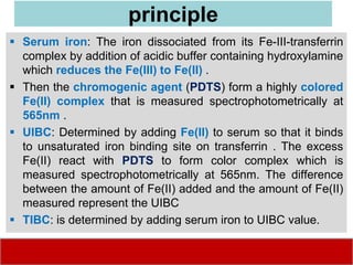
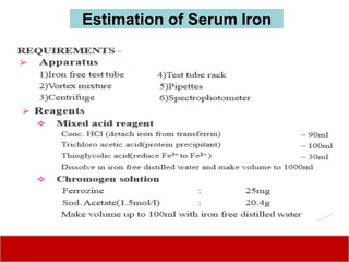
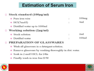
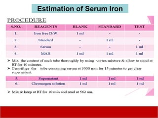
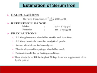
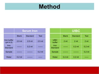
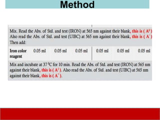
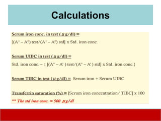
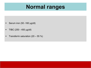
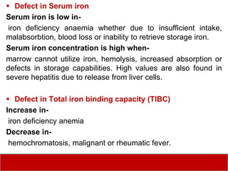
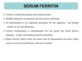
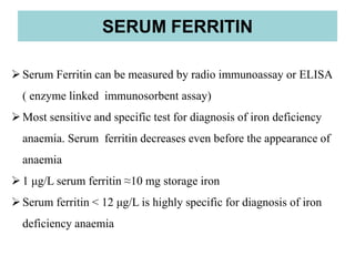
INSTITUTE OF PUBLIC HEALTH, DHAKA
Department of Laboratory Medicine
BSc in Health Technology (Laboratory)
Haematology
Estimation of Serum Iron (Fc)
By
Sk. MIZANUR RAHMAN
Assistant Bacteriologist,
PhD Candidate
MS in BGE; MS in Microbiology; MPH in Epidemiology
Objectives
 To determine the normal level of serum iron.
 To establish SOPs (Introduction, Procedure, QA, QC
etc.)
 To correlate different reference value with interpretation
 To determine the use of this test in diagnosis of anemia
(iron deficiency).
• Iron studies should be performed on fasting blood samples because
recent ingestion of iron results in a falsely high serum iron
concentration and, therefore, transferrin saturation level.
Special Notes
• The normal range for transferrin is 204 to 360 mg/dl. If you have a
higher amount, you may have iron-deficiency anemia. If you have a
lower level, you may have another problem, such as liver disease and
hemolytic anemia.
• Transferrin may also be measured using a value called total iron-
binding capacity (TIBC)
• Transferrin is a blood-plasma glycoprotein, which plays a central role
in iron metabolism and is responsible for ferric-ion delivery.
• It functions as the most critical ferric pool in the body.
• It transports iron through the blood to various tissues such as the liver,
spleen and bone marrow.
Iron in the body
 Iron is the metal component of
haemoglobin, myoglobin,
cytochromes and some proteins
of the electron transport chain.
 The total iron of an adult male is
4-5g and of a female is 3-4g.
•Iron deficiency is usually detected on the basis
of the amount of iron bound to transferrin in the
plasma (serum iron) and the total amount of iron
that can be bound to the plasma transferrin in
vitro
Normal values
Serum iron – 80-180 μg/dl
TIBC – 250 – 370 μg/dl
Serum Iron and Total Iron Binding Capacity
principle
 Serum iron: The iron dissociated from its Fe-III-transferrin
complex by addition of acidic buffer containing hydroxylamine
which reduces the Fe(III) to Fe(II) .
 Then the chromogenic agent (PDTS) form a highly colored
Fe(II) complex that is measured spectrophotometrically at
565nm .
 UIBC: Determined by adding Fe(II) to serum so that it binds
to unsaturated iron binding site on transferrin . The excess
Fe(II) react with PDTS to form color complex which is
measured spectrophotometrically at 565nm. The difference
between the amount of Fe(II) added and the amount of Fe(II)
measured represent the UIBC
 TIBC: is determined by adding serum iron to UIBC value.
principle
The serum iron gives a measure of the iron supply to the
tissues at the time of sampling.
Chromogenic Method
Principle
• When serum iron is treated with Mixed Acid Reagent, the
protein bound iron is detached by the action of conc. HCl
& reduced to ferrous state by thioglycolic acid & proteins
are precipitated by the action of TCA.
• This ferrous iron is then treated with chromogen solution &
it forms a colored complex which is then measured at 562
nm.
Estimation of Serum Iron
6. There should be no BT during last 20 days & no iron supplements taken
by the patient
Estimation of Serum Iron
Method
Serum Iron
Blank Standard Test
Iron buffer
(pH 4.5)
2.5 ml 2.5 ml 2.5 ml
Iron
Standard
------- 0.2 ml -------
Sample -------- -------- 0.2 ml
Water 0.2 ml -------- ------
UIBC
Blank Standard Test
UIBC
buffer
2 ml 2 ml 2 ml
Iron
Standard
------- 0.2 ml 0.2 ml
Sample -------- -------- 0.2 ml
Water 0.4 ml 0.2 ml ------
Method
Calculations
Normal ranges
 Serum iron (50 -160 μg/dl)
 TIBC (250 - 450 μg/dl)
 Transferrin saturation (20 – 55 %)
 Defect in Serum iron
Serum iron is low in-
iron deficiency anaemia whether due to insufficient intake,
malabsorbtion, blood loss or inability to retrieve storage iron.
Serum iron concentration is high when-
marrow cannot utilize iron, hemolysis, increased absorption or
defects in storage capabilities. High values are also found in
severe hepatitis due to release from liver cells.
 Defect in Total iron binding capacity (TIBC)
Increase in-
iron deficiency anemia
Decrease in-
hemochromatosis, malignant or rheumatic fever.
 Ferritin is found intracellular and in blood stream.
 Reliable parameter to determine the iron stores in the body.
 Its determination is an important parameter for the diagnosis and therapy
control of the iron deficiency .
 Ferritin measurement is recommended for risk group like blood donors,
pregnant woman, hemodialysis patient and infants.
 Serum ferritin reflects body iron stores and its measurement has been widely
used as a test for iron deficiency and iron overload
SERUM FERRITIN
Serum Ferritin can be measured by radio immunoassay or ELISA
( enzyme linked immunosorbent assay)
Most sensitive and specific test for diagnosis of iron deficiency
anaemia. Serum ferritin decreases even before the appearance of
anaemia
1 μg/L serum ferritin ≈10 mg storage iron
Serum ferritin < 12 μg/L is highly specific for diagnosis of iron
deficiency anaemia
SERUM FERRITIN
 Anti human Ferritin Ab are bound to micro-wells.
 Ferritin present in diluted serum or patient plasma bind to the respective Ab.
 Washing of micro-wells removes unspecific serum and plasma components.
 HRP conjugate anti human ferritin immunologically detect the bound patient
ferritin forming a conjugate/ferritin/Ab complex.
PRINCIPLE
 Washing of the micro-wells removes unbound conjugate.
 An enzyme substrate in the presence of bound conjugate hydrolyses to form blue
color, the addition of an acid stops the reaction forming yellow end product.
 The intensity of the yellow color is measured photometrically at 450 nm. The
amount of the color is directly proportional to the amount of ferritin present in
original sample.
PRINCIPLE
MATERIALS
APPARATUS
1) Microplate
2) Multichannel dispenser
3) Laboratory timing device
4) Distilled or deionized water
5) Graduated cylinder 100 and 1000 ml
6) Plastic container for the storage of wash solution.
7) Graph paper.
1) Prepare a sufficient no of micro plate wells to accommodate control and patient
samples.
2) Pipette 25 µl of calibrator, controls and patient samples in duplicate into the wells.
3) Add 100µl sample buffer to each wells.
4) Incubate for 30 min at 25⁰-28⁰ C.
5) Discard the content of micro-wells and wash 3 times with 300 µl of wash solution or
use automatic ELISA plate washer.
6) Dispense 100µl of enzyme conjugate into each well.
7) Incubate for 15 min at 25⁰-28⁰ C.
8) Discard the content of micro-well and wash
9) Dispense 100 µl of TMB substrate solution to each well
10) Incubate for 15 min at 25⁰-28⁰ C .
11) Add 100 µl stop solution to each well and incubate for 5 min at RT
12) Read the OD at 450 nm and calculate the result
PROCEDURE
Vitamin B12 and Serum Folate
Test Indications:
Useful in detecting vitamin B12 deficiency anemia. Helps diagnose the
cause of anemia, especially when the RBC’s are described as macrocytic in
non-neonates. Helps diagnose the cause of dementia or other CNS
symptoms.
Notes
• Vitamin B12 and serum folate are no longer performed on elderly patients in
the acute setting unless there is a specific indication e.g. macrocytosis +/-
anaemia, pancytopenia, or blood film report comment.
• This test will not be repeated if it has been carried out within the last 3 months.
• This test can be added on to a patient request if the laboratory has an
appropriate sample that is less than 3 days old.
• Grossly haemolysed, lipaemic or icteric samples may give erroneous results
and will not be processed
• Patient Preparation: For Folate: Patients should be fasting (8 hours
recommended) and should not have recently received methotrexate or other
folic acid antagonist.
Vitamin B12 and Serum Folate
The Vitamin B12 assay principle is for the Roche Modular P chemistry analyzer.
• The test is based on the principle of β-complementation of the enzyme β-galactosidase and
the competition between an enzyme donor-vitamin B12 conjugate, an anti-vitamin B12
protein (Intrinsic Factor) and the Vitamin B12 content of a serum sample.
• Samples with higher Vitamin B12 concentrations produce higher β-galactosidase activities
and vice versa.
• A nitro-phenyl-β-galactoside derivative is used as the enzyme substrate. The reaction's
product has maximum absorbance at 415 nm.
• The Vitamin B12 concentration of a sample is proportional to the measured β-galactosidase
activity.
Vitamin B12-Assay Principle
Serum Folate
• Serum folate falls rapidly with an inadequate diet despite sufficient
body stores . It is however more reliably analysed than Red Cell folate
and is therefore used to screen for folate deficiency.
• The common causes of deficiency are inadequate diet, malabsorption,
anti-folate drugs (especially anticonvulsants) and occasionally in
association with any disease that causes increase cell turnover, e.g.
exfoliation.
Result Interpretation
Vitamin B12
Interpretation is difficult. The following observations are offered.
• In clear cut deficiency, levels of B12 are nearly always <150 pg/ml and usually
<100 pg/ml.
• Pernicious anaemia is the cause of the majority of severe deficiencies in adults.
Around 50% of patients with pernicious anaemia have intrinsic factor antibodies
while this antibody is rarely seen in normal controls. Parietal cell antibodies are
present in 90% of patients but are also seen in 1 - 2% of normal controls.
• Patients with B12 in the borderline range (150 - 180 pg/ml) may have either early
B12 deficiency or be healthy "low normal".
• Neurological disease or glossitis may occur without anaemia or macrocytosis and
may be irreversible.
• Interpretation of early megaloblastic change in the marrow is difficult and cannot
be reliably distinguished from myelodysplasia.
Result Interpretation
Vitamin B 12:
Patients taking vitamin B12 supplementation may have misleading results. Many other
conditions are known to cause an increase or decrease in the serum vitamin B 12
concentration including:
Increases: Ingestion of vitamin C, ingestion of estrogens, ingestion of vitamin A,
hepatocellular injury, myeloproliferative disorder, uremia.
Decreases: Pregnancy, aspirin, anticonvulsants, colchicine, ethanol ingestion,
contraceptive hormones, smoking, hemodialysis, multiple myeloma. The evaluation of
macrocytic anemia requires measurement of both vitamin B 12 and folate levels;
ideally they should be measured simultaneously.
Limitations
Folate:
Patients with combined deficiency of folate and iron may not demonstrate the
erythrocyte macrocytosis otherwise typical of folate deficiency anemia. In these
patients, however, the red cell distribution width (RDW) will typically be elevated. A
non-fasting specimen results in falsely elevated results.
Limitations

Estimation of iron profile

  • 1.
    INSTITUTE OF PUBLICHEALTH, DHAKA Department of Laboratory Medicine BSc in Health Technology (Laboratory) Haematology Estimation of Serum Iron (Fc) By Sk. MIZANUR RAHMAN Assistant Bacteriologist, PhD Candidate MS in BGE; MS in Microbiology; MPH in Epidemiology
  • 2.
    Objectives  To determinethe normal level of serum iron.  To establish SOPs (Introduction, Procedure, QA, QC etc.)  To correlate different reference value with interpretation  To determine the use of this test in diagnosis of anemia (iron deficiency).
  • 9.
    • Iron studiesshould be performed on fasting blood samples because recent ingestion of iron results in a falsely high serum iron concentration and, therefore, transferrin saturation level. Special Notes • The normal range for transferrin is 204 to 360 mg/dl. If you have a higher amount, you may have iron-deficiency anemia. If you have a lower level, you may have another problem, such as liver disease and hemolytic anemia. • Transferrin may also be measured using a value called total iron- binding capacity (TIBC) • Transferrin is a blood-plasma glycoprotein, which plays a central role in iron metabolism and is responsible for ferric-ion delivery. • It functions as the most critical ferric pool in the body. • It transports iron through the blood to various tissues such as the liver, spleen and bone marrow.
  • 10.
    Iron in thebody  Iron is the metal component of haemoglobin, myoglobin, cytochromes and some proteins of the electron transport chain.  The total iron of an adult male is 4-5g and of a female is 3-4g.
  • 12.
    •Iron deficiency isusually detected on the basis of the amount of iron bound to transferrin in the plasma (serum iron) and the total amount of iron that can be bound to the plasma transferrin in vitro Normal values Serum iron – 80-180 μg/dl TIBC – 250 – 370 μg/dl Serum Iron and Total Iron Binding Capacity
  • 13.
    principle  Serum iron:The iron dissociated from its Fe-III-transferrin complex by addition of acidic buffer containing hydroxylamine which reduces the Fe(III) to Fe(II) .  Then the chromogenic agent (PDTS) form a highly colored Fe(II) complex that is measured spectrophotometrically at 565nm .  UIBC: Determined by adding Fe(II) to serum so that it binds to unsaturated iron binding site on transferrin . The excess Fe(II) react with PDTS to form color complex which is measured spectrophotometrically at 565nm. The difference between the amount of Fe(II) added and the amount of Fe(II) measured represent the UIBC  TIBC: is determined by adding serum iron to UIBC value.
  • 14.
  • 15.
    The serum irongives a measure of the iron supply to the tissues at the time of sampling. Chromogenic Method Principle • When serum iron is treated with Mixed Acid Reagent, the protein bound iron is detached by the action of conc. HCl & reduced to ferrous state by thioglycolic acid & proteins are precipitated by the action of TCA. • This ferrous iron is then treated with chromogen solution & it forms a colored complex which is then measured at 562 nm. Estimation of Serum Iron
  • 16.
  • 17.
  • 18.
  • 19.
    6. There shouldbe no BT during last 20 days & no iron supplements taken by the patient Estimation of Serum Iron
  • 20.
    Method Serum Iron Blank StandardTest Iron buffer (pH 4.5) 2.5 ml 2.5 ml 2.5 ml Iron Standard ------- 0.2 ml ------- Sample -------- -------- 0.2 ml Water 0.2 ml -------- ------ UIBC Blank Standard Test UIBC buffer 2 ml 2 ml 2 ml Iron Standard ------- 0.2 ml 0.2 ml Sample -------- -------- 0.2 ml Water 0.4 ml 0.2 ml ------
  • 21.
  • 22.
  • 23.
    Normal ranges  Serumiron (50 -160 μg/dl)  TIBC (250 - 450 μg/dl)  Transferrin saturation (20 – 55 %)
  • 24.
     Defect inSerum iron Serum iron is low in- iron deficiency anaemia whether due to insufficient intake, malabsorbtion, blood loss or inability to retrieve storage iron. Serum iron concentration is high when- marrow cannot utilize iron, hemolysis, increased absorption or defects in storage capabilities. High values are also found in severe hepatitis due to release from liver cells.  Defect in Total iron binding capacity (TIBC) Increase in- iron deficiency anemia Decrease in- hemochromatosis, malignant or rheumatic fever.
  • 25.
     Ferritin isfound intracellular and in blood stream.  Reliable parameter to determine the iron stores in the body.  Its determination is an important parameter for the diagnosis and therapy control of the iron deficiency .  Ferritin measurement is recommended for risk group like blood donors, pregnant woman, hemodialysis patient and infants.  Serum ferritin reflects body iron stores and its measurement has been widely used as a test for iron deficiency and iron overload SERUM FERRITIN
  • 26.
    Serum Ferritin canbe measured by radio immunoassay or ELISA ( enzyme linked immunosorbent assay) Most sensitive and specific test for diagnosis of iron deficiency anaemia. Serum ferritin decreases even before the appearance of anaemia 1 μg/L serum ferritin ≈10 mg storage iron Serum ferritin < 12 μg/L is highly specific for diagnosis of iron deficiency anaemia SERUM FERRITIN
  • 27.
     Anti humanFerritin Ab are bound to micro-wells.  Ferritin present in diluted serum or patient plasma bind to the respective Ab.  Washing of micro-wells removes unspecific serum and plasma components.  HRP conjugate anti human ferritin immunologically detect the bound patient ferritin forming a conjugate/ferritin/Ab complex. PRINCIPLE
  • 28.
     Washing ofthe micro-wells removes unbound conjugate.  An enzyme substrate in the presence of bound conjugate hydrolyses to form blue color, the addition of an acid stops the reaction forming yellow end product.  The intensity of the yellow color is measured photometrically at 450 nm. The amount of the color is directly proportional to the amount of ferritin present in original sample. PRINCIPLE
  • 29.
  • 30.
    APPARATUS 1) Microplate 2) Multichanneldispenser 3) Laboratory timing device 4) Distilled or deionized water 5) Graduated cylinder 100 and 1000 ml 6) Plastic container for the storage of wash solution. 7) Graph paper.
  • 31.
    1) Prepare asufficient no of micro plate wells to accommodate control and patient samples. 2) Pipette 25 µl of calibrator, controls and patient samples in duplicate into the wells. 3) Add 100µl sample buffer to each wells. 4) Incubate for 30 min at 25⁰-28⁰ C. 5) Discard the content of micro-wells and wash 3 times with 300 µl of wash solution or use automatic ELISA plate washer. 6) Dispense 100µl of enzyme conjugate into each well. 7) Incubate for 15 min at 25⁰-28⁰ C. 8) Discard the content of micro-well and wash 9) Dispense 100 µl of TMB substrate solution to each well 10) Incubate for 15 min at 25⁰-28⁰ C . 11) Add 100 µl stop solution to each well and incubate for 5 min at RT 12) Read the OD at 450 nm and calculate the result PROCEDURE
  • 34.
    Vitamin B12 andSerum Folate Test Indications: Useful in detecting vitamin B12 deficiency anemia. Helps diagnose the cause of anemia, especially when the RBC’s are described as macrocytic in non-neonates. Helps diagnose the cause of dementia or other CNS symptoms.
  • 35.
    Notes • Vitamin B12and serum folate are no longer performed on elderly patients in the acute setting unless there is a specific indication e.g. macrocytosis +/- anaemia, pancytopenia, or blood film report comment. • This test will not be repeated if it has been carried out within the last 3 months. • This test can be added on to a patient request if the laboratory has an appropriate sample that is less than 3 days old. • Grossly haemolysed, lipaemic or icteric samples may give erroneous results and will not be processed • Patient Preparation: For Folate: Patients should be fasting (8 hours recommended) and should not have recently received methotrexate or other folic acid antagonist. Vitamin B12 and Serum Folate
  • 36.
    The Vitamin B12assay principle is for the Roche Modular P chemistry analyzer. • The test is based on the principle of β-complementation of the enzyme β-galactosidase and the competition between an enzyme donor-vitamin B12 conjugate, an anti-vitamin B12 protein (Intrinsic Factor) and the Vitamin B12 content of a serum sample. • Samples with higher Vitamin B12 concentrations produce higher β-galactosidase activities and vice versa. • A nitro-phenyl-β-galactoside derivative is used as the enzyme substrate. The reaction's product has maximum absorbance at 415 nm. • The Vitamin B12 concentration of a sample is proportional to the measured β-galactosidase activity. Vitamin B12-Assay Principle
  • 37.
    Serum Folate • Serumfolate falls rapidly with an inadequate diet despite sufficient body stores . It is however more reliably analysed than Red Cell folate and is therefore used to screen for folate deficiency. • The common causes of deficiency are inadequate diet, malabsorption, anti-folate drugs (especially anticonvulsants) and occasionally in association with any disease that causes increase cell turnover, e.g. exfoliation. Result Interpretation
  • 38.
    Vitamin B12 Interpretation isdifficult. The following observations are offered. • In clear cut deficiency, levels of B12 are nearly always <150 pg/ml and usually <100 pg/ml. • Pernicious anaemia is the cause of the majority of severe deficiencies in adults. Around 50% of patients with pernicious anaemia have intrinsic factor antibodies while this antibody is rarely seen in normal controls. Parietal cell antibodies are present in 90% of patients but are also seen in 1 - 2% of normal controls. • Patients with B12 in the borderline range (150 - 180 pg/ml) may have either early B12 deficiency or be healthy "low normal". • Neurological disease or glossitis may occur without anaemia or macrocytosis and may be irreversible. • Interpretation of early megaloblastic change in the marrow is difficult and cannot be reliably distinguished from myelodysplasia. Result Interpretation
  • 39.
    Vitamin B 12: Patientstaking vitamin B12 supplementation may have misleading results. Many other conditions are known to cause an increase or decrease in the serum vitamin B 12 concentration including: Increases: Ingestion of vitamin C, ingestion of estrogens, ingestion of vitamin A, hepatocellular injury, myeloproliferative disorder, uremia. Decreases: Pregnancy, aspirin, anticonvulsants, colchicine, ethanol ingestion, contraceptive hormones, smoking, hemodialysis, multiple myeloma. The evaluation of macrocytic anemia requires measurement of both vitamin B 12 and folate levels; ideally they should be measured simultaneously. Limitations
  • 40.
    Folate: Patients with combineddeficiency of folate and iron may not demonstrate the erythrocyte macrocytosis otherwise typical of folate deficiency anemia. In these patients, however, the red cell distribution width (RDW) will typically be elevated. A non-fasting specimen results in falsely elevated results. Limitations