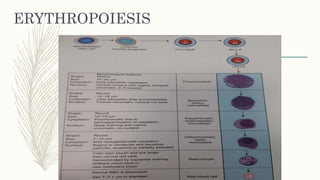
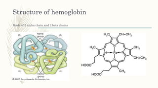
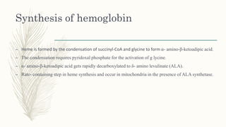
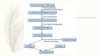
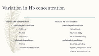
HEMOGLOBIN
SYNTHESIS
AND
FUNCTIONS
Ms. Shama Praveen
Physiology department
ERYTHROPOIESIS
RBCs destruction
– Normal lifespan- 120 days
– Rigid & less active cell trapped by cells of reticuloendothelial systems especially by
macrophage in the liver & spleen.
– Engulf cell by phagocytosis and release hemoglobin.
– 2 mechanism of red cell destruction: Intravascular and Extravascular destruction.
– Intravascular destruction- for example:-paroxysmal nocturnal hemoglobin, complement
complexes create holes in the red cell membrane that promotes intravascular hemolysis.
– Extravascular destruction- two important changes occur in older red cells.
1. decreased deformability.
2. alteration in surface properties.
–
Fate of intravascular destruction
– Destruction of red cells
Fate of extravascular destruction
Hb in plasma
Binds with haptoglobin
Hb + haptoglobin complex
Heme of Hb
iron biliverdin
(heme oxygenase)
CO
bilirubin
(biliverdin reductase)
globin
Amino acid
bilirubin excreted through bile into GI tract
Urobilinogen by bacterial reduction
stercobilinogen
Excreted in stool
HEMOGLOBIN
– Oxygen binding protein present in cytoplasm of red cells.
– Largest contributor to buffering capacity of blood.
– Normal value:- 18-22gm/dl in new born baby
– 12-16gm/dl in females
– 14-18gm/dl in males
Structure of hemoglobin
– Globular molecule comprising four subunits.
– Each subunit consisting of a polypeptide chain and heme(iron containing porphyrin).
– Four polypeptide chains together constitute the protein called globin
– 1gm Hb- 3.4mg of iron and 1.34ml of oxygen.
– Iron present in ferrous form.
Structure of hemoglobin
Made of 2 alpha chain and 2 beta chains
Synthesis of hemoglobin
– Heme is formed by the condensation of succinyl-CoA and glycine to form α- amino-β-ketoadipic acid.
– The condensation requires pyridoxal phosphate for the activation of g lycine.
– α- amino-β-ketoadipic acid gets rapidly decarboxylated to δ- amino levulinate (ALA).
– Rate- containing step in heme synthesis and occur in mitochondria in the presence of ALA synthetase.
Functions of hemoglobin
– Transports oxygen from lungs and carbon dioxide to lungs by forming oxyhemoglobin and
carbaminohemoglobin.
– When reduced hemoglobin is exposed to oxygen at increased pressure, oxygen is taken up at the iron
atom until each molecule of hemoglobin has bound four oxygen molecules, one molecule at each iron
atom.
– Hemoglobin acts as a buffer in maintaining blood pH.
– Hb serves to destroy physiologically important nitric oxide molecule.
– It imparts red colour to hemoglobin.
Variation in Hb concentration
– Decrease Hb concentration Increase Hb concentration
– Physiological conditions physiological conditions
– Children high altitude
– Women newborn baby
– Pregnancy excessive sweating
– Pathological conditions pathological conditions
– Anemia diarrhea, vomiting
– Excessive ADH secretion hypoxia, congenital heart
disease, emphysema etc.
–Thank you

Hemoglobin

  • 1.
  • 2.
  • 3.
    RBCs destruction – Normallifespan- 120 days – Rigid & less active cell trapped by cells of reticuloendothelial systems especially by macrophage in the liver & spleen. – Engulf cell by phagocytosis and release hemoglobin. – 2 mechanism of red cell destruction: Intravascular and Extravascular destruction. – Intravascular destruction- for example:-paroxysmal nocturnal hemoglobin, complement complexes create holes in the red cell membrane that promotes intravascular hemolysis. – Extravascular destruction- two important changes occur in older red cells. 1. decreased deformability. 2. alteration in surface properties.
  • 4.
  • 5.
    Fate of intravasculardestruction – Destruction of red cells Fate of extravascular destruction Hb in plasma Binds with haptoglobin Hb + haptoglobin complex Heme of Hb iron biliverdin (heme oxygenase) CO bilirubin (biliverdin reductase) globin Amino acid bilirubin excreted through bile into GI tract Urobilinogen by bacterial reduction stercobilinogen Excreted in stool
  • 6.
    HEMOGLOBIN – Oxygen bindingprotein present in cytoplasm of red cells. – Largest contributor to buffering capacity of blood. – Normal value:- 18-22gm/dl in new born baby – 12-16gm/dl in females – 14-18gm/dl in males Structure of hemoglobin – Globular molecule comprising four subunits. – Each subunit consisting of a polypeptide chain and heme(iron containing porphyrin). – Four polypeptide chains together constitute the protein called globin – 1gm Hb- 3.4mg of iron and 1.34ml of oxygen. – Iron present in ferrous form.
  • 7.
    Structure of hemoglobin Madeof 2 alpha chain and 2 beta chains
  • 8.
    Synthesis of hemoglobin –Heme is formed by the condensation of succinyl-CoA and glycine to form α- amino-β-ketoadipic acid. – The condensation requires pyridoxal phosphate for the activation of g lycine. – α- amino-β-ketoadipic acid gets rapidly decarboxylated to δ- amino levulinate (ALA). – Rate- containing step in heme synthesis and occur in mitochondria in the presence of ALA synthetase.
  • 10.
    Functions of hemoglobin –Transports oxygen from lungs and carbon dioxide to lungs by forming oxyhemoglobin and carbaminohemoglobin. – When reduced hemoglobin is exposed to oxygen at increased pressure, oxygen is taken up at the iron atom until each molecule of hemoglobin has bound four oxygen molecules, one molecule at each iron atom. – Hemoglobin acts as a buffer in maintaining blood pH. – Hb serves to destroy physiologically important nitric oxide molecule. – It imparts red colour to hemoglobin.
  • 11.
    Variation in Hbconcentration – Decrease Hb concentration Increase Hb concentration – Physiological conditions physiological conditions – Children high altitude – Women newborn baby – Pregnancy excessive sweating – Pathological conditions pathological conditions – Anemia diarrhea, vomiting – Excessive ADH secretion hypoxia, congenital heart disease, emphysema etc.
  • 12.