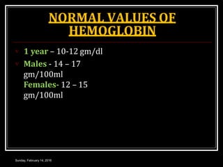
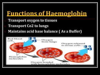
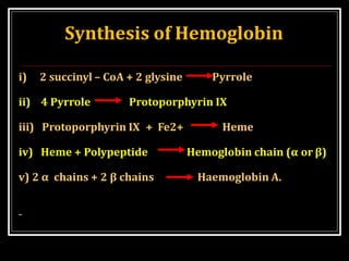
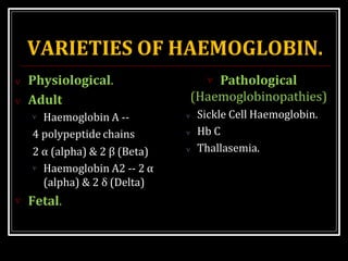
presented
HAEMOGLOBIN
PRESENTED BY
MISS TASMIA ZEB
HAEMOGLOBIN
It is a Red pigment
Present in RBC of Blood.
It is a conjugatedprotein,
& Chromoprotein.
It is made up of Iron and
Protein
It’s molecular weight is
68000.
STRUCTURE OF HAEMOGLOBIN.
 Iron containing pigment
called Haem attached
with protein – Globin.
 Haeme is Iron –
porphyrin complex
called IRON-
PROTOPORPHYRIN IX.
 Globin – Protein.
Sunday, February 14, 2016
STRUCTURE OF HAEME
IRON-PROTOPORPHYRIN IX.
 IRON
 Ferrous form (Fe2+).
 Iron attached to
nitrogen atom of each
pyrrole ring.
 On iron loose bond for


Oxygen
Carbon monoxide.
Sunday, February 14, 2016
STRUCTURE OF HAEME
IRON-PROTOPORPHYRIN IX.
 Porphyrin nucleus.
 4 Pyrrole Rings
(Tetrapyrrole)
 Bridges – Methine (CH)
 Side chains – 8
 Methyl (CH3) - 4
 Vinyl (CH.CH2) - 2
 Propionic acid - 2
(CH2.CH2.COOH)
Sunday, February 14, 2016
Structure of Globin.
 Made up of 4
polypeptide chains.
 Globin is HbA
 2 alpha chains ( ) –
141 amino acids
 2 Beta chains ( ) – 146
amino acids.
Sunday, February 14, 2016
Attachment of Haeme to
Globin.
 4 units of Haeme
attached to 1 unit of
Globin.
 So 1 Haemoglobin
molecules contains 4
Iron Atoms which
carry 4 molecules of
oxygen.
Sunday, February 14, 2016
NORMAL VALUES OF
HEMOGLOBIN
 The Normal Hb level:
 Fetus – 16-18 gm/dl
 Newborn – 20-24
gm/dl.
 Transfusion from
placenta
 Haemoconcentration
NORMAL VALUES OF
HEMOGLOBIN
Sunday, February 14, 2016
 1 year – 10-12 gm/dl
 Males - 14 – 17
gm/100ml
Females- 12 – 15
gm/100ml
Functions of Haemoglobin
 Transport oxygen to tissues
 Transport Co2 to lungs
 Maintains acid base balance ( As a Buffer)
Disadvantages if haemoglobin
present in plasma.
 Increase viscosity.
 Increase osmotic
pressure.
 Rapid destruction by
reticuloendothelial
system.
Haemoglobinuria
( excretion through
kidney)
Sunday, February 14, 2016
Synthesis of Hemoglobin
i) 2 succinyl – CoA + 2 glysine Pyrrole
ii) 4 Pyrrole Protoporphyrin IX
iii) Protoporphyrin IX + Fe2+ Heme
iv) Heme + Polypeptide Hemoglobin chain (α or β)
v) 2 α chains + 2 β chains Haemoglobin A.
Factors controlling
Haemoglobin formation.
Sunday, February 14, 2016
 Role Of Proteins – First class proteins provide
amino acids.
 Most imp – food of animal origin, liver, spleen,
kidney & heart
 Intermediate value – muscles
 Least – cereals, dairy products, veg & fruits.
ROLE OF IRON.
Sunday, February 14, 2016
 Important for formation of Haeme part of
Haemoglobin.
 Sources of iron – Dietary iron
 Other sources – Iron released from degradation of
RBC.
Role of other metals
Sunday, February 14, 2016
 Copper – Promotes
Absorption, Mobilization
& Utilization of iron.
 Cobalt – Increases
production of
Erythropoietin.
& subsequent utilization.
 Role of vitamins.
 Vit B12, Folic acid help in
synthesis of nucleic acid.
 & vit C helps in
absorption of iron from
gut. (Fe3+ to Fe2+)
 Role of bile salts.
 Calcium – conserve iron  Imp for proper
absorption of copper &
nickel.
VARIETIES OF HAEMOGLOBIN.
 Pathological
(Haemoglobinopathies)
 Sickle Cell Haemoglobin.
 Hb C
 Thallasemia.
 Physiological.
 Adult
 Haemoglobin A --
4 polypeptide chains
2 α (alpha) & 2 β (Beta)
 Haemoglobin A2 -- 2 α
(alpha) & 2 δ (Delta)
 Fetal.
FETAL HAEMOGLOBIN.
 Present in fetal RBC &
disappear in 2-3 months
after birth.
 Structure
 4 polypeptide chains
2 α(alpha) & 2 γ (gamma)
 Characteristics.
 Affinity for oxygen – more
 Resistance to action of alkalies
 Life span – less.
Sunday, February 14, 2016
PATHOLOGICAL
(HAEMOGLOBINOPATHIES)
 Sickle cell
haemoglobin.(HbS)
 Substitution of Valine
for Glutamic Acid at 6th
position in beta chain.
 When HbS is reduced (in
low O2 tension)
precipitate into crystals
in RBC changes shape
become Sickle shaped.
Sunday, February 14, 2016
EFFECTS OF SICKLE CELL SHAPE.
Sunday, February 14, 2016
 Less flexible – blockage
of microcirculation.
 Increases blood
viscosity.
 More fragile – More
Hemolysis – Anaemia.
Pathological
(Haemoglobinopathies)
Sunday, February 14, 2016
 Hemoglobin c
(abbreviated as HbC) is an abnormal hemoglobin
in which glutamic acid residue at the 6th position
of the β-globin chain is replaced with a lysine
residue due to a point mutation in the HBB gene.
It produces sickle cell trait but not the disease, as
it causes only mild sickling of the RBCs.
Pathological
(Haemoglobinopathies)
 Other varieties are HbE, HbI, HbJ,
HbM
 Thalassaemia
 Defect in synthesis of polypeptide chain.
 Types
 Major
 Mino
DIFFERENCE IN THALASSAEMIA
MAJOR & MINOR.
Sunday, February 14, 2016







Less common
Homozygous transmission
Complete absence of beta
chain synthesis.
Anemia – moderate to
severe
HbF – markedly increased
Life span – short
Cooley’s Anaemia
β Thalassaemia Major β Thalassaemia Minor.






More common.
Heterozygous
transmission.
Partial Absense.
Anemia- mild.
HbF – slightly elevated.
Life span – comparatively
longer.
FATE OF HAEMOGLOBIN
Sunday, February 14, 2016
Tetra pyrrole straight chain with
Globin & Iron
THANK
YOU

Haemoglobin

  • 1.
  • 2.
    HAEMOGLOBIN It is aRed pigment Present in RBC of Blood. It is a conjugatedprotein, & Chromoprotein. It is made up of Iron and Protein It’s molecular weight is 68000.
  • 3.
    STRUCTURE OF HAEMOGLOBIN. Iron containing pigment called Haem attached with protein – Globin.  Haeme is Iron – porphyrin complex called IRON- PROTOPORPHYRIN IX.  Globin – Protein. Sunday, February 14, 2016
  • 4.
    STRUCTURE OF HAEME IRON-PROTOPORPHYRINIX.  IRON  Ferrous form (Fe2+).  Iron attached to nitrogen atom of each pyrrole ring.  On iron loose bond for   Oxygen Carbon monoxide. Sunday, February 14, 2016
  • 5.
    STRUCTURE OF HAEME IRON-PROTOPORPHYRINIX.  Porphyrin nucleus.  4 Pyrrole Rings (Tetrapyrrole)  Bridges – Methine (CH)  Side chains – 8  Methyl (CH3) - 4  Vinyl (CH.CH2) - 2  Propionic acid - 2 (CH2.CH2.COOH) Sunday, February 14, 2016
  • 6.
    Structure of Globin. Made up of 4 polypeptide chains.  Globin is HbA  2 alpha chains ( ) – 141 amino acids  2 Beta chains ( ) – 146 amino acids. Sunday, February 14, 2016
  • 7.
    Attachment of Haemeto Globin.  4 units of Haeme attached to 1 unit of Globin.  So 1 Haemoglobin molecules contains 4 Iron Atoms which carry 4 molecules of oxygen. Sunday, February 14, 2016
  • 8.
    NORMAL VALUES OF HEMOGLOBIN The Normal Hb level:  Fetus – 16-18 gm/dl  Newborn – 20-24 gm/dl.  Transfusion from placenta  Haemoconcentration
  • 9.
    NORMAL VALUES OF HEMOGLOBIN Sunday,February 14, 2016  1 year – 10-12 gm/dl  Males - 14 – 17 gm/100ml Females- 12 – 15 gm/100ml
  • 10.
    Functions of Haemoglobin Transport oxygen to tissues  Transport Co2 to lungs  Maintains acid base balance ( As a Buffer)
  • 11.
    Disadvantages if haemoglobin presentin plasma.  Increase viscosity.  Increase osmotic pressure.  Rapid destruction by reticuloendothelial system. Haemoglobinuria ( excretion through kidney) Sunday, February 14, 2016
  • 12.
    Synthesis of Hemoglobin i)2 succinyl – CoA + 2 glysine Pyrrole ii) 4 Pyrrole Protoporphyrin IX iii) Protoporphyrin IX + Fe2+ Heme iv) Heme + Polypeptide Hemoglobin chain (α or β) v) 2 α chains + 2 β chains Haemoglobin A.
  • 13.
    Factors controlling Haemoglobin formation. Sunday,February 14, 2016  Role Of Proteins – First class proteins provide amino acids.  Most imp – food of animal origin, liver, spleen, kidney & heart  Intermediate value – muscles  Least – cereals, dairy products, veg & fruits.
  • 14.
    ROLE OF IRON. Sunday,February 14, 2016  Important for formation of Haeme part of Haemoglobin.  Sources of iron – Dietary iron  Other sources – Iron released from degradation of RBC.
  • 15.
    Role of othermetals Sunday, February 14, 2016  Copper – Promotes Absorption, Mobilization & Utilization of iron.  Cobalt – Increases production of Erythropoietin. & subsequent utilization.  Role of vitamins.  Vit B12, Folic acid help in synthesis of nucleic acid.  & vit C helps in absorption of iron from gut. (Fe3+ to Fe2+)  Role of bile salts.  Calcium – conserve iron  Imp for proper absorption of copper & nickel.
  • 16.
    VARIETIES OF HAEMOGLOBIN. Pathological (Haemoglobinopathies)  Sickle Cell Haemoglobin.  Hb C  Thallasemia.  Physiological.  Adult  Haemoglobin A -- 4 polypeptide chains 2 α (alpha) & 2 β (Beta)  Haemoglobin A2 -- 2 α (alpha) & 2 δ (Delta)  Fetal.
  • 17.
    FETAL HAEMOGLOBIN.  Presentin fetal RBC & disappear in 2-3 months after birth.  Structure  4 polypeptide chains 2 α(alpha) & 2 γ (gamma)  Characteristics.  Affinity for oxygen – more  Resistance to action of alkalies  Life span – less. Sunday, February 14, 2016
  • 18.
    PATHOLOGICAL (HAEMOGLOBINOPATHIES)  Sickle cell haemoglobin.(HbS) Substitution of Valine for Glutamic Acid at 6th position in beta chain.  When HbS is reduced (in low O2 tension) precipitate into crystals in RBC changes shape become Sickle shaped. Sunday, February 14, 2016
  • 19.
    EFFECTS OF SICKLECELL SHAPE. Sunday, February 14, 2016  Less flexible – blockage of microcirculation.  Increases blood viscosity.  More fragile – More Hemolysis – Anaemia.
  • 20.
    Pathological (Haemoglobinopathies) Sunday, February 14,2016  Hemoglobin c (abbreviated as HbC) is an abnormal hemoglobin in which glutamic acid residue at the 6th position of the β-globin chain is replaced with a lysine residue due to a point mutation in the HBB gene. It produces sickle cell trait but not the disease, as it causes only mild sickling of the RBCs.
  • 21.
    Pathological (Haemoglobinopathies)  Other varietiesare HbE, HbI, HbJ, HbM  Thalassaemia  Defect in synthesis of polypeptide chain.  Types  Major  Mino
  • 22.
    DIFFERENCE IN THALASSAEMIA MAJOR& MINOR. Sunday, February 14, 2016        Less common Homozygous transmission Complete absence of beta chain synthesis. Anemia – moderate to severe HbF – markedly increased Life span – short Cooley’s Anaemia β Thalassaemia Major β Thalassaemia Minor.       More common. Heterozygous transmission. Partial Absense. Anemia- mild. HbF – slightly elevated. Life span – comparatively longer.
  • 23.
    FATE OF HAEMOGLOBIN Sunday,February 14, 2016 Tetra pyrrole straight chain with Globin & Iron
  • 24.