This document discusses haemoglobin and methods for its determination. It provides information on:
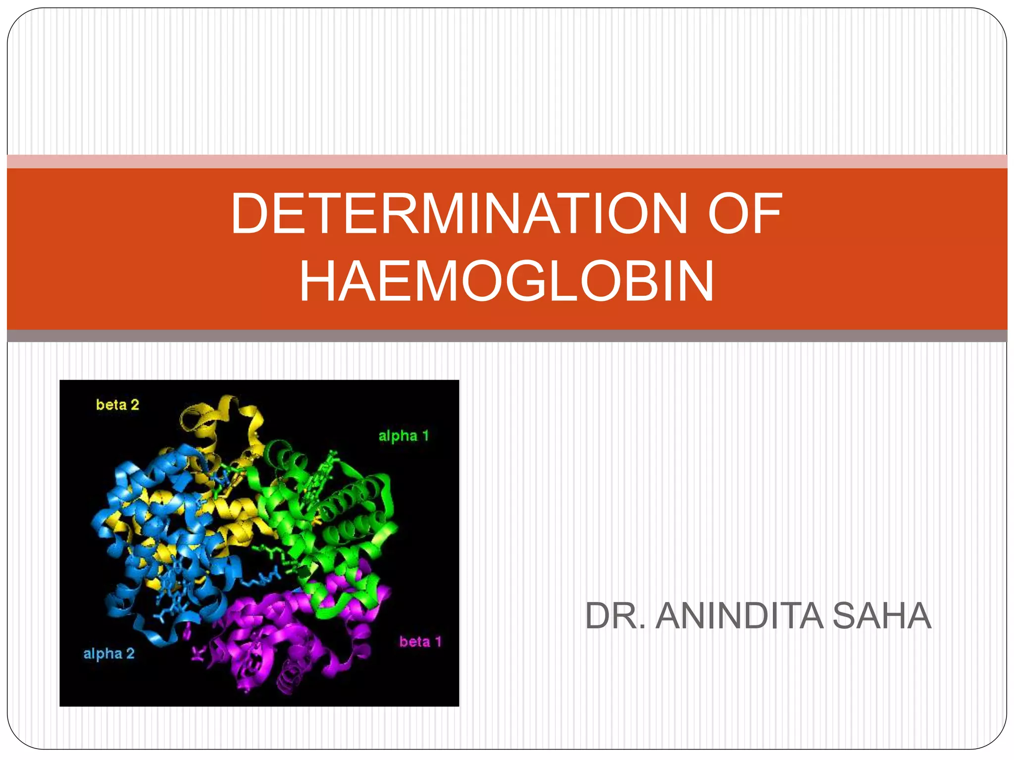
1) The structure and function of haemoglobin as the iron-containing protein in red blood cells that carries oxygen from the lungs to tissues.
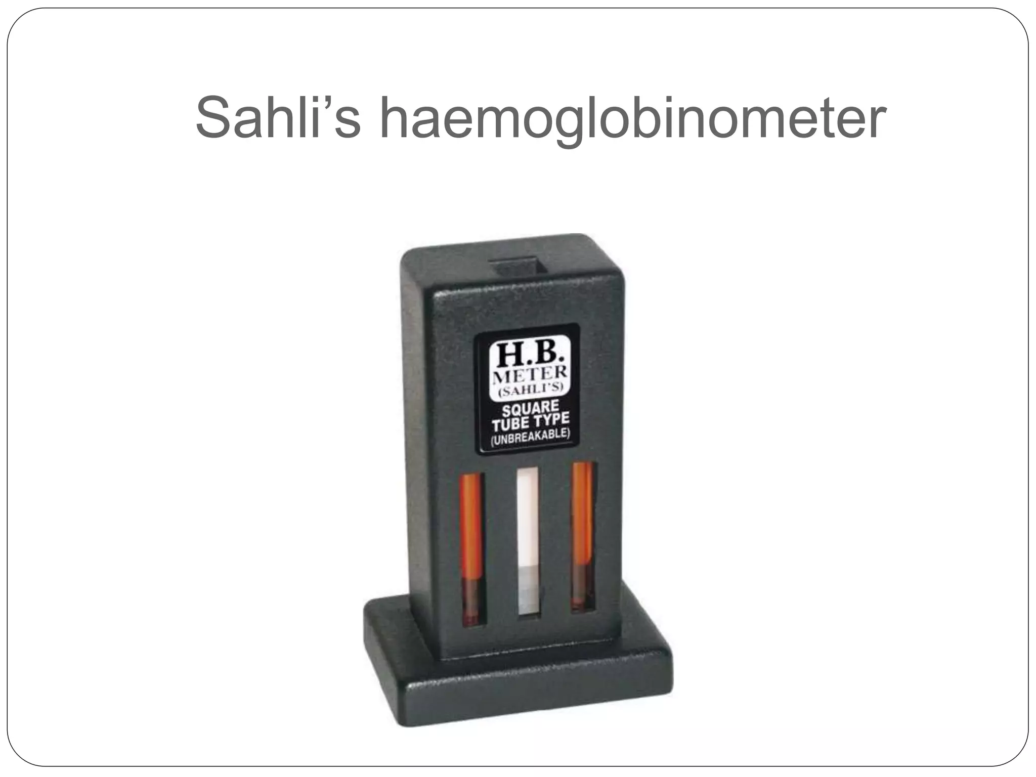
2) Common methods to estimate haemoglobin concentration including the colorimetric Sahli's method and electronic counter methods.
3) Normal haemoglobin values vary by age and sex, and haemoglobin levels may be decreased in conditions like anaemia or increased in situations like dehydration.


![Erythrocyte [ESR]](https://cdn.slidesharecdn.com/ss_thumbnails/8bayhnvlrsiykjj5ehvg-signature-34298b855e83ad5b1d037284f880734bababf6eacd0b448fe71ebe2b3f18666d-poli-160326210134-thumbnail.jpg?width=640&height=640&fit=bounds)


