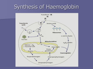
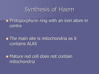
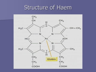
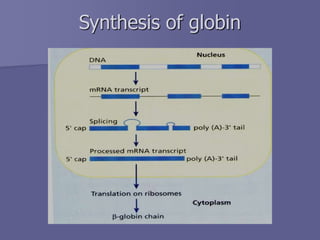
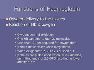
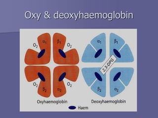
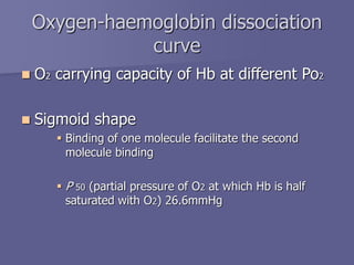
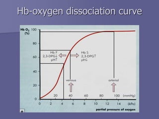
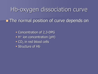
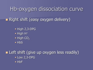
Structure and function of
Haemoglobin
Dr. Tariq M Roshan
Department of Hematology
PPSP
 The main function of red blood cell
 Transfer of O2 from lungs to tissue
 Transfer of CO2 from tissue to lungs
 To accomplish this function red cells has
haemoglobin (Hb)
 Each red cell has 640 million molecules of
Hb
Introduction
Introduction
 Haemoglobin (Hb), protein constituting 1/3 of
the red blood cells
 Synthesis begins in proerythroblast
 65% at erythroblast stage
 35% at reticulocyte stage
 Two parts
 Haem
 Globin
Synthesis of Haemoglobin (Hb)
 Haem & globin produced at two different
sites in the cells
 Haem in mitochondria
 Globin in polyribosomes
 Well synchronized
Synthesis of Haemoglobin
Synthesis of Haem
 Protoporphyrin ring with an iron atom in
centre
 The main site is mitochondria as it
contains ALAS
 Mature red cell does not contain
mitochondria
Structure of Haem
Synthesis of globin
Synthesis of globin
 Various types of globin combines with
haem to from different haemoglobin
 Eight functional globin chains, arranged in
two clusters the
 b- cluster (b, g, d and e globin genes) on the short
arm of chromosome 11
 a- cluster (a and z globin genes) on the short arm
of chromosome 16
Globin gene clusters
Globin synthesis, starts at 3rd week of
gestation
 Embryonic
Haemoglobin Gower I ( z2e2)
Haemoglobin Portland ( z2g2)
Haemoglobin Gower II (a2e2)
 Fetal : HbF (a2g2), HbA (a2b2)
 Adult : HbA, HbA2 ( a2d2), HbF.
Synthesis of globin
Globin chain switch
Alpha & beta chains
Hb A Hb A2 Hb F
structure a2b2 a2d2 a2g2
Normal % 96-98 % 1.5-3.2 % 0.5-0.8 %
Adult haemoblobin
Functions of Haemoglobin
 Oxygen delivery to the tissues
 Reaction of Hb & oxygen
 Oxygenation not oxidation
 One Hb can bind to four O2 molecules
 Less than .01 sec required for oxygenation
 b chain move closer when oxygenated
 When oxygenated 2,3-DPG is pushed out
 b chains are pulled apart when O2 is unloaded,
permitting entry of 2,3-DPG resulting in lower
affinity of O2
Oxy & deoxyhaemoglobin
Oxygen-haemoglobin dissociation
curve
 O2 carrying capacity of Hb at different Po2
 Sigmoid shape
 Binding of one molecule facilitate the second
molecule binding
 P 50 (partial pressure of O2 at which Hb is half
saturated with O2) 26.6mmHg
Hb-oxygen dissociation curve
 The normal position of curve depends on
 Concentration of 2,3-DPG
 H+ ion concentration (pH)
 CO2 in red blood cells
 Structure of Hb
Hb-oxygen dissociation curve
 Right shift (easy oxygen delivery)
 High 2,3-DPG
 High H+
 High CO2
 HbS
 Left shift (give up oxygen less readily)
 Low 2,3-DPG
 HbF
Hb-oxygen dissociation curve
Summary
 Normal structure including the proportion of
globin chains are necessary for the normal
function of haemoglobin
 Reduced haemoglobin in the red blood cells due
to any abnormality of any of its constituents
result into a clinical situation called anaemia
 Metabolic & other abnormalities result into
abnormal oxygen supply to the tissue

Structure and function of Haemoglobin.ppt

  • 1.
    Structure and functionof Haemoglobin Dr. Tariq M Roshan Department of Hematology PPSP
  • 2.
     The mainfunction of red blood cell  Transfer of O2 from lungs to tissue  Transfer of CO2 from tissue to lungs  To accomplish this function red cells has haemoglobin (Hb)  Each red cell has 640 million molecules of Hb Introduction
  • 3.
    Introduction  Haemoglobin (Hb),protein constituting 1/3 of the red blood cells  Synthesis begins in proerythroblast  65% at erythroblast stage  35% at reticulocyte stage  Two parts  Haem  Globin
  • 4.
    Synthesis of Haemoglobin(Hb)  Haem & globin produced at two different sites in the cells  Haem in mitochondria  Globin in polyribosomes  Well synchronized
  • 5.
  • 6.
    Synthesis of Haem Protoporphyrin ring with an iron atom in centre  The main site is mitochondria as it contains ALAS  Mature red cell does not contain mitochondria
  • 7.
  • 8.
  • 9.
    Synthesis of globin Various types of globin combines with haem to from different haemoglobin  Eight functional globin chains, arranged in two clusters the  b- cluster (b, g, d and e globin genes) on the short arm of chromosome 11  a- cluster (a and z globin genes) on the short arm of chromosome 16
  • 10.
  • 11.
    Globin synthesis, startsat 3rd week of gestation  Embryonic Haemoglobin Gower I ( z2e2) Haemoglobin Portland ( z2g2) Haemoglobin Gower II (a2e2)  Fetal : HbF (a2g2), HbA (a2b2)  Adult : HbA, HbA2 ( a2d2), HbF. Synthesis of globin
  • 12.
  • 13.
  • 14.
    Hb A HbA2 Hb F structure a2b2 a2d2 a2g2 Normal % 96-98 % 1.5-3.2 % 0.5-0.8 % Adult haemoblobin
  • 15.
    Functions of Haemoglobin Oxygen delivery to the tissues  Reaction of Hb & oxygen  Oxygenation not oxidation  One Hb can bind to four O2 molecules  Less than .01 sec required for oxygenation  b chain move closer when oxygenated  When oxygenated 2,3-DPG is pushed out  b chains are pulled apart when O2 is unloaded, permitting entry of 2,3-DPG resulting in lower affinity of O2
  • 16.
  • 17.
    Oxygen-haemoglobin dissociation curve  O2carrying capacity of Hb at different Po2  Sigmoid shape  Binding of one molecule facilitate the second molecule binding  P 50 (partial pressure of O2 at which Hb is half saturated with O2) 26.6mmHg
  • 18.
  • 19.
     The normalposition of curve depends on  Concentration of 2,3-DPG  H+ ion concentration (pH)  CO2 in red blood cells  Structure of Hb Hb-oxygen dissociation curve
  • 20.
     Right shift(easy oxygen delivery)  High 2,3-DPG  High H+  High CO2  HbS  Left shift (give up oxygen less readily)  Low 2,3-DPG  HbF Hb-oxygen dissociation curve
  • 21.
    Summary  Normal structureincluding the proportion of globin chains are necessary for the normal function of haemoglobin  Reduced haemoglobin in the red blood cells due to any abnormality of any of its constituents result into a clinical situation called anaemia  Metabolic & other abnormalities result into abnormal oxygen supply to the tissue