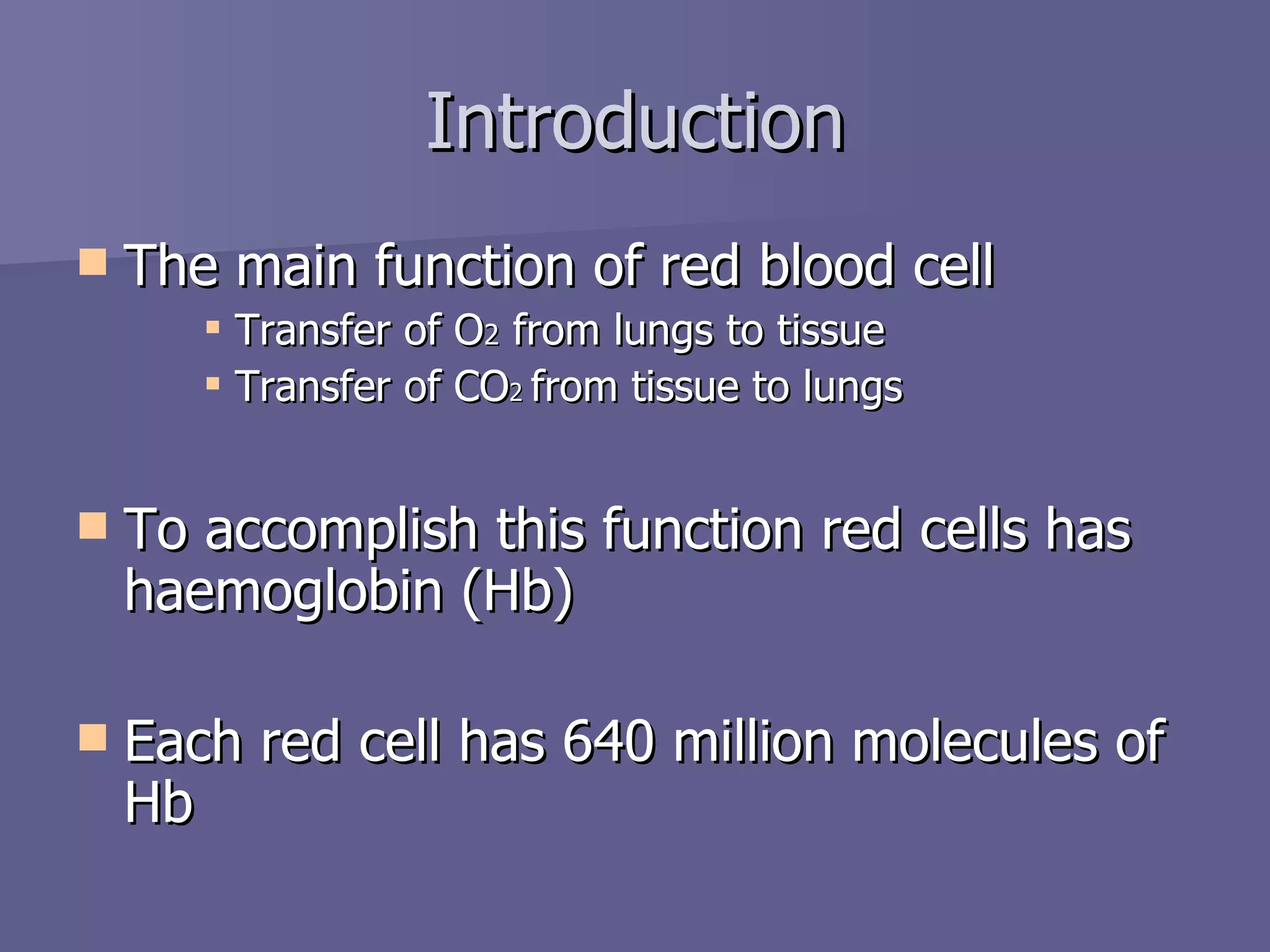
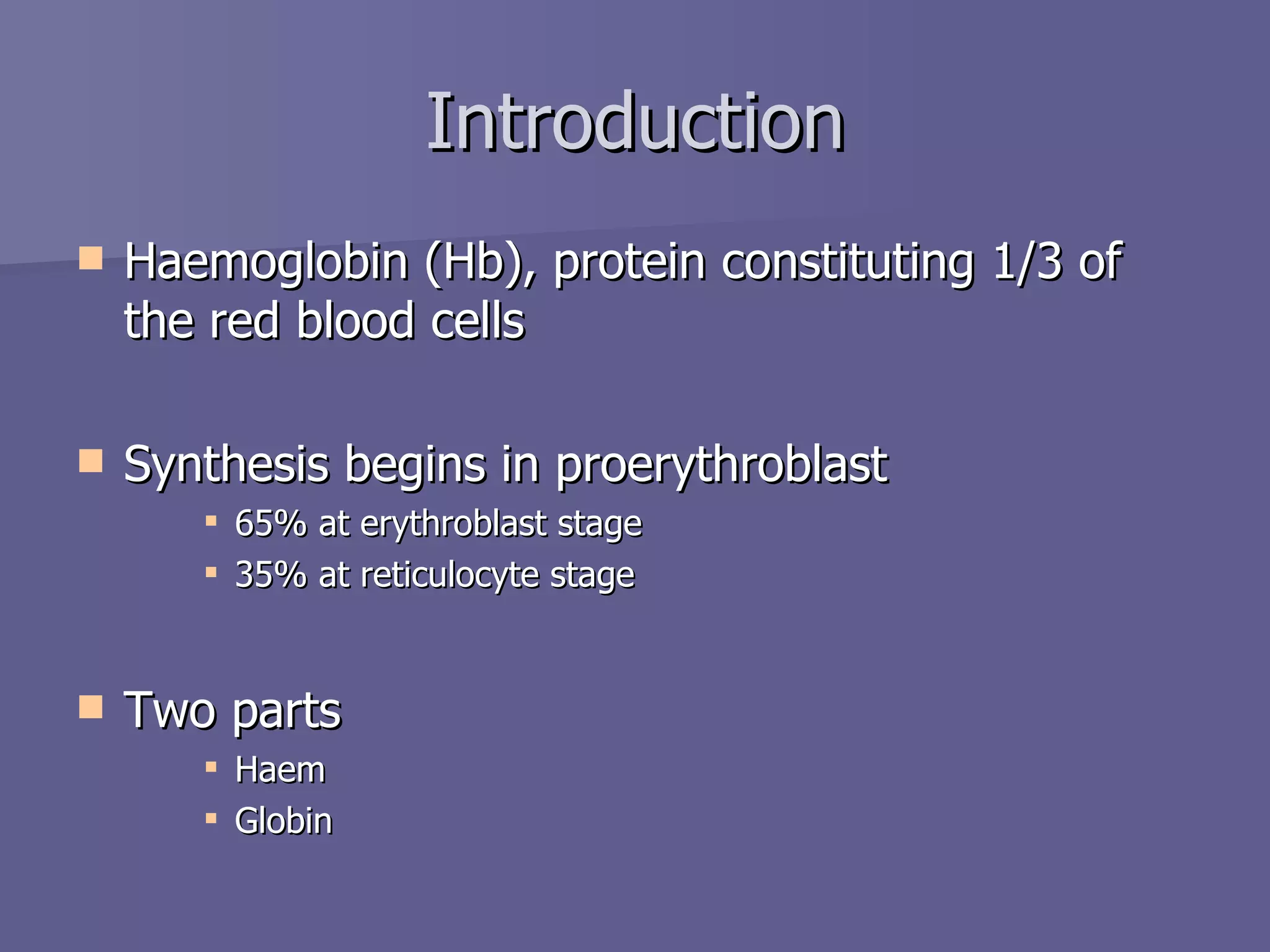
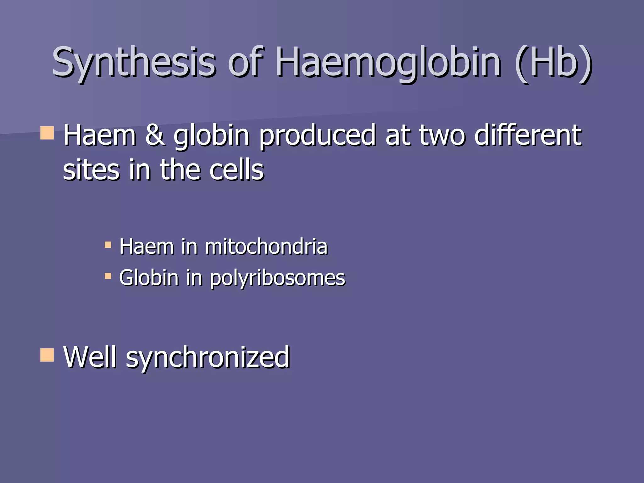
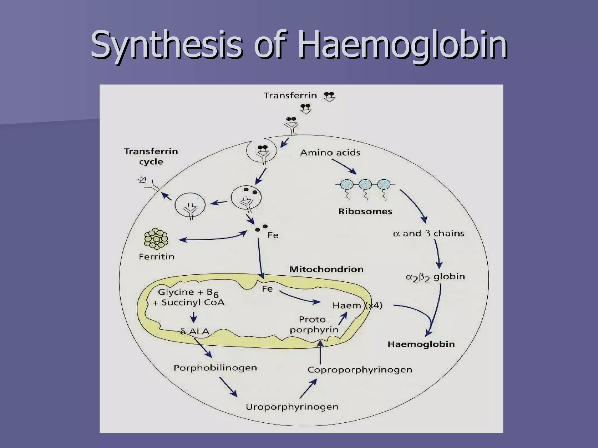
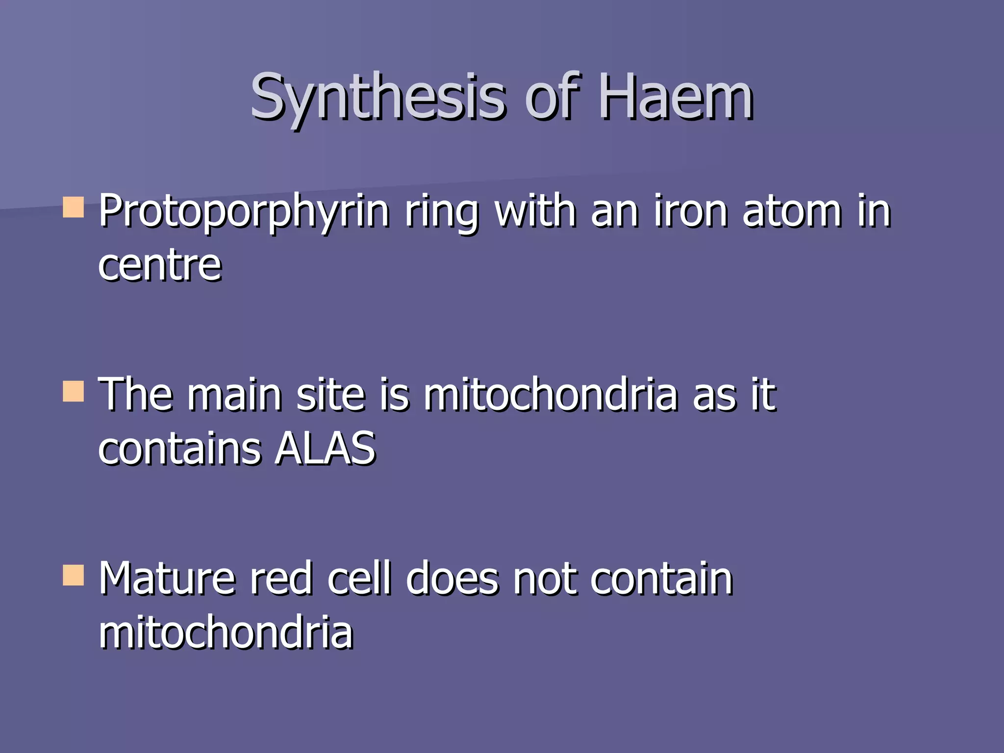
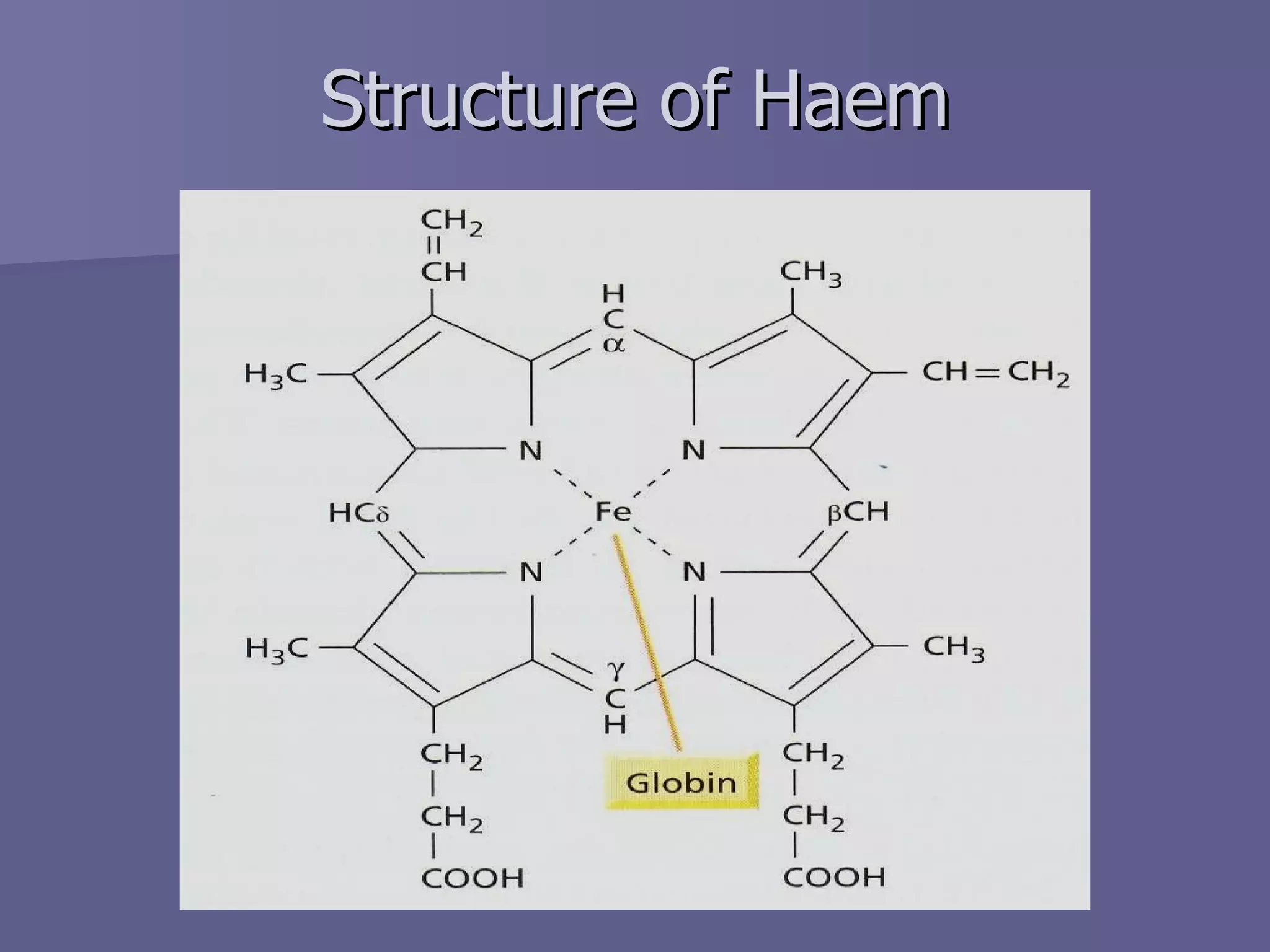
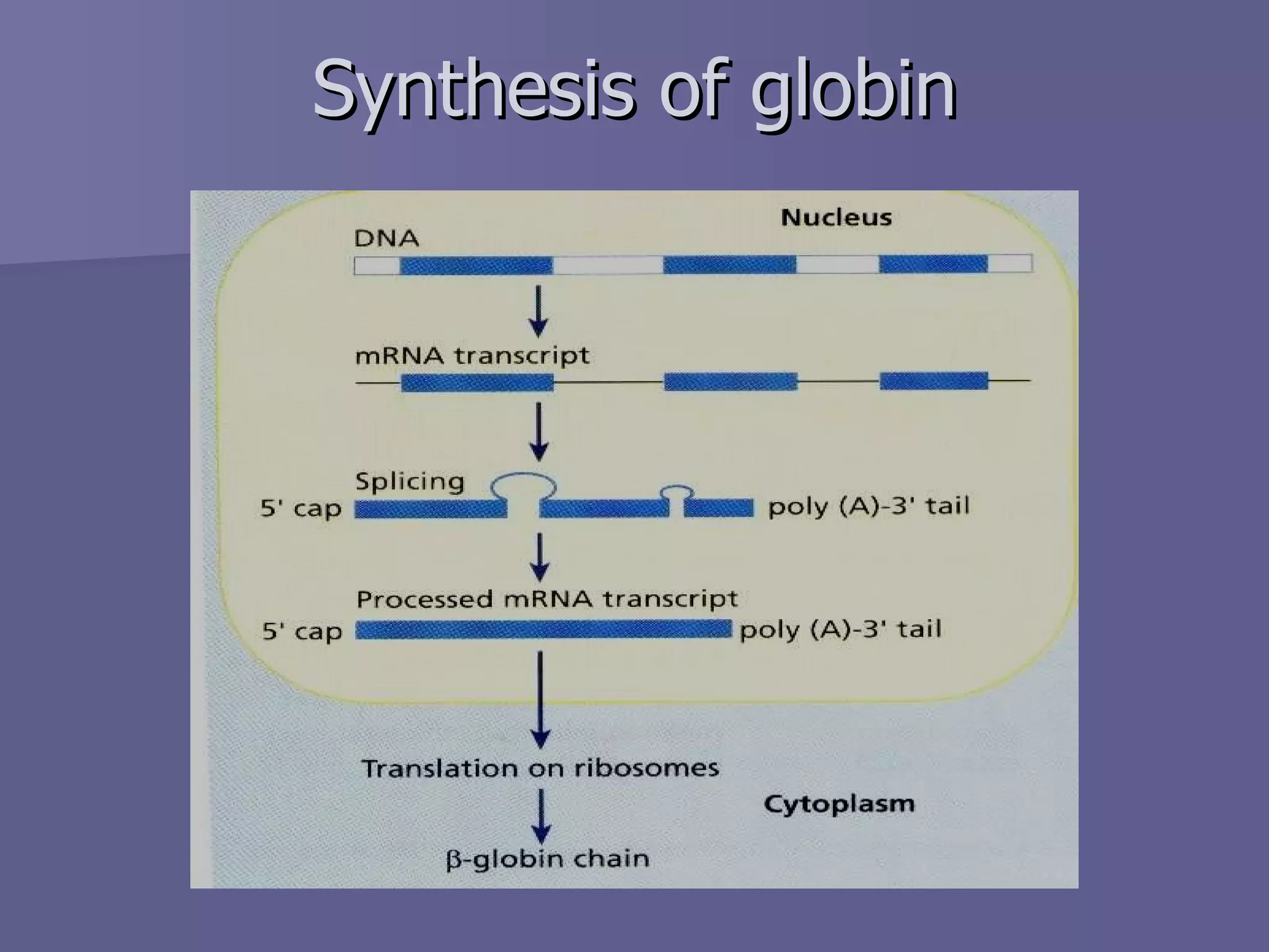
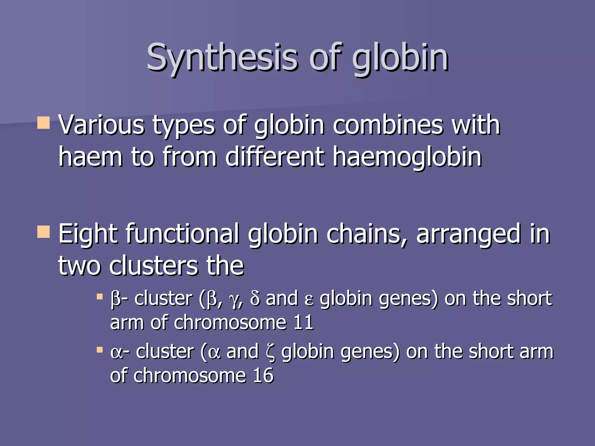
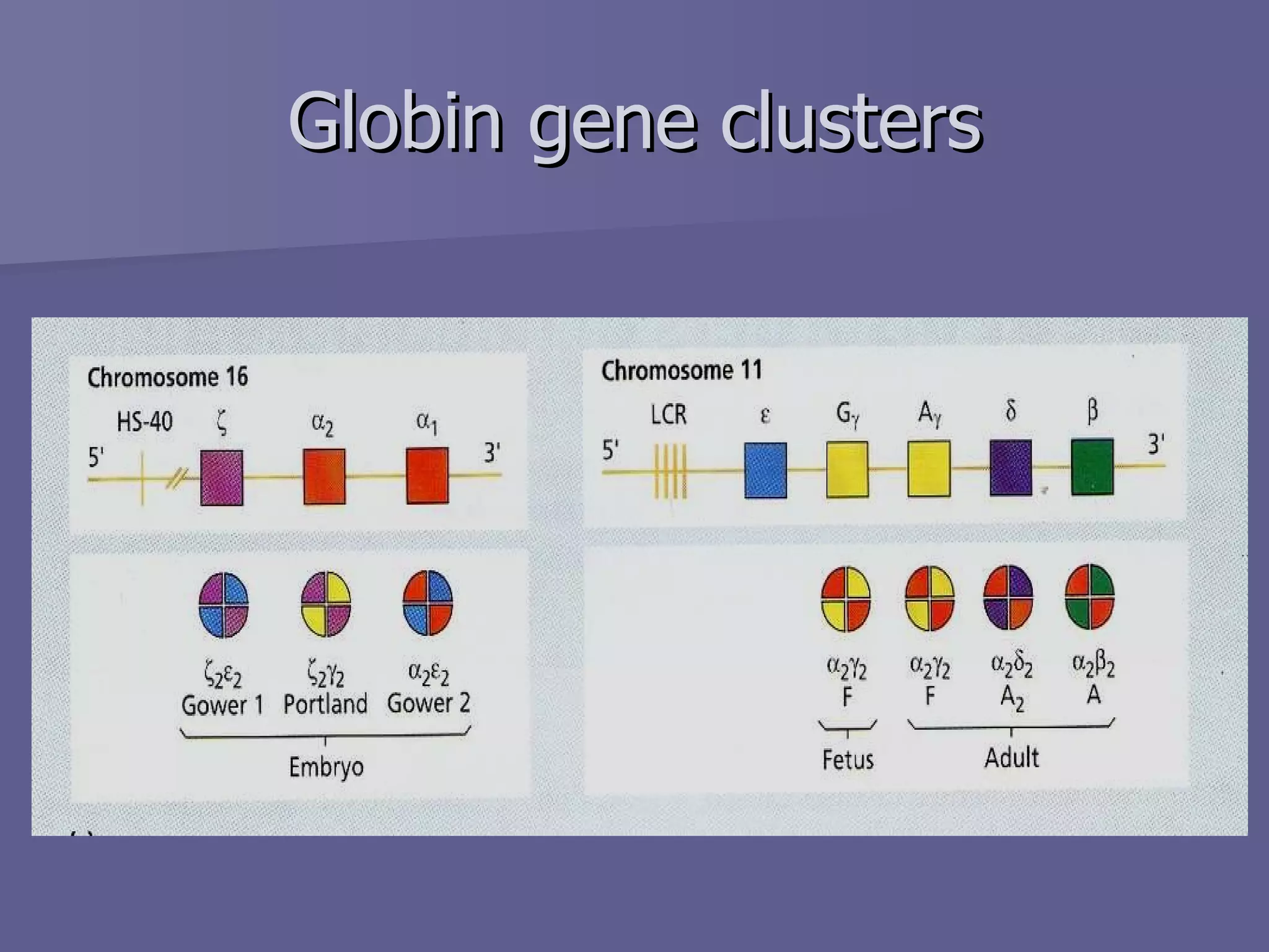
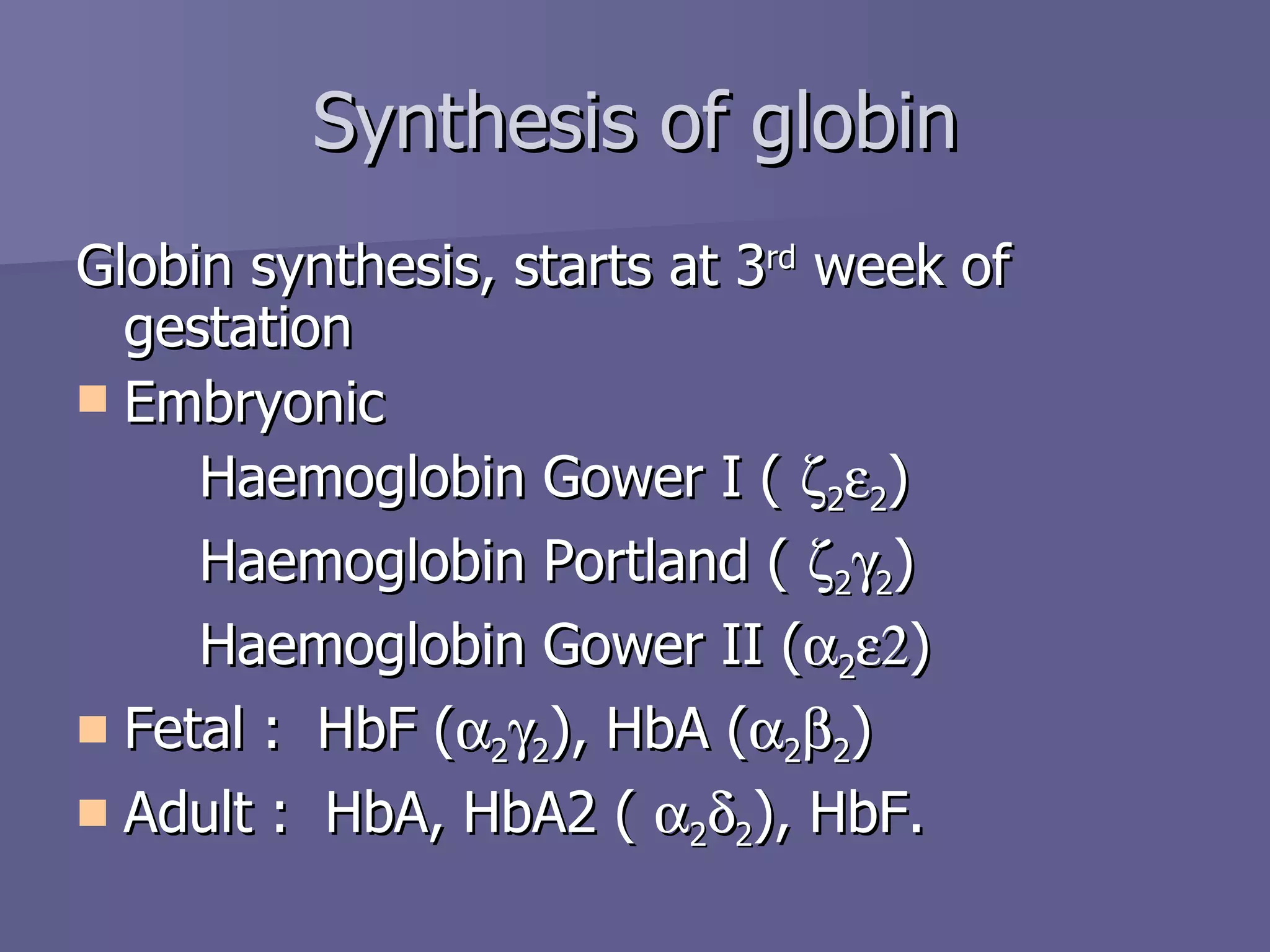
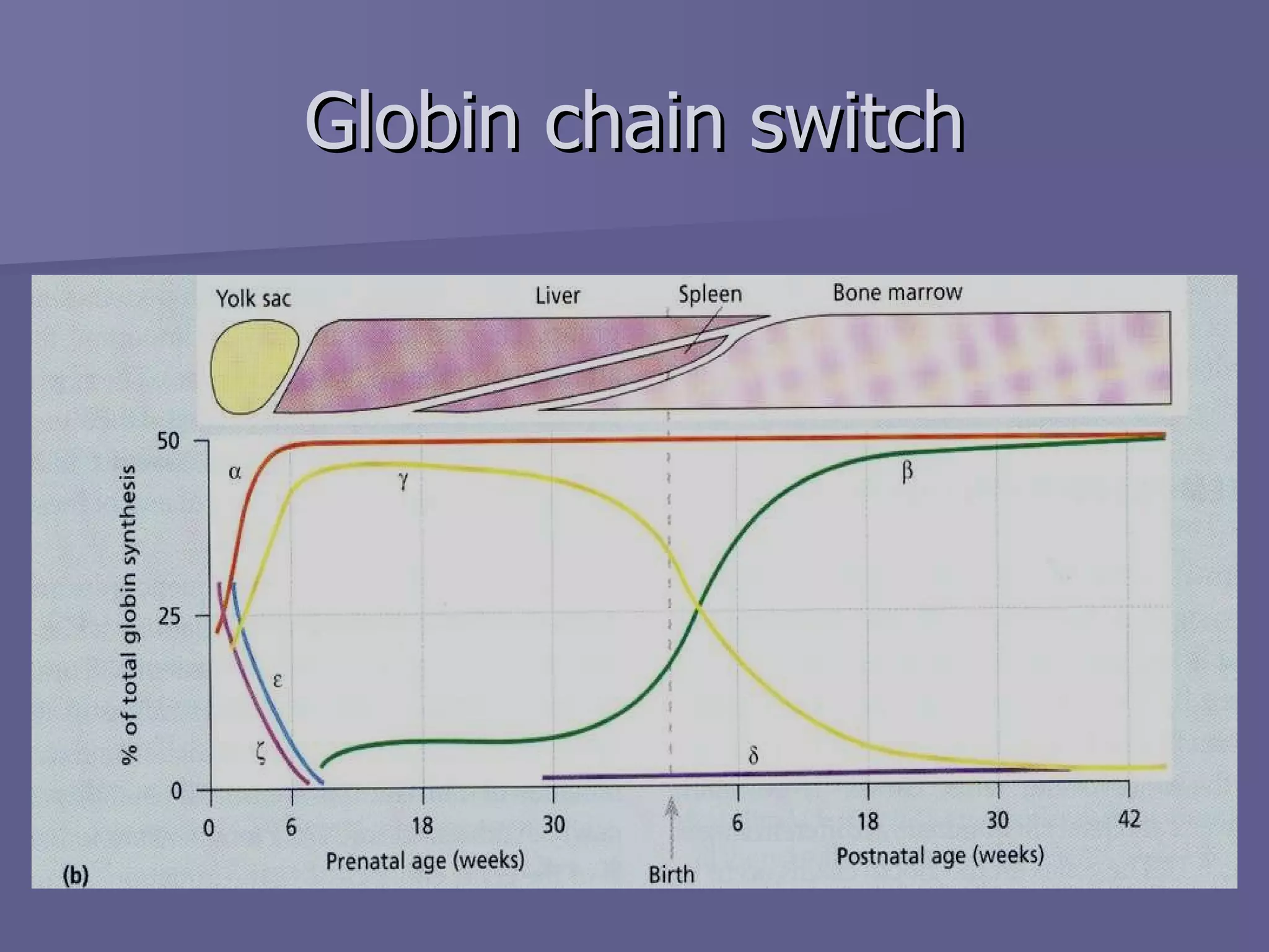
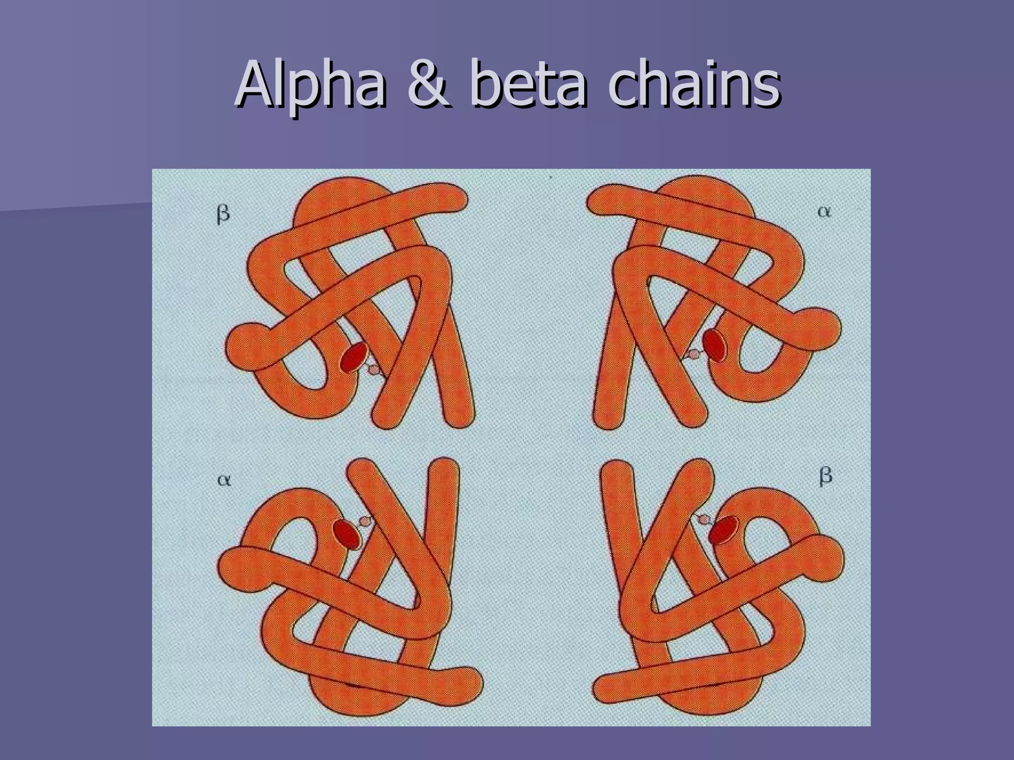
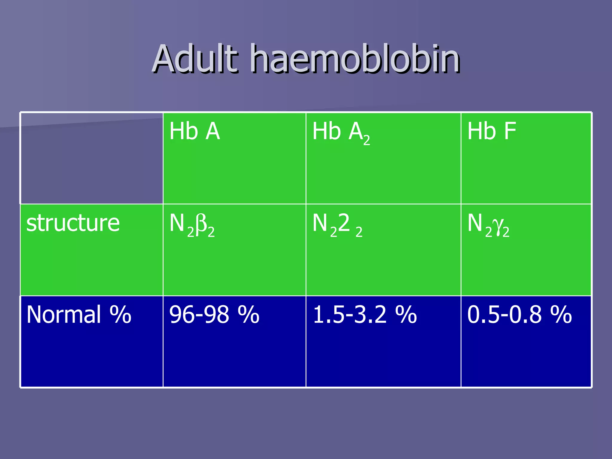
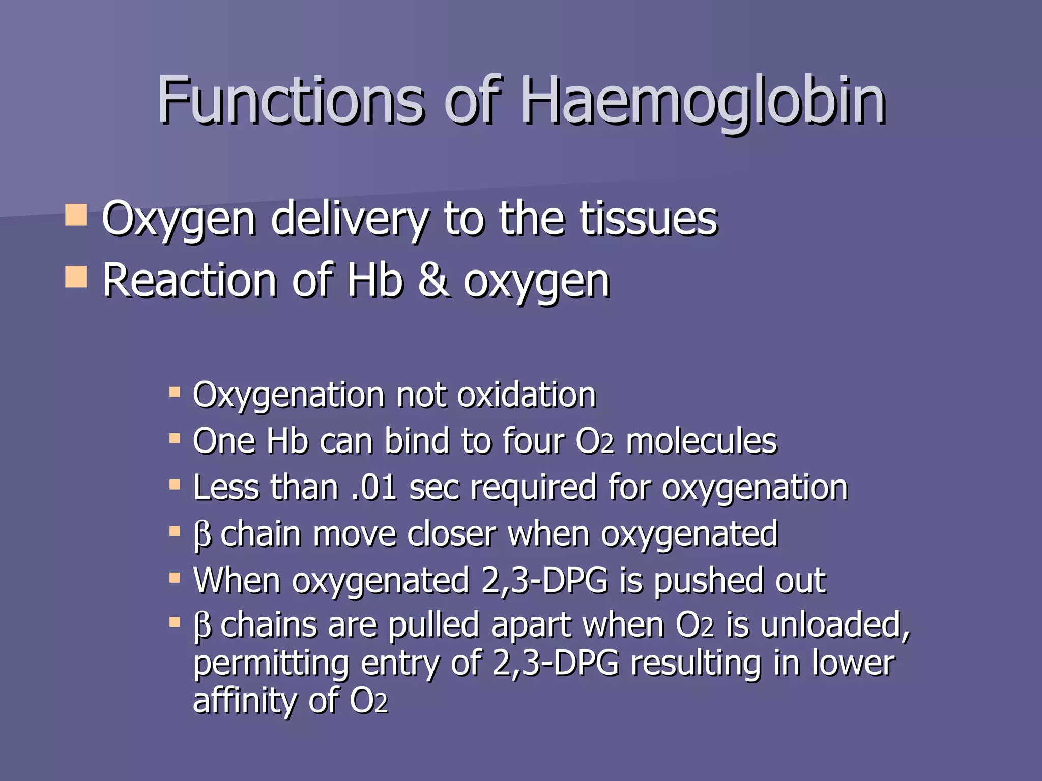
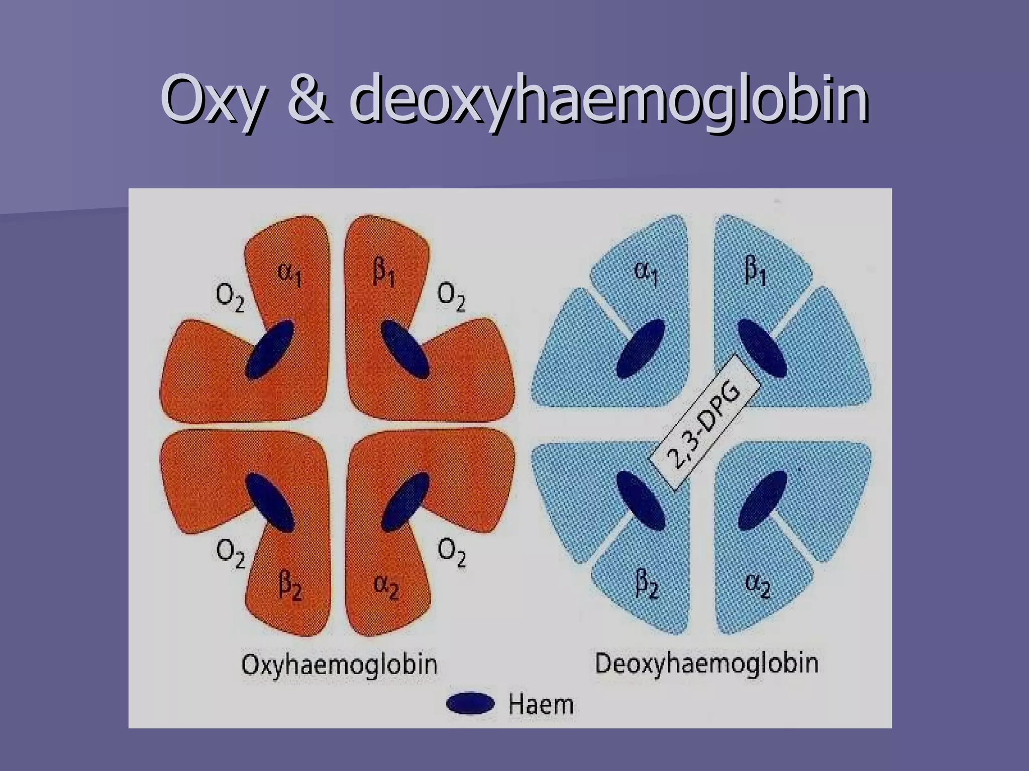
Structure and function of Haemoglobin Dr. Tariq M Roshan Department of Hematology PPSP
The main function of red blood cell Transfer of O 2  from lungs to tissue Transfer of CO 2  from tissue to lungs To accomplish this function red cells has haemoglobin (Hb) Each red cell has 640 million molecules of Hb Introduction
Introduction Haemoglobin (Hb), protein constituting 1/3 of the red blood cells Synthesis begins in proerythroblast 65% at erythroblast stage 35% at reticulocyte stage Two parts Haem Globin
Synthesis of Haemoglobin (Hb) Haem & globin produced at two different sites in the cells Haem in mitochondria Globin in polyribosomes Well synchronized
Synthesis of Haemoglobin
Synthesis of Haem Protoporphyrin ring with an iron atom in centre The main site is mitochondria as it contains ALAS Mature red cell does not contain mitochondria
Structure of Haem
Synthesis of globin
Synthesis of globin Various types of globin combines with haem to from different haemoglobin Eight functional globin chains, arranged in two clusters the   - cluster (  ,   ,    and    globin genes) on the short arm of chromosome 11   - cluster (   and    globin genes) on the short arm of chromosome 16
Globin gene clusters
Globin synthesis, starts at 3 rd  week of gestation Embryonic Haemoglobin Gower I (   2  2 ) Haemoglobin Portland (   2  2 ) Haemoglobin Gower II (  2  ) Fetal :  HbF (  2  2 ), HbA (  2  2 ) Adult :  HbA, HbA2 (   2  2 ), HbF. Synthesis of globin
Globin chain switch
Alpha & beta chains
Adult haemoblobin 0.5-0.8 % 1.5-3.2 % 96-98 % Normal %  2  2  2  2  2  2 structure Hb F Hb A 2 Hb A
Functions of Haemoglobin Oxygen delivery to the tissues Reaction of Hb & oxygen Oxygenation not oxidation One Hb can bind to four O 2  molecules Less than .01 sec required for oxygenation  chain move closer when oxygenated When oxygenated 2,3-DPG is pushed out  chains are pulled apart when O 2  is unloaded, permitting entry of 2,3-DPG resulting in lower affinity of O 2
Oxy & deoxyhaemoglobin
Oxygen-haemoglobin dissociation curve O 2  carrying capacity of Hb at different Po 2 Sigmoid shape Binding of one molecule facilitate the second molecule binding P  50  (partial pressure of O 2  at which Hb is half saturated with O 2 ) 26.6mmHg
Hb-oxygen dissociation curve
The normal position of curve depends on Concentration of 2,3-DPG H +  ion concentration (pH) CO 2  in red blood cells Structure of Hb Hb-oxygen dissociation curve
Right shift (easy oxygen delivery) High 2,3-DPG High H + High CO 2 HbS Left shift (give up oxygen less readily) Low 2,3-DPG HbF Hb-oxygen dissociation curve
Summary Normal structure including the proportion of globin chains are necessary for the normal function of haemoglobin Reduced haemoglobin in the red blood cells due to any abnormality of any of its constituents result into a clinical situation called anaemia Metabolic & other abnormalities result into abnormal oxygen supply to the tissue

Structure And Function Of Haemoglobin

  • 1.
    Structure and functionof Haemoglobin Dr. Tariq M Roshan Department of Hematology PPSP
  • 2.
    The main functionof red blood cell Transfer of O 2 from lungs to tissue Transfer of CO 2 from tissue to lungs To accomplish this function red cells has haemoglobin (Hb) Each red cell has 640 million molecules of Hb Introduction
  • 3.
    Introduction Haemoglobin (Hb),protein constituting 1/3 of the red blood cells Synthesis begins in proerythroblast 65% at erythroblast stage 35% at reticulocyte stage Two parts Haem Globin
  • 4.
    Synthesis of Haemoglobin(Hb) Haem & globin produced at two different sites in the cells Haem in mitochondria Globin in polyribosomes Well synchronized
  • 5.
  • 6.
    Synthesis of HaemProtoporphyrin ring with an iron atom in centre The main site is mitochondria as it contains ALAS Mature red cell does not contain mitochondria
  • 7.
  • 8.
  • 9.
    Synthesis of globinVarious types of globin combines with haem to from different haemoglobin Eight functional globin chains, arranged in two clusters the  - cluster (  ,  ,  and  globin genes) on the short arm of chromosome 11  - cluster (  and  globin genes) on the short arm of chromosome 16
  • 10.
  • 11.
    Globin synthesis, startsat 3 rd week of gestation Embryonic Haemoglobin Gower I (  2  2 ) Haemoglobin Portland (  2  2 ) Haemoglobin Gower II (  2  ) Fetal : HbF (  2  2 ), HbA (  2  2 ) Adult : HbA, HbA2 (  2  2 ), HbF. Synthesis of globin
  • 12.
  • 13.
  • 14.
    Adult haemoblobin 0.5-0.8% 1.5-3.2 % 96-98 % Normal %  2  2  2  2  2  2 structure Hb F Hb A 2 Hb A
  • 15.
    Functions of HaemoglobinOxygen delivery to the tissues Reaction of Hb & oxygen Oxygenation not oxidation One Hb can bind to four O 2 molecules Less than .01 sec required for oxygenation  chain move closer when oxygenated When oxygenated 2,3-DPG is pushed out  chains are pulled apart when O 2 is unloaded, permitting entry of 2,3-DPG resulting in lower affinity of O 2
  • 16.
  • 17.
    Oxygen-haemoglobin dissociation curveO 2 carrying capacity of Hb at different Po 2 Sigmoid shape Binding of one molecule facilitate the second molecule binding P 50 (partial pressure of O 2 at which Hb is half saturated with O 2 ) 26.6mmHg
  • 18.
  • 19.
    The normal positionof curve depends on Concentration of 2,3-DPG H + ion concentration (pH) CO 2 in red blood cells Structure of Hb Hb-oxygen dissociation curve
  • 20.
    Right shift (easyoxygen delivery) High 2,3-DPG High H + High CO 2 HbS Left shift (give up oxygen less readily) Low 2,3-DPG HbF Hb-oxygen dissociation curve
  • 21.
    Summary Normal structureincluding the proportion of globin chains are necessary for the normal function of haemoglobin Reduced haemoglobin in the red blood cells due to any abnormality of any of its constituents result into a clinical situation called anaemia Metabolic & other abnormalities result into abnormal oxygen supply to the tissue