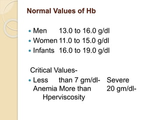
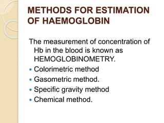
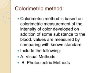
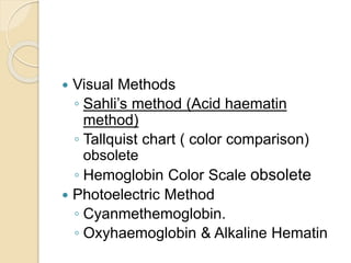
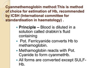
Hemoglobin Estimation
 (RBC) contains two pairs of
polypeptide chains α2 β2 and four
haem groups each having an atom of
ferrous iron(Fe+2).
Hb is the main component of RBCs
and accounts for approximately 34%
of the RBCs by weight.
Iron content of HB is 3.47 mg /100 g.
Main role to transport oxygen.
Various forms of Hb
 Oxyhemoglobin (Hb O2)
 Carboxy hemoglobin (Hb CO)
 Sulfhemoglobin (SHb)
 Methaemoglobin -Fe3+ (ferric)
Variants of Hb
 Normal: Hb F ,Hb A, Hb A2-delta
Abnormal: HbS, HbC, HbD & HbE
 Embryo are two zeta and two epsilon
chains
 Fetal hemoglobin Haemoglobin F
(α2γ2) .
 adult -Haemoglobin A (α2β2) – Has
two alpha chains and two beta chains,
 Haemoglobin A2 (α2δ2). 97% HBA
adult
 Samples used for Hb estimation
 Capillary blood from finger prick.
 Intravenous sample—It should be well
anticoagulated, preferably in EDTA.
Liquid anticoagulants should not be
used at all as these dilute and
decrease Hb concentration.
Normal Values of Hb
 Men 13.0 to 16.0 g/dl
 Women 11.0 to 15.0 g/dl
 Infants 16.0 to 19.0 g/dl
Critical Values-
 Less than 7 gm/dl- Severe
Anemia More than 20 gm/dl-
Hperviscosity
METHODS FOR ESTIMATION
OF HAEMOGLOBIN
The measurement of concentration of
Hb in the blood is known as
HEMOGLOBINOMETRY.
 Colorimetric method
 Gasometric method.
 Specific gravity method
 Chemical method.
Colorimetric method:
 Colorimetric method is based on
colorimetric measurement of the
intensity of color developed on
addition of some substance to the
blood. values are measured by
comparing with known standard.
 Include the following:
 A. Visual Methods
 B. Photoelectric Methods
 Visual Methods
◦ Sahli’s method (Acid haematin
method)
◦ Tallquist chart ( color comparison)
obsolete
◦ Hemoglobin Color Scale obsolete
 Photoelectric Method
◦ Cyanmethemoglobin.
◦ Oxyhaemoglobin & Alkaline Hematin
Commonly used methods
Sahli’s Method – A Color Based Method
 Visual method.
 Principle :
◦ Hb is converted into acid hematin with the
action of dilute hydrochloric acid (N/10 HCl).

◦ The acid hematin is brown in color and its
intensity is matched with a standard brown
glass comparator in a visual colorimeter
called Sahli’s colorimeter.
 Equipment:
◦ Sahli’s Hemaglobinometer consisting of :
 Graduated hemoglobin tube
 Comparator box with a brown glass
standard
◦ Hb Pipette
◦ Stirrer
◦ Dropper (dropping pipette)
 REAGENTS:
 a. N/10 HCl b. Distilled Water
 Sample:
 EDTA anticoagulated venous blood.
 Blood obtained by skin puncture
Sahli’s Method
 Procedure
 Fill Sahli’s Hb tube upto mark 2 with N/10 HCl.
 Deliver 20 μl (0.02 ml) of blood from a Hb pipette into it.
„
 Stir with a stirrer and wait for 10 minutes. „
 Add distilled water drop by drop and stir till color
matches with the comparator.
 Take the reading at upper meniscus.
 ADVANTAGES:
◦ Simple bedside test no colorimeter is
required.
◦ Reagents and apparatus are cheap.
 DISADVANTAGES
◦ There can be visual error.
◦ Carboxy, met and sulfhaemoglobins cannot
be converted to acid hematin.
◦ Comparator can fade over the years.
◦ Color appearing of acid hematin takes long
time and also fades quickly.
◦ Source of light (day light or artificial)
influences the color comparison.
Hemoglobin Color Scale
 This is rapid, simple, inexpensive and
reliable.
 Procedure:
 A drop of blood is placed on strip of
chromatography paper and the color
developed is matched visually against
the printed color scale.
 Color scale consists of printed set of
colors
 corresponding to Hb values.
 Tallquist Hb Chart
 This chart consists of a series of
lithographic colors said to correspond
to Hb values ranging from 10% to
100%. It is obsolete.
 B. Photoelectric Method
 Alkaline haematin
 Oxyhaemoglobin
 Cyanmethemoglobin
Alkaline haematin
 N/10 NaOH solution is used to convert
alkaline haematin read under
Spectrometer
 Advantages-
Stable solution reched.
Disadvantage-
Not reliable in infant and newborn.
Alkalin resistant. Less accurate.
Oxyhaemoglobin method
 Hb is converted into oxyhaemoglobin
by 0.04% ammonia solution .
Measured by spectrophotometer at
540 nm.
 Advantage-
 Reading done in few seconds. And
stable for 3 days.
 Disadvantage - derivatives other than
oxyHb are not measured.
Cyanmethemoglobin method:This is method
of choice for estimation of Hb, recommended
by ICSH (International committee for
standardisation in haematology) .
 Principle – Blood is diluted in a
solution called drabkin’s fluid
containing
 Pot. Ferricyanide converts Hb to
methamoglobin.
 Methamoglobin reacts with Pot.
Cyanide to form cyanmetHb.
 All forms are converted except SULF-
Hb.
 Sample: Blood from Skin Puncture or
EDTA anti-coagulated blood
◦ Take 5 ml of Drabkin solution in two test
tubes each.
◦ Add 20 micro liter of blood and mix well
◦ Allow the tube to stand for at least 5 min.
or more.
◦ Read in Spectrometer at 540 nm.
 Advantage-
 Less error,
 all forms of hemoglobin estimated
except sulphaemoglobin.
 Disadvantage-
 Hyperbilirubinemia affect value.
 Turbidity effect.
Gasometric method
 It is indirect method, which estimates
the amount of haemoglobin from the
amount of oxygen it will absorb. It is
accurate method
but too complicated for clinical work.
Specific Gravity Method
 This method gives approximate value of Hb.
 Method
 A drop of blood is allowed to fall in copper
sulphate solution of specific gravity 1.053
from a height of 1 cm.
 Specific gravity of 1.053 is equivalent to Hb of
12.5 grams.
 If drop sinks within this time its specific
gravity is higher.
 If it floats- specific gravity is low hence low
Hb.
• Normal specific gravity of blood ranges from
1.048-1.066.
Hemoglobin estimation dr sandeep

Hemoglobin estimation dr sandeep

  • 1.
  • 2.
     (RBC) containstwo pairs of polypeptide chains α2 β2 and four haem groups each having an atom of ferrous iron(Fe+2). Hb is the main component of RBCs and accounts for approximately 34% of the RBCs by weight. Iron content of HB is 3.47 mg /100 g. Main role to transport oxygen.
  • 4.
    Various forms ofHb  Oxyhemoglobin (Hb O2)  Carboxy hemoglobin (Hb CO)  Sulfhemoglobin (SHb)  Methaemoglobin -Fe3+ (ferric)
  • 5.
    Variants of Hb Normal: Hb F ,Hb A, Hb A2-delta Abnormal: HbS, HbC, HbD & HbE  Embryo are two zeta and two epsilon chains  Fetal hemoglobin Haemoglobin F (α2γ2) .  adult -Haemoglobin A (α2β2) – Has two alpha chains and two beta chains,  Haemoglobin A2 (α2δ2). 97% HBA adult
  • 6.
     Samples usedfor Hb estimation  Capillary blood from finger prick.  Intravenous sample—It should be well anticoagulated, preferably in EDTA. Liquid anticoagulants should not be used at all as these dilute and decrease Hb concentration.
  • 7.
    Normal Values ofHb  Men 13.0 to 16.0 g/dl  Women 11.0 to 15.0 g/dl  Infants 16.0 to 19.0 g/dl Critical Values-  Less than 7 gm/dl- Severe Anemia More than 20 gm/dl- Hperviscosity
  • 8.
    METHODS FOR ESTIMATION OFHAEMOGLOBIN The measurement of concentration of Hb in the blood is known as HEMOGLOBINOMETRY.  Colorimetric method  Gasometric method.  Specific gravity method  Chemical method.
  • 9.
    Colorimetric method:  Colorimetricmethod is based on colorimetric measurement of the intensity of color developed on addition of some substance to the blood. values are measured by comparing with known standard.  Include the following:  A. Visual Methods  B. Photoelectric Methods
  • 10.
     Visual Methods ◦Sahli’s method (Acid haematin method) ◦ Tallquist chart ( color comparison) obsolete ◦ Hemoglobin Color Scale obsolete  Photoelectric Method ◦ Cyanmethemoglobin. ◦ Oxyhaemoglobin & Alkaline Hematin
  • 11.
    Commonly used methods Sahli’sMethod – A Color Based Method  Visual method.  Principle : ◦ Hb is converted into acid hematin with the action of dilute hydrochloric acid (N/10 HCl).  ◦ The acid hematin is brown in color and its intensity is matched with a standard brown glass comparator in a visual colorimeter called Sahli’s colorimeter.
  • 12.
     Equipment: ◦ Sahli’sHemaglobinometer consisting of :  Graduated hemoglobin tube  Comparator box with a brown glass standard ◦ Hb Pipette ◦ Stirrer ◦ Dropper (dropping pipette)  REAGENTS:  a. N/10 HCl b. Distilled Water  Sample:  EDTA anticoagulated venous blood.  Blood obtained by skin puncture
  • 13.
    Sahli’s Method  Procedure Fill Sahli’s Hb tube upto mark 2 with N/10 HCl.  Deliver 20 μl (0.02 ml) of blood from a Hb pipette into it. „  Stir with a stirrer and wait for 10 minutes. „  Add distilled water drop by drop and stir till color matches with the comparator.  Take the reading at upper meniscus.
  • 16.
     ADVANTAGES: ◦ Simplebedside test no colorimeter is required. ◦ Reagents and apparatus are cheap.  DISADVANTAGES ◦ There can be visual error. ◦ Carboxy, met and sulfhaemoglobins cannot be converted to acid hematin. ◦ Comparator can fade over the years. ◦ Color appearing of acid hematin takes long time and also fades quickly. ◦ Source of light (day light or artificial) influences the color comparison.
  • 17.
    Hemoglobin Color Scale This is rapid, simple, inexpensive and reliable.  Procedure:  A drop of blood is placed on strip of chromatography paper and the color developed is matched visually against the printed color scale.  Color scale consists of printed set of colors  corresponding to Hb values.
  • 19.
     Tallquist HbChart  This chart consists of a series of lithographic colors said to correspond to Hb values ranging from 10% to 100%. It is obsolete.  B. Photoelectric Method  Alkaline haematin  Oxyhaemoglobin  Cyanmethemoglobin
  • 20.
    Alkaline haematin  N/10NaOH solution is used to convert alkaline haematin read under Spectrometer  Advantages- Stable solution reched. Disadvantage- Not reliable in infant and newborn. Alkalin resistant. Less accurate.
  • 21.
    Oxyhaemoglobin method  Hbis converted into oxyhaemoglobin by 0.04% ammonia solution . Measured by spectrophotometer at 540 nm.  Advantage-  Reading done in few seconds. And stable for 3 days.  Disadvantage - derivatives other than oxyHb are not measured.
  • 22.
    Cyanmethemoglobin method:This ismethod of choice for estimation of Hb, recommended by ICSH (International committee for standardisation in haematology) .  Principle – Blood is diluted in a solution called drabkin’s fluid containing  Pot. Ferricyanide converts Hb to methamoglobin.  Methamoglobin reacts with Pot. Cyanide to form cyanmetHb.  All forms are converted except SULF- Hb.
  • 23.
     Sample: Bloodfrom Skin Puncture or EDTA anti-coagulated blood ◦ Take 5 ml of Drabkin solution in two test tubes each. ◦ Add 20 micro liter of blood and mix well ◦ Allow the tube to stand for at least 5 min. or more. ◦ Read in Spectrometer at 540 nm.
  • 24.
     Advantage-  Lesserror,  all forms of hemoglobin estimated except sulphaemoglobin.  Disadvantage-  Hyperbilirubinemia affect value.  Turbidity effect.
  • 25.
    Gasometric method  Itis indirect method, which estimates the amount of haemoglobin from the amount of oxygen it will absorb. It is accurate method but too complicated for clinical work.
  • 26.
    Specific Gravity Method This method gives approximate value of Hb.  Method  A drop of blood is allowed to fall in copper sulphate solution of specific gravity 1.053 from a height of 1 cm.  Specific gravity of 1.053 is equivalent to Hb of 12.5 grams.  If drop sinks within this time its specific gravity is higher.  If it floats- specific gravity is low hence low Hb. • Normal specific gravity of blood ranges from 1.048-1.066.