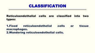
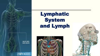
Reticuloendothelial System,
Tissue Macrophages
By
Dr. Faraza Javaid
Reticuloendothelial system
Reticuloendothelial system or tissue macrophage system is the
system of primitive phagocytic cells, which play an important
role in defense mechanism of the body.
The reticuloendothelial cells are found in the following
structures:
1. Endothelial lining of vascular and lymph channels.
2. Connective tissue and some organs like spleen, liver, lungs,
lymph nodes, bone marrow, etc.
Reticular cells in these tissues form the tissue macrophage
system.
CLASSIFICATION
Reticuloendothelial cells are classified into two
types:
1.Fixed reticuloendothelial cells or tissue
macrophages.
2.Wandering reticuloendothelial cells.
FIXED RETICULOENDOTHELIAL CELLS –
TISSUE MACROPHAGES
Fixed reticuloendothelial cells are also called the tissue
macrophages or fixed histiocytes because, these cells are usually
located in the tissues.
Connective Tissue
Endothelium of Blood Sinusoid
Central Nervous System
Lungs
Subcutaneous Tissue
WANDERING RETICULOENDOTHELIAL
CELLS AND TISSUE MACROPHAGES
Wandering reticuloendothelial cells are also called free
histiocytes. There are two types of wandering reticuloendothelial
cells:
1. Free Histiocytes of Blood
Macrophages migrate to the site of injury or infection.
2. Free Histiocytes of Solid Tissue
During emergency, the fixed histiocytes from connective tissue
and other organs
FUNCTIONS OF
RETICULOENDOTHELIAL SYSTEM
Reticuloendothelial system plays an important role in the
defense mechanism of the body.
 Phagocytic Function
 Secretion of Bactericidal Agents
 Secretion of Interleukins
 Secretion of Tumor Necrosis Factors
 Secretion of Transforming Growth Factor
 Secretion of Platelet-derived Growth Factor
 Destruction of Hemoglobin
Spleen
Spleen is the largest lymphoid organ in
the body and it is highly vascular. It is
situated in the upper left part of the
abdomen, behind the stomach and just
below the diaphragm.
Spleen is covered by an outer serous
coat and an inner fibromuscular
capsule.
From the capsule, the trabeculae and
trabecular network arise.
All the three structures, viz. capsule, trabeculae and
trabecular network contain collagen fibers, elastic fibers and
smooth muscle fibers.
The spleen is divided into red and white pulp.
„
RED PULP
Red pulp consists of venous sinus and cords of structures like
blood cells, macrophages and mesenchymal cells.
„
WHITE PULP
The structure of white pulp is similar to that of lymphoid tissue.
It has a central artery that are formed by lymphatic sheath
containing lymphocytes and macrophages.
Function of Spleen
 FORMATION OF BLOOD CELLS
 DESTRUCTION OF BLOOD CELLS
 BLOOD RESERVOIR FUNCTION
 ROLE IN DEFENSE OF BODY
Pathologies Related to Spleen
SPLENOMEGALY AND HYPERSPLENISM
Splenomegaly refers to enlargement of spleen. Increase in the
activities of spleen is called hypersplenism.
Diseases which cause splenomegaly:
1. Infectious diseases such as malaria, typhoid and tuberculosis
2. Inflammatory diseases like rheumatoid arthritis
4. Liver diseases
HYPOSPLENISM AND ASPLENIA
 Hyposplenism or hyposplenia refers to diminished
functioning of spleen. It occurs after partial removal of
spleen due to trauma or cyst.
 Asplenia means absence of spleen.
Lymphatic
System
and Lymph
LYMPHATIC SYSTEM
Lymphatic system is a closed
system of lymph channels or
lymph vessels, through which
lymph flows. It is a one-way
system and allows the lymph
flow from tissue spaces toward
the blood.
Lymphatic system arises from
tissue spaces as a meshwork of
delicate vessels. These vessels
are called lymph capillaries.
DRAINAGE OF LYMPHATIC SYSTEM
Larger lymph vessels
ultimately form the
right lymphatic duct
and thoracic duct.
Right lymphatic duct
opens into right
subclavian vein and
the thoracic duct
opens into left
subclavian vein.
Lymph vessels are situated in the following
regions:
1. Deeper layers of skin
2. Subcutaneous tissues
3. Diaphragm
4. Wall of abdominal cavity
5. Linings of respiratory, digestive, urinary tract
6. Liver, Heart
Lymph vessels are not present in the following structures
1. Superficial layers of skin
2. Central nervous system
3. Cornea
4. Bones
5. Alveoli of lungs.
STRUCTUTRE OF LYMPH NODES
The structures are
arranged in three
layers namely:
 Cortex
 Paracortex
 Medulla
FUNCTIONS OF LYMPH NODES
1. When lymph passes through the lymph nodes, it is
filtered, i.e. the water and electrolytes are removed.
But, the proteins and lipids are retained in the lymph.
2. Bacteria and other toxic substances are destroyed by
macrophages of lymph nodes. Because of this, lymph
nodes are called DEFENSE BARRIERS.
COMPOSITION OF LYMPH
FUNCTIONS OF LYMPH
 It is responsible for redistribution of fluid in the body.
 Bacteria, toxins and other foreign bodies are removed
from tissues via lymph.
 Lymph flow is responsible for the maintenance of
structural and functional integrity of tissue.
 It plays an important role in immunity by transport of
lymphocytes.
Tissue Fluid and Edema
Tissue fluid is the medium in which cells are bathed. It is
otherwise known as interstitial fluid.
„FUNCTIONS OF TISSUE FLUID
 Because of the capillary membrane, there is no direct
contact between blood and cells. And, tissue fluid acts as a
medium for exchange of various substances between the
cells and blood.
 Oxygen and nutritive substances diffuse from the arterial
end of capillary through the tissue fluid and reach the cells.
 Carbon dioxide and waste materials diffuse from the cells
into the venous end of capillary through this fluid.
FORMATION OF TISSUE FLUID
Formation of tissue fluid involves two processes:
1. Filtration.
2. Reabsorption.
Tissue fluid is formed by the process of filtration. Normally, the
blood pressure (also called hydrostatic pressure) in arterial end
of the capillary is about 30 mm Hg.
This hydrostatic pressure is the driving force for filtration of
water and other substances from blood into tissue spaces.
Along the course of the capillary, the pressure falls gradually
and it is about 15 mm Hg at the venous end.
Starling Hypothesis
Starling hypothesis states that the net filtration through
capillary membrane is proportional to the hydrostatic pressure
difference across the membrane minus the oncotic pressure
difference. These pressures are called Starling forces.
Hydrostatic pressure is the force that pushes the fluid
out of blood capillaries whereas, oncotic pressure is
the force that pushes the fluid into the blood
capillaries.
REABSORPTION
 Fluid filtered at the arterial end of capillaries is reabsorbed
back into the blood at the venous end of capillaries. Here also,
the pressure gradient plays an important role.
 Thus, the process of filtration at the arterial end of the
capillaries helps in the formation of tissue fluids and the
process of reabsorption at the venous end helps to maintain
the volume of tissue fluid..
EDEMA
Edema is defined as the swelling caused by excessive
accumulation of fluid in the tissues. It may be generalized
or local.
TYPES OF EDEMA
1. Intracellular edema
2. Extracellular edema.
INTRACELLULAR EDEMA
Intracellular edema is the accumulation of fluid inside the
cell. It occurs because of three reasons:
1. Malnutrition
2. Poor metabolism
3. Inflammation of the tissues
EXTRACELLULAR EDEMA
Extracellular edema is defined as the accumulation of fluid
outside the cell.
Conditions which lead to extracellular edema
1. Heart failure.
2. Renal disease.
3. Decreased amount of plasma proteins.
4. Lymphatic obstruction.
5. Increased endothelial permeability.
Elephantiasis
Elephantiasis is a
disorder of lymphatic
system, characterized
by thickening of skin
and extreme
enlargement of the
affected area, most
commonly limbs (legs),
certain areas of trunk
and parts of head.
PITTING AND NON-PITTING EDEMA
Introduction to
Digestive System
INTRODUCTION
Digestive system is made up of gastrointestinal tract (GI tract)
or alimentary canal and accessory organs, which help in the
process of digestion and absorption. GI tract is a tubular
structure extending from the mouth up to anus, with a length of
about 30 feet.
Digestion
Digestion is defined as the mechanical process by which food
is broken down into simple chemical substances that can be
absorbed and used as nutrients by the body.
A normal young healthy adult consumes about 1 kg of solid diet
and about 1 to 2 liter of liquid diet every day.
Muscular propulsion of materials into
the esophagus
Transport of materials to the
stomach
Major Organs of
the Digestive Tract
Oral Cavity (Mouth)
Pharynx
Esophagus
Stomach
Chemical breakdown of materials by
acid
and enzymes; mechanical processing
through muscular contractions
Small Intestine
Enzymatic digestion and absorption of
water, organic substrates, vitamins, and
ions
Large Intestine
Dehydration and compaction of
indigestible
materials in preparation for elimination
Anus
Ingestion, mechanical processing with
accessory organs (teeth and tongue),
moistening, mixing with salivary secretions
Accessory Organs of
the Digestive System
Mechanical processing by
chewing (mastication)
Teeth
Assists mechanical processing
with teeth, sensory analysis
Tongue
Secretion of lubricating fluid
containing enzymes that
break down carbohydrates
Salivary Glands
Secretion of bile (important
for lipid digestion), storage of
nutrients, many other vital
functions
Liver
Storage and concentration of
bile
Gallbladder
Exocrine cells secrete buffers
and digestive enzymes;
Endocrine cells secrete
hormones
Pancreas
Movement of the
nutrient molecules
from the external
environment to the
internal
environment
Function of Digestive Tract
The Digestive Tract
Histological Organization of the Digestive
Tract
Four major layers of the digestive tract
1. Mucosa
(Digestion, Absorption and Secretion)
2. Submucosa
(Glands and Blood Supply)
3. Muscularis externa
(Circular/Longitudinal muscular Tissue)
4. Serosa (Connective Tissues)
NERVE SUPPLY TO
GASTROINTESTINAL TRACT
GI tract has two types of nerve supply:
I. Intrinsic nerve supply
II. Extrinsic nerve supply.
INTRINSIC NERVE SUPPLY –
ENTERIC NERVOUS SYSTEM
Intrinsic nerves to GI tract form the enteric nervous system that
controls all the secretions and movements of GI tract.
Enteric nervous system is present within the wall of GI tract
from esophagus to anus.
Nerve fibers of this system are interconnected and form two
major networks called:
1. Auerbach plexus
2. Meissner plexus
Auerbach Plexus/ Myenteric Nerve Plexus
Major function of this plexus is to regulate the movements of
GI tract.
Some nerve fibers of this plexus accelerate the movements by
secreting the excitatory neurotransmitter substances like
acetylcholine, serotonin and substance P.
Other fibers of this plexus inhibit the GI motility by secreting
the inhibitory neurotransmitters such as vasoactive intestinal
polypeptide (VIP), neurotensin and enkephalin.
Meissner Nerve Plexus
Function of Meissner plexus is the regulation of secretory
functions of GI tract.
EXTRINSIC NERVE SUPPLY
Extrinsic nerves that control the enteric nervous system are from
autonomic nervous system. Both sympathetic and parasympathetic
divisions of autonomic nervous system innervate the GI tract.
Sympathetic Nerve Fibers
Sympathetic nerve fibers inhibit the movements and decrease the
secretions of GI tract by secreting the neurotransmitter noradrenaline.
Parasympathetic Nerve Fibers
Parasympathetic nerve fibers accelerate the movements and increase
the secretions of GI tract.
The neurotransmitter secreted by the parasympathetic nerve fibers is
acetylcholine (Ach).
THANK YOU

Reticuloendothelial System, Tissue Macrophages, Lymphatic System.pptx

  • 1.
  • 2.
    Reticuloendothelial system Reticuloendothelial systemor tissue macrophage system is the system of primitive phagocytic cells, which play an important role in defense mechanism of the body. The reticuloendothelial cells are found in the following structures: 1. Endothelial lining of vascular and lymph channels. 2. Connective tissue and some organs like spleen, liver, lungs, lymph nodes, bone marrow, etc. Reticular cells in these tissues form the tissue macrophage system.
  • 3.
    CLASSIFICATION Reticuloendothelial cells areclassified into two types: 1.Fixed reticuloendothelial cells or tissue macrophages. 2.Wandering reticuloendothelial cells.
  • 4.
    FIXED RETICULOENDOTHELIAL CELLS– TISSUE MACROPHAGES Fixed reticuloendothelial cells are also called the tissue macrophages or fixed histiocytes because, these cells are usually located in the tissues. Connective Tissue Endothelium of Blood Sinusoid Central Nervous System Lungs Subcutaneous Tissue
  • 5.
    WANDERING RETICULOENDOTHELIAL CELLS ANDTISSUE MACROPHAGES Wandering reticuloendothelial cells are also called free histiocytes. There are two types of wandering reticuloendothelial cells: 1. Free Histiocytes of Blood Macrophages migrate to the site of injury or infection. 2. Free Histiocytes of Solid Tissue During emergency, the fixed histiocytes from connective tissue and other organs
  • 6.
    FUNCTIONS OF RETICULOENDOTHELIAL SYSTEM Reticuloendothelialsystem plays an important role in the defense mechanism of the body.  Phagocytic Function  Secretion of Bactericidal Agents  Secretion of Interleukins  Secretion of Tumor Necrosis Factors  Secretion of Transforming Growth Factor  Secretion of Platelet-derived Growth Factor  Destruction of Hemoglobin
  • 7.
    Spleen Spleen is thelargest lymphoid organ in the body and it is highly vascular. It is situated in the upper left part of the abdomen, behind the stomach and just below the diaphragm. Spleen is covered by an outer serous coat and an inner fibromuscular capsule. From the capsule, the trabeculae and trabecular network arise.
  • 8.
    All the threestructures, viz. capsule, trabeculae and trabecular network contain collagen fibers, elastic fibers and smooth muscle fibers. The spleen is divided into red and white pulp.
  • 9.
    „ RED PULP Red pulpconsists of venous sinus and cords of structures like blood cells, macrophages and mesenchymal cells. „ WHITE PULP The structure of white pulp is similar to that of lymphoid tissue. It has a central artery that are formed by lymphatic sheath containing lymphocytes and macrophages.
  • 10.
    Function of Spleen FORMATION OF BLOOD CELLS  DESTRUCTION OF BLOOD CELLS  BLOOD RESERVOIR FUNCTION  ROLE IN DEFENSE OF BODY
  • 11.
    Pathologies Related toSpleen SPLENOMEGALY AND HYPERSPLENISM Splenomegaly refers to enlargement of spleen. Increase in the activities of spleen is called hypersplenism. Diseases which cause splenomegaly: 1. Infectious diseases such as malaria, typhoid and tuberculosis 2. Inflammatory diseases like rheumatoid arthritis 4. Liver diseases
  • 12.
    HYPOSPLENISM AND ASPLENIA Hyposplenism or hyposplenia refers to diminished functioning of spleen. It occurs after partial removal of spleen due to trauma or cyst.  Asplenia means absence of spleen.
  • 13.
  • 14.
    LYMPHATIC SYSTEM Lymphatic systemis a closed system of lymph channels or lymph vessels, through which lymph flows. It is a one-way system and allows the lymph flow from tissue spaces toward the blood. Lymphatic system arises from tissue spaces as a meshwork of delicate vessels. These vessels are called lymph capillaries.
  • 15.
    DRAINAGE OF LYMPHATICSYSTEM Larger lymph vessels ultimately form the right lymphatic duct and thoracic duct. Right lymphatic duct opens into right subclavian vein and the thoracic duct opens into left subclavian vein.
  • 16.
    Lymph vessels aresituated in the following regions: 1. Deeper layers of skin 2. Subcutaneous tissues 3. Diaphragm 4. Wall of abdominal cavity 5. Linings of respiratory, digestive, urinary tract 6. Liver, Heart
  • 17.
    Lymph vessels arenot present in the following structures 1. Superficial layers of skin 2. Central nervous system 3. Cornea 4. Bones 5. Alveoli of lungs.
  • 18.
    STRUCTUTRE OF LYMPHNODES The structures are arranged in three layers namely:  Cortex  Paracortex  Medulla
  • 19.
    FUNCTIONS OF LYMPHNODES 1. When lymph passes through the lymph nodes, it is filtered, i.e. the water and electrolytes are removed. But, the proteins and lipids are retained in the lymph. 2. Bacteria and other toxic substances are destroyed by macrophages of lymph nodes. Because of this, lymph nodes are called DEFENSE BARRIERS.
  • 20.
  • 21.
    FUNCTIONS OF LYMPH It is responsible for redistribution of fluid in the body.  Bacteria, toxins and other foreign bodies are removed from tissues via lymph.  Lymph flow is responsible for the maintenance of structural and functional integrity of tissue.  It plays an important role in immunity by transport of lymphocytes.
  • 22.
    Tissue Fluid andEdema Tissue fluid is the medium in which cells are bathed. It is otherwise known as interstitial fluid. „FUNCTIONS OF TISSUE FLUID  Because of the capillary membrane, there is no direct contact between blood and cells. And, tissue fluid acts as a medium for exchange of various substances between the cells and blood.  Oxygen and nutritive substances diffuse from the arterial end of capillary through the tissue fluid and reach the cells.  Carbon dioxide and waste materials diffuse from the cells into the venous end of capillary through this fluid.
  • 23.
    FORMATION OF TISSUEFLUID Formation of tissue fluid involves two processes: 1. Filtration. 2. Reabsorption. Tissue fluid is formed by the process of filtration. Normally, the blood pressure (also called hydrostatic pressure) in arterial end of the capillary is about 30 mm Hg. This hydrostatic pressure is the driving force for filtration of water and other substances from blood into tissue spaces. Along the course of the capillary, the pressure falls gradually and it is about 15 mm Hg at the venous end.
  • 24.
    Starling Hypothesis Starling hypothesisstates that the net filtration through capillary membrane is proportional to the hydrostatic pressure difference across the membrane minus the oncotic pressure difference. These pressures are called Starling forces. Hydrostatic pressure is the force that pushes the fluid out of blood capillaries whereas, oncotic pressure is the force that pushes the fluid into the blood capillaries.
  • 25.
    REABSORPTION  Fluid filteredat the arterial end of capillaries is reabsorbed back into the blood at the venous end of capillaries. Here also, the pressure gradient plays an important role.  Thus, the process of filtration at the arterial end of the capillaries helps in the formation of tissue fluids and the process of reabsorption at the venous end helps to maintain the volume of tissue fluid..
  • 26.
    EDEMA Edema is definedas the swelling caused by excessive accumulation of fluid in the tissues. It may be generalized or local. TYPES OF EDEMA 1. Intracellular edema 2. Extracellular edema.
  • 27.
    INTRACELLULAR EDEMA Intracellular edemais the accumulation of fluid inside the cell. It occurs because of three reasons: 1. Malnutrition 2. Poor metabolism 3. Inflammation of the tissues
  • 28.
    EXTRACELLULAR EDEMA Extracellular edemais defined as the accumulation of fluid outside the cell. Conditions which lead to extracellular edema 1. Heart failure. 2. Renal disease. 3. Decreased amount of plasma proteins. 4. Lymphatic obstruction. 5. Increased endothelial permeability.
  • 29.
    Elephantiasis Elephantiasis is a disorderof lymphatic system, characterized by thickening of skin and extreme enlargement of the affected area, most commonly limbs (legs), certain areas of trunk and parts of head.
  • 30.
  • 31.
  • 32.
    INTRODUCTION Digestive system ismade up of gastrointestinal tract (GI tract) or alimentary canal and accessory organs, which help in the process of digestion and absorption. GI tract is a tubular structure extending from the mouth up to anus, with a length of about 30 feet. Digestion Digestion is defined as the mechanical process by which food is broken down into simple chemical substances that can be absorbed and used as nutrients by the body. A normal young healthy adult consumes about 1 kg of solid diet and about 1 to 2 liter of liquid diet every day.
  • 33.
    Muscular propulsion ofmaterials into the esophagus Transport of materials to the stomach Major Organs of the Digestive Tract Oral Cavity (Mouth) Pharynx Esophagus Stomach Chemical breakdown of materials by acid and enzymes; mechanical processing through muscular contractions Small Intestine Enzymatic digestion and absorption of water, organic substrates, vitamins, and ions Large Intestine Dehydration and compaction of indigestible materials in preparation for elimination Anus Ingestion, mechanical processing with accessory organs (teeth and tongue), moistening, mixing with salivary secretions
  • 34.
    Accessory Organs of theDigestive System Mechanical processing by chewing (mastication) Teeth Assists mechanical processing with teeth, sensory analysis Tongue Secretion of lubricating fluid containing enzymes that break down carbohydrates Salivary Glands Secretion of bile (important for lipid digestion), storage of nutrients, many other vital functions Liver Storage and concentration of bile Gallbladder Exocrine cells secrete buffers and digestive enzymes; Endocrine cells secrete hormones Pancreas
  • 35.
    Movement of the nutrientmolecules from the external environment to the internal environment Function of Digestive Tract
  • 36.
    The Digestive Tract HistologicalOrganization of the Digestive Tract Four major layers of the digestive tract 1. Mucosa (Digestion, Absorption and Secretion) 2. Submucosa (Glands and Blood Supply) 3. Muscularis externa (Circular/Longitudinal muscular Tissue) 4. Serosa (Connective Tissues)
  • 37.
    NERVE SUPPLY TO GASTROINTESTINALTRACT GI tract has two types of nerve supply: I. Intrinsic nerve supply II. Extrinsic nerve supply.
  • 38.
    INTRINSIC NERVE SUPPLY– ENTERIC NERVOUS SYSTEM Intrinsic nerves to GI tract form the enteric nervous system that controls all the secretions and movements of GI tract. Enteric nervous system is present within the wall of GI tract from esophagus to anus. Nerve fibers of this system are interconnected and form two major networks called: 1. Auerbach plexus 2. Meissner plexus
  • 39.
    Auerbach Plexus/ MyentericNerve Plexus Major function of this plexus is to regulate the movements of GI tract. Some nerve fibers of this plexus accelerate the movements by secreting the excitatory neurotransmitter substances like acetylcholine, serotonin and substance P. Other fibers of this plexus inhibit the GI motility by secreting the inhibitory neurotransmitters such as vasoactive intestinal polypeptide (VIP), neurotensin and enkephalin. Meissner Nerve Plexus Function of Meissner plexus is the regulation of secretory functions of GI tract.
  • 40.
    EXTRINSIC NERVE SUPPLY Extrinsicnerves that control the enteric nervous system are from autonomic nervous system. Both sympathetic and parasympathetic divisions of autonomic nervous system innervate the GI tract. Sympathetic Nerve Fibers Sympathetic nerve fibers inhibit the movements and decrease the secretions of GI tract by secreting the neurotransmitter noradrenaline. Parasympathetic Nerve Fibers Parasympathetic nerve fibers accelerate the movements and increase the secretions of GI tract. The neurotransmitter secreted by the parasympathetic nerve fibers is acetylcholine (Ach).
  • 41.