The document summarizes the cardiac cycle, including:
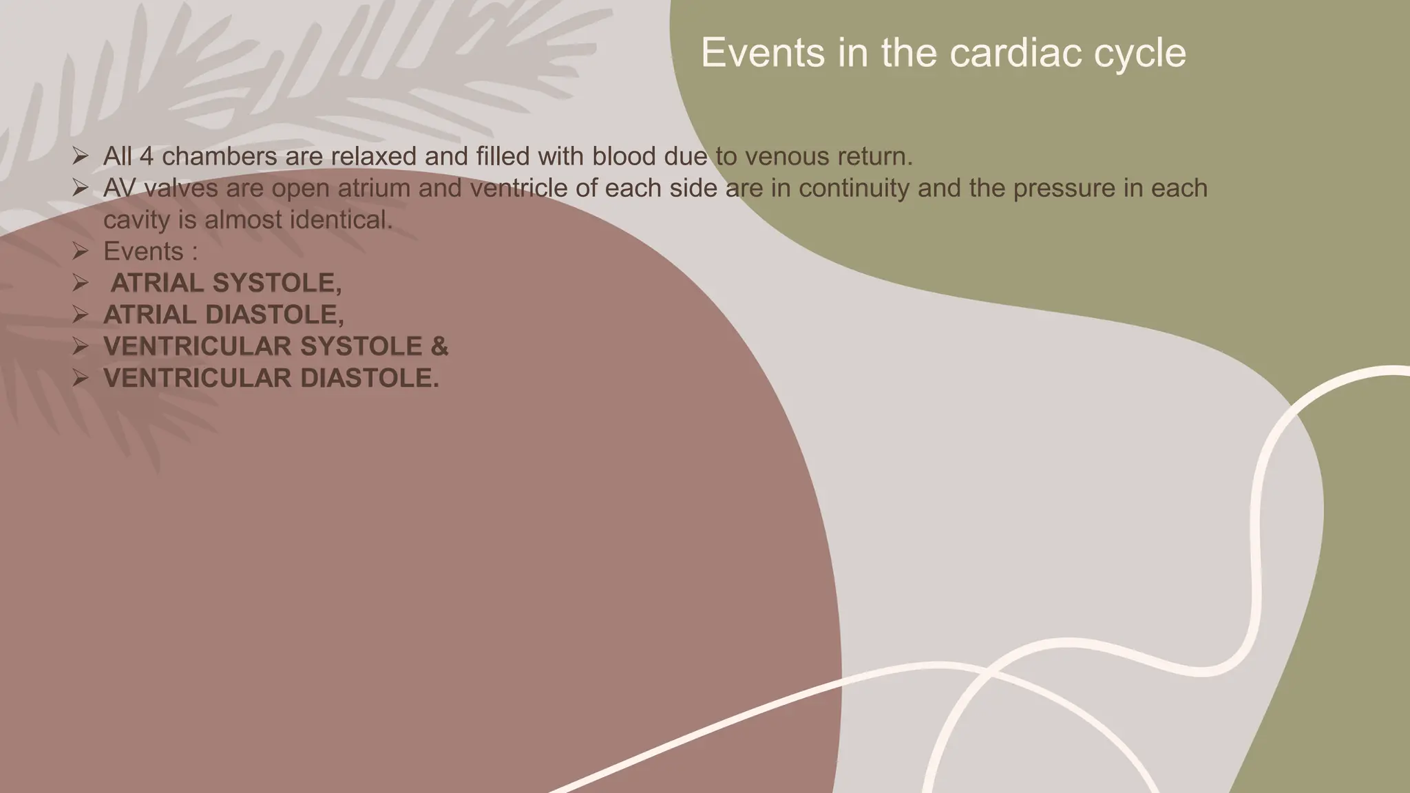
(1) It describes the four main events - atrial systole, ventricular systole, ventricular diastole, and atrial diastole.
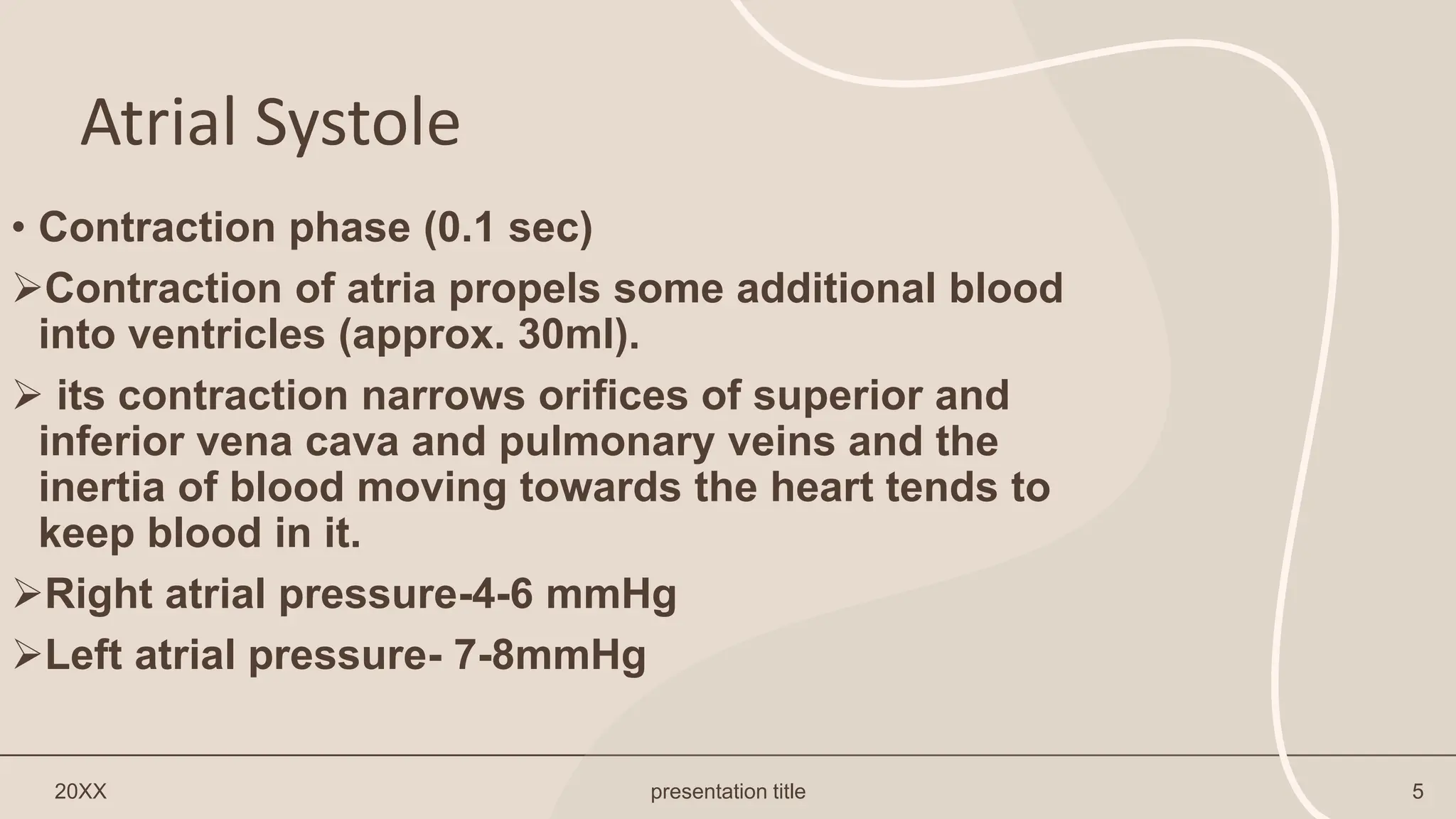
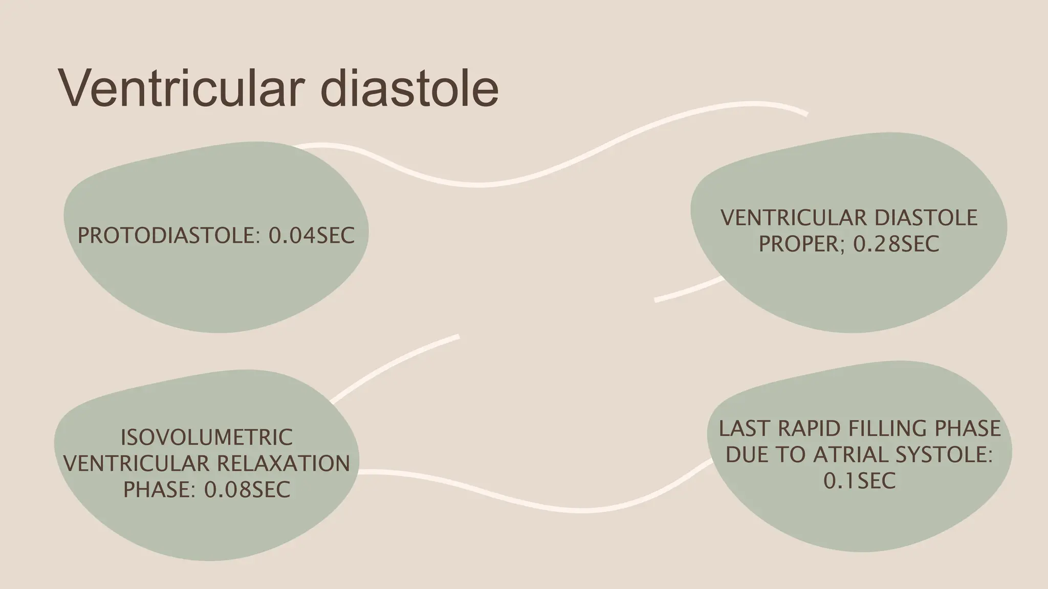
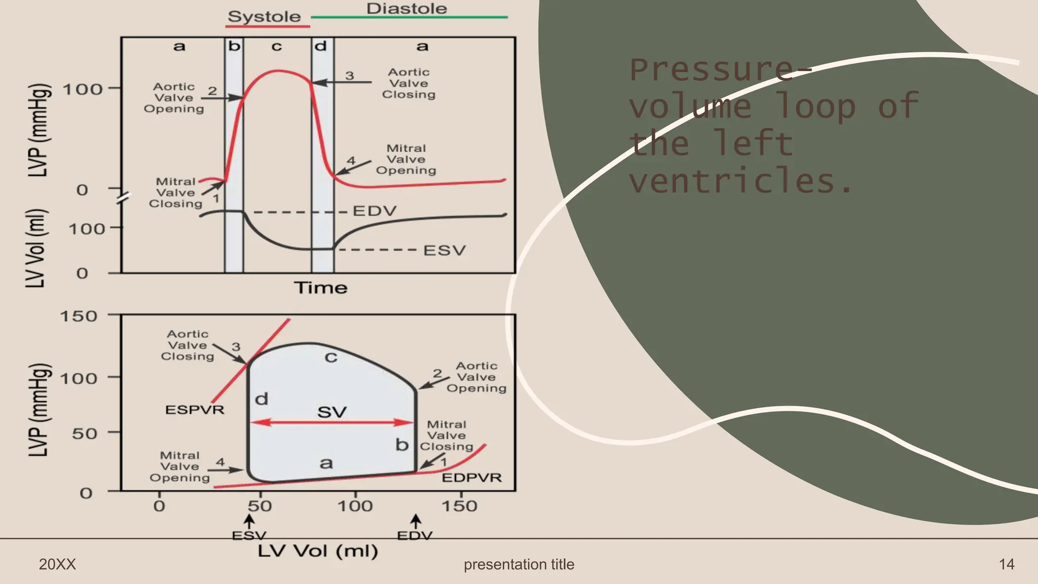
(2) It outlines the pressure and blood flow changes that occur in the heart chambers and valves during each phase of the cycle.
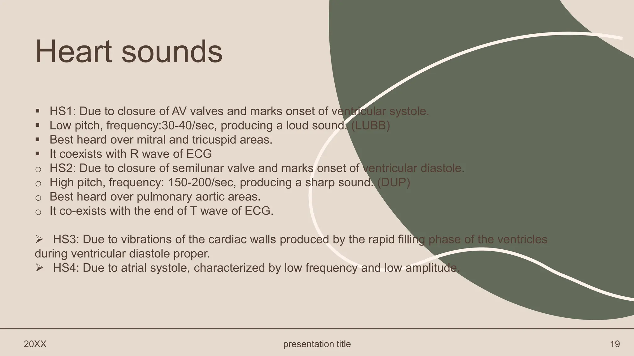
(3) It examines the corresponding electrocardiogram changes and heart sounds that occur with each phase of the cardiac cycle.


![Transport of gases carbon dioxide and oxygen [Autosaved].pptx](https://cdn.slidesharecdn.com/ss_thumbnails/transportofgasesautosaved-250324055730-f2aabc7a-thumbnail.jpg?width=640&height=640&fit=bounds)


