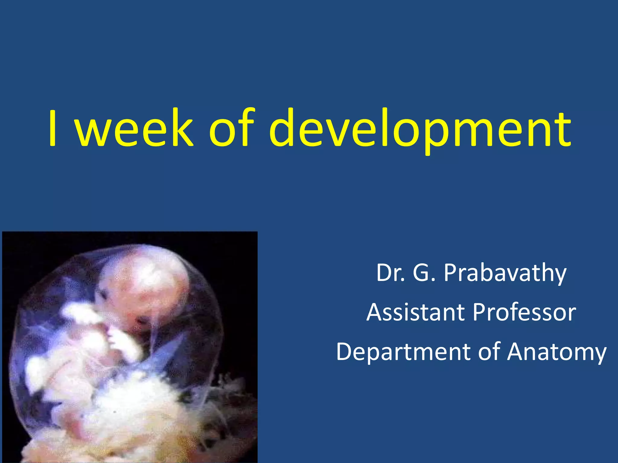
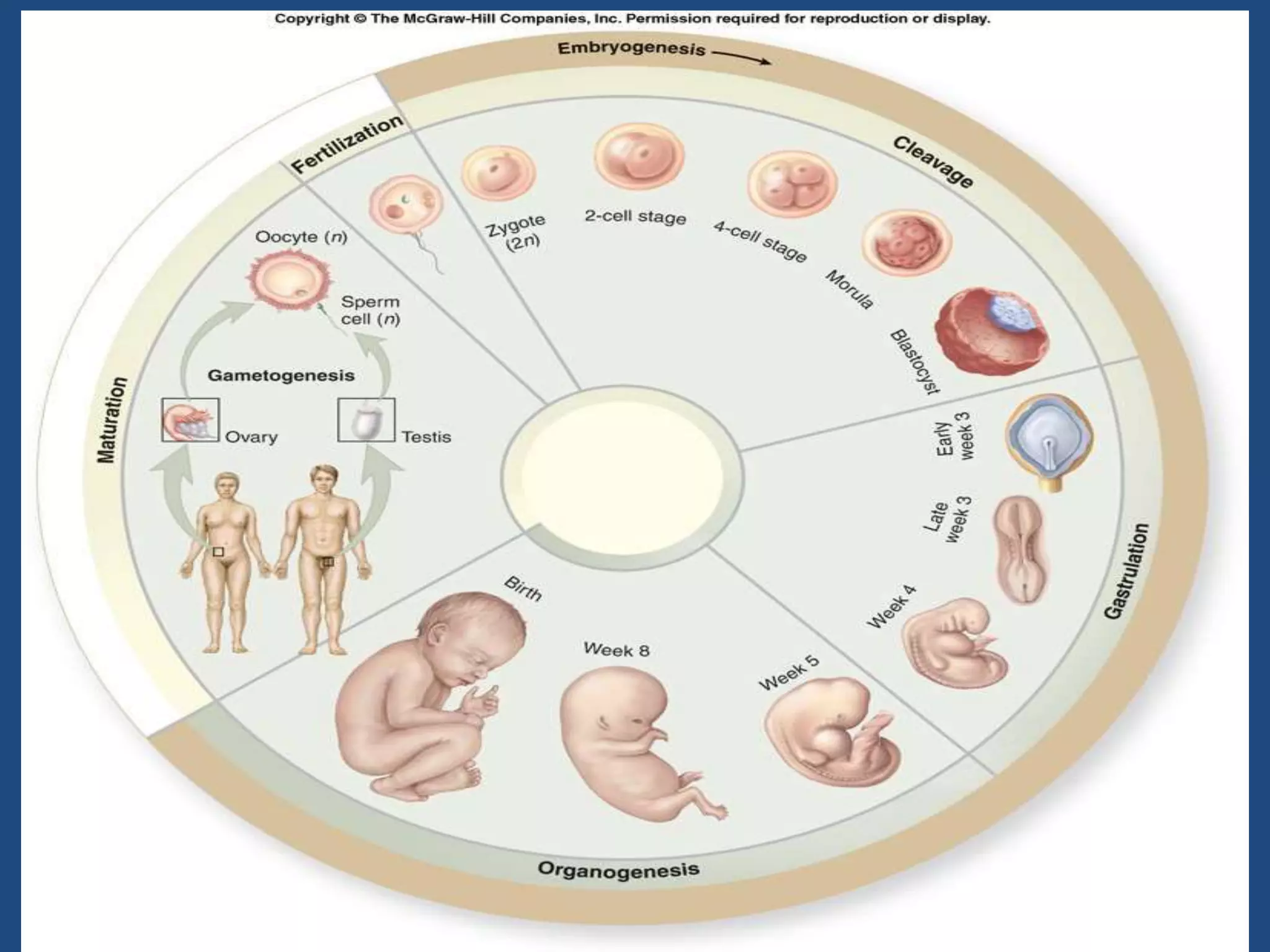
1) The document discusses the key stages in the first week of human development including fertilization, cleavage, formation of the morula and blastocyst, and implantation.
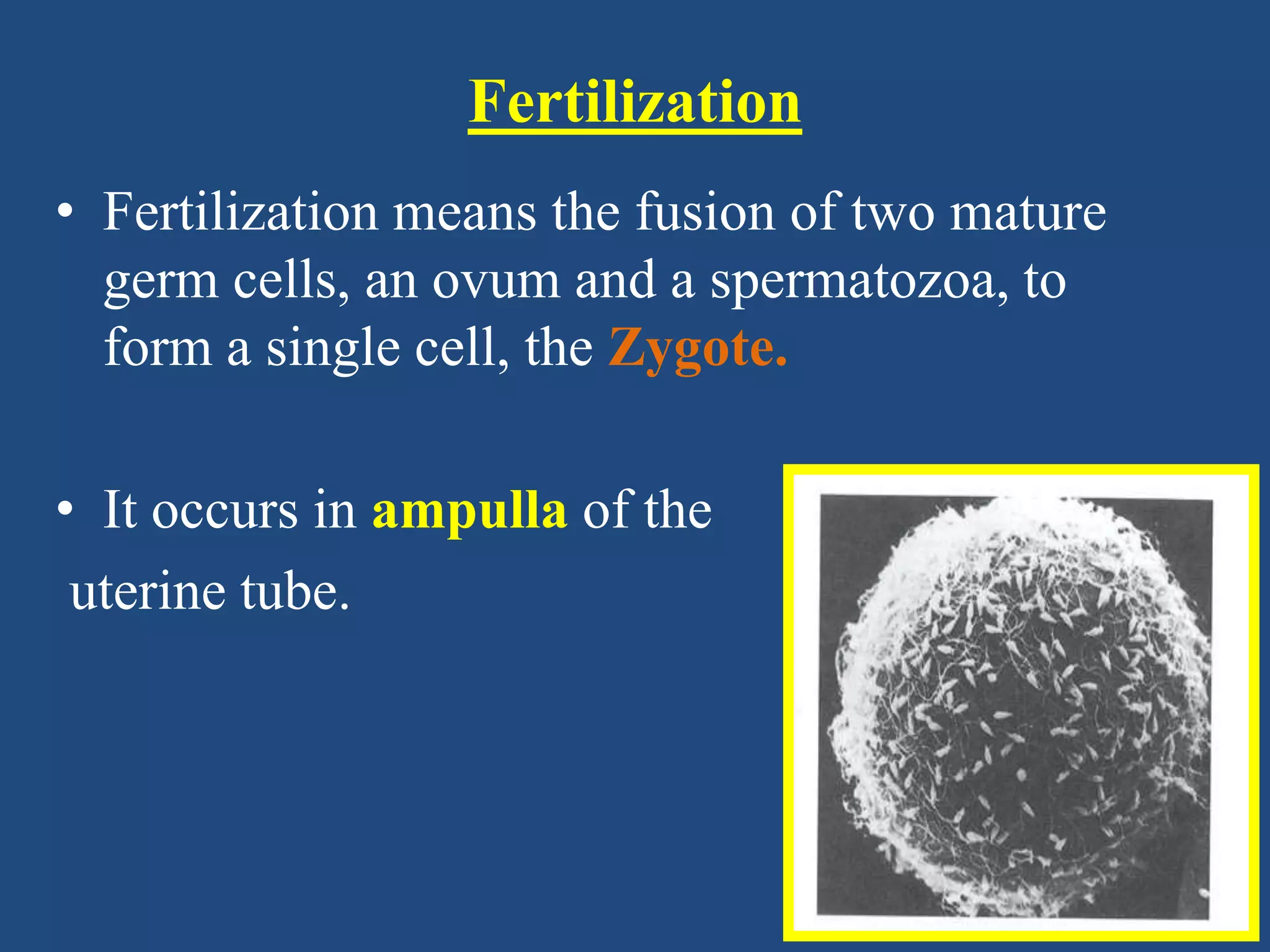
2) Fertilization involves the fusion of an ovum and spermatozoa to form a zygote, which occurs in the fallopian tube. The zygote then undergoes cleavage divisions as it moves through the uterine tube.
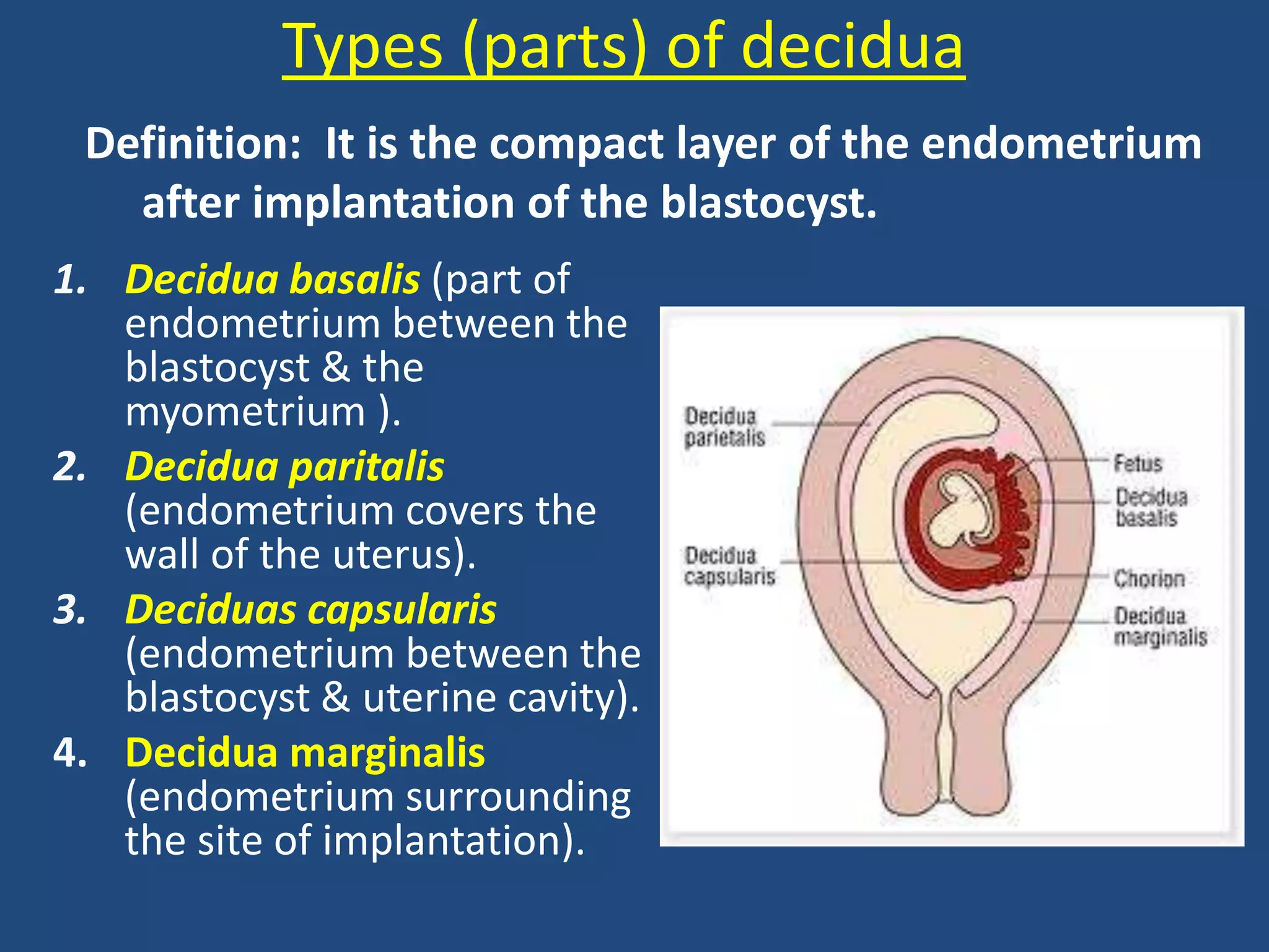
3) By day 5-6, the blastocyst has formed with an inner cell mass and outer trophoblast layer. The blastocyst implants in the endometrium around day 7, initiating formation of the placenta and decidua. Abnormal implantation can result in


![4-EMBRYOLOGICAL_DEVELOPMENT_OF_BODY_TISSUES,_ORGANS_AND_SYSTEMS.[1].pptx](https://cdn.slidesharecdn.com/ss_thumbnails/4-embryologicaldevelopmentofbodytissuesorgansandsystems-230811134542-e6d1c32e-thumbnail.jpg?width=640&height=640&fit=bounds)


