Sinus tachycardia
•Download as PPTX, PDF•
11 likes•21,521 views

Sinus tachycardia is characterized by a heart rate over 100 beats per minute originating from the sinus node. It can be a normal response to exercise or stress or indicate underlying conditions like heart failure. Symptoms may occur if the heart rate is very fast or the patient has heart disease. Treatment involves addressing the underlying cause and reducing stress or anxiety.

Recommended
More Related Content
What's hot
What's hot (20)
Viewers also liked
Viewers also liked (20)
Similar to Sinus tachycardia
Similar to Sinus tachycardia (20)
More from Ann Bentley
More from Ann Bentley (20)
Sinus tachycardia
- 2. Sinus Tachycardia • In an adult is characterized by a sinus rate of more than 100 beats/minute • Rate rarely exceeds 160 beats/minute except during strenuous exercise • Each impulse follows the normal pathway of conduction resulting in atrial and ventricular depolarization
- 3. Sinus Tachycardia • How it happens – Depends on the underlying cause • May be of no clinical significance – May be the body’s response to exercise – May be the body’s response to high emotional state • May also occur with hypovolemia, hemorrhage, or pain – When the stimulus for the tachycardia is removed, the arrhythmia spontaneously resolves
- 4. Sinus Tachycardia • Causes – Normal response to • Exercise, pain, stress, fever, or strong emotions – Certain cardiac conditions • Heart failure – Medications • Epinephrine and atropine – Substances • Caffeine, nicotine, and cocaine – Other conditions • Anemia, respiratory distress, pulmonary embolism, sepsis, and hyperthyroidism
- 5. Sinus Tachycardia • Hard on the heart – Not good for those with heart conditions already – Considered a poor prognostic sign if follows MI • Is associated with massive heart damage – Persistent tachycardia may signal impending heart failure or cardiogenic shock – Consequences • Bring on an episode of chest pain in patients with CAD
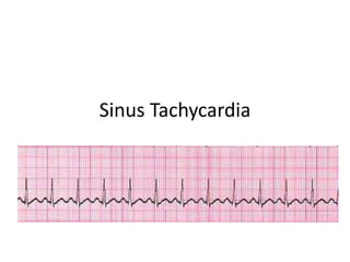
- 6. Sinus Tachycardia • What to look for – Look for a pulse rate of more than 100 beats/minute – Rhythm is regular
- 7. Symptomatic Tachycardia • Pulse rate of more than 100 beats/minute but with regular rhythm – Usually patient is asymptomatic
- 8. Symptomatic Tachycardia • If cardiac output falls and compensatory mechanisms fail – Will experience symptoms • Hypotension • Syncope • Blurred vision • Chest pain and palpitations • Nervousness or anxiety • Heart failure – JVD – crackles
- 9. Symptomatic Tachycardia • Steps to take – Prompt recognition is vital so treatment can be started – Provide the patient with a calm environment; help to reduce fear and anxiety which can fuel the arrhythmia – Tachycardia is commonly the first sign of pulmonary embolism
- 10. Symptomatic Tachycardia • When to call for help and what to do until help arrives – Look at the patient and ask how they are doing – Call for help if heart rate is too fast and/or symptomatic • Compare it their normal heart rate and rhythm – Stay with the patient – If the patient is not breathing and does not respond • Call code • ABCs/CPR
- 11. Sinus Tachycardia • What to look for – Look for a pulse rate of more than 100 beats/minute – Rhythm is regular
- 12. Sinus Tachycardia • Normal – P wave preceding each QRS complex – PR interval – QRS complex – T wave – QT interval
- 13. Sinus Tachycardia • P wave – Normal size and shape and precedes each QRS, but it may increase in amplitude – As the heart rate increases, the P wave may be superimposed on the preceding T wave and difficult to identify
- 14. Sinus Tachycardia • PR interval – Normal indicating that the impulse is following normal conduction pathways • 0.12-0.20 seconds
- 15. Sinus Tachycardia • QRS complex • Normal duration representing normal ventricular impulse conduction and recovery – Less than 0.12 seconds
- 16. Sinus Tachycardia • T wave – Upright in lead II, confirming that normal repolarization has taken place
- 17. Sinus Tachycardia • QT interval – Within normal limits • 0.36 to 0.44 seconds • QT normally shortens with tachycardia