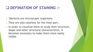
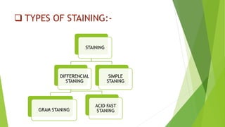
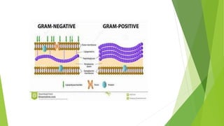
BACTERIAL STANING
PRESENTED BY- LALIT DHAKAR
B.PHARM (7TH SEM)
BANSAL COLLEGE OF PHARMACY, BHOPAL
CONTENTS
 DEFINATION OF STANING
 TECHNIQUES
 TYPES OF STANING
 DEFINATION OF STAINING :-
 Bacteria are microscopic organisms.
 They are also colorless for the most part.
 In order to visualize them to study their structure,
shape and other structural characteristics, it
becomes necessary to make them more easily
visible
 TECHNIQUE :-
 HANGING DROP TECHNIQUE:-
 Hanging drop technique enables viewing of size
shape, arrangement and motility of live microorganisms in fluid media
FIXATION :-
 Before staining it is essential to fix the bacterial sample on
to the slide
 (i) With a wire loop place a small drop of the broth
culture or a loop full of bacteria on a clean slide.
 (ii) Place a drop of water over it.
 (iii) Spread the culture so as to form a thin film.
 (iv) Allow slide to dry in the air or by holding it above a
bunsen flame.
 (v) Avoid excess heating.
 TYPES OF STAINING:-
STAINING
DIFFERENCIAL
STANING
GRAM STANING
ACID FAST
STANING
SIMPLE
STANING
 SIMPLE STANING :-
 The use of a single stain to colour a bacterial organism is commonly
referred as simple staining.
 Dyes-crystal violet,carbol fuschin
Differential stanning :-
a) Gram stanning:-
Developed by Christian Gram in 1884
 using this procedure, bacteria are subdivided by their reaction to this
stain into those which retain it, termed Gram-positive, and those
which are decolourized, termed Gram-negative.
Gram staining requires 4 different solutions:-
(i) A Basic Dye: crystal violet
(ii) A Mordant: It increases the affinity or attraction
between the cell and the dye, e.g. Iodine.
(iii)Decolorising agent: It removes the dye from a stained
cell, e.g., alcohol, acetone or ether.
(iv) Counter stain: It is a basic dye of a different colour
than the initial one, e.g., Safranin.
 GRAM +VE AND GRAM –VE BACTERIA:-
 Gram negative bacteria have cell wall generally thinner than those of gram positive bacteria.
 Gram negative ones have higher phospholipid content than the gram positive bacteria.
 During staining of gram positive bacteria, the alcohol treatment extracts the lipid, which
results in increased porosity or permeability of the cell wall.
 The CV-I (crystal violet iodine) complex can be extracted and the gram negative organism is
decolorized.
 These cells subsequently take the colour of the safranin a Counter stain.
 The cell walls of gram positive bacteria, due to their different composition (lower lipid
content) become dehydrated during treatment with alcohol presumably causes diminution in
the pore diameter of the walls of peptioglycan and CV-I complex is trapped in the wall
following ethanol treatment.
 The pore size decreases, permeability is reduced, and CV-I complex cannot be extracted.
Hence these cells remain purple violet.
 EXAMPLES-Gram Positive Bacteria: Actinomyces, Bacillus, Clostridium, Corynebacterium,
Enterococcus, Gardnerella, Lactobacillus, Listeria, ,etc.
Gram Negative Bacteria: Escherichia coli (E. coli), Salmonella, Shigella, and other
Enterobacteriaceae, Pseudomonas,Moraxella, HelicobacterR etc
Procedure
 1. Prepare smear on a clean slide.
 2. Stain with crystal violet for 30 seconds.
 3. Rinse with water.
 4. Flood the film with Grams iodine and allow it to act for 30 sec.
 5. Rinse with water.
 6. Decolorise with 95% alcohol.
 7. Rinse with water.
 8. Counter stain with safranin for 20-30 sec.
 9. Rinse with water and blot dry.
 10. Examine under oil immersion objective.

 Acid fast stain :-
 The Ziel-Nehlson method employs carbol fuchsin, acid alcohol and a blue or
green counter stain.
 Types of bacteria where they resist the process of de-colorization.
 These bacteria also resist staining and it may require heat and concentrated
staining to colorize them.
 Once this colorization has taken place, it becomes difficult to decolorize
using acid, or stain them with another color unless the heat and
concentration technique is used.
 This is why the term given to them is 'acid fast' just like one would use the
term 'color fast’.
 The most common type of acid fast bacteria is mycobacteria. A strain of
mycobacteria is responsible for the disease tuberculosis.
THANK YOU

Bacterial staning by LALIT DHAKAR

  • 1.
    BACTERIAL STANING PRESENTED BY-LALIT DHAKAR B.PHARM (7TH SEM) BANSAL COLLEGE OF PHARMACY, BHOPAL
  • 2.
    CONTENTS  DEFINATION OFSTANING  TECHNIQUES  TYPES OF STANING
  • 3.
     DEFINATION OFSTAINING :-  Bacteria are microscopic organisms.  They are also colorless for the most part.  In order to visualize them to study their structure, shape and other structural characteristics, it becomes necessary to make them more easily visible
  • 4.
     TECHNIQUE :- HANGING DROP TECHNIQUE:-  Hanging drop technique enables viewing of size shape, arrangement and motility of live microorganisms in fluid media
  • 5.
    FIXATION :-  Beforestaining it is essential to fix the bacterial sample on to the slide  (i) With a wire loop place a small drop of the broth culture or a loop full of bacteria on a clean slide.  (ii) Place a drop of water over it.  (iii) Spread the culture so as to form a thin film.  (iv) Allow slide to dry in the air or by holding it above a bunsen flame.  (v) Avoid excess heating.
  • 6.
     TYPES OFSTAINING:- STAINING DIFFERENCIAL STANING GRAM STANING ACID FAST STANING SIMPLE STANING
  • 7.
     SIMPLE STANING:-  The use of a single stain to colour a bacterial organism is commonly referred as simple staining.  Dyes-crystal violet,carbol fuschin
  • 8.
    Differential stanning :- a)Gram stanning:- Developed by Christian Gram in 1884  using this procedure, bacteria are subdivided by their reaction to this stain into those which retain it, termed Gram-positive, and those which are decolourized, termed Gram-negative.
  • 9.
    Gram staining requires4 different solutions:- (i) A Basic Dye: crystal violet (ii) A Mordant: It increases the affinity or attraction between the cell and the dye, e.g. Iodine. (iii)Decolorising agent: It removes the dye from a stained cell, e.g., alcohol, acetone or ether. (iv) Counter stain: It is a basic dye of a different colour than the initial one, e.g., Safranin.
  • 11.
     GRAM +VEAND GRAM –VE BACTERIA:-  Gram negative bacteria have cell wall generally thinner than those of gram positive bacteria.  Gram negative ones have higher phospholipid content than the gram positive bacteria.  During staining of gram positive bacteria, the alcohol treatment extracts the lipid, which results in increased porosity or permeability of the cell wall.  The CV-I (crystal violet iodine) complex can be extracted and the gram negative organism is decolorized.  These cells subsequently take the colour of the safranin a Counter stain.  The cell walls of gram positive bacteria, due to their different composition (lower lipid content) become dehydrated during treatment with alcohol presumably causes diminution in the pore diameter of the walls of peptioglycan and CV-I complex is trapped in the wall following ethanol treatment.  The pore size decreases, permeability is reduced, and CV-I complex cannot be extracted. Hence these cells remain purple violet.  EXAMPLES-Gram Positive Bacteria: Actinomyces, Bacillus, Clostridium, Corynebacterium, Enterococcus, Gardnerella, Lactobacillus, Listeria, ,etc. Gram Negative Bacteria: Escherichia coli (E. coli), Salmonella, Shigella, and other Enterobacteriaceae, Pseudomonas,Moraxella, HelicobacterR etc
  • 12.
    Procedure  1. Preparesmear on a clean slide.  2. Stain with crystal violet for 30 seconds.  3. Rinse with water.  4. Flood the film with Grams iodine and allow it to act for 30 sec.  5. Rinse with water.  6. Decolorise with 95% alcohol.  7. Rinse with water.  8. Counter stain with safranin for 20-30 sec.  9. Rinse with water and blot dry.  10. Examine under oil immersion objective. 
  • 13.
     Acid faststain :-  The Ziel-Nehlson method employs carbol fuchsin, acid alcohol and a blue or green counter stain.  Types of bacteria where they resist the process of de-colorization.  These bacteria also resist staining and it may require heat and concentrated staining to colorize them.  Once this colorization has taken place, it becomes difficult to decolorize using acid, or stain them with another color unless the heat and concentration technique is used.  This is why the term given to them is 'acid fast' just like one would use the term 'color fast’.  The most common type of acid fast bacteria is mycobacteria. A strain of mycobacteria is responsible for the disease tuberculosis.
  • 14.