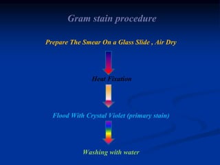
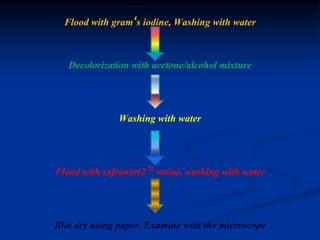
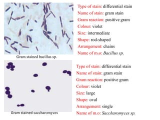
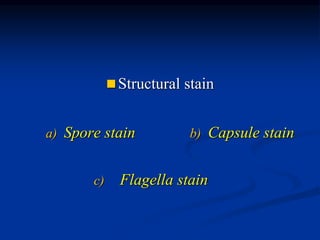
Bacterial staining 
Made by 
Sameh aly gad 
Demonstrator of microbiology and immunology 
College of pharmacy 
Minia university
outlines 
Dyes(stains) 
Negative staining 
Structural staining 
Spore stain 
Capsule stain 
Simple staining 
Differential staining 
Gram stain 
Acid-fast stain
Dyes 
 Why we need to stain bacteria? 
 Bacteria are transparent and colorless , so they would be 
invisible to naked eye if observed under a microscope 
thus 
bacteria should be stained with certain dyes in order to 
visualize bacterial cell or their internal structures using 
the light microscope.
Dyes (continued) 
 Dye (stain): 
colored organic compound in the form of salt, 
composed of positive and negative ion, one of these ions 
is responsible for colour called chromogen. 
 Types of Dyes: 
 Basic dyes 
 Acidic dyes
Dyes (continued) 
 Basic dyes: 
 In which chromogen is the positive ion (cation). 
 Basic dye has the form: dye+ Cl- 
 examples include crystal violet, methylene blue and safranin. 
 Acidic dyes: 
 In which chromogen is the negative ion (anion). 
 Acidic dye has the form: Na+dye- 
 Examples include nigrosin and India ink.
Simple staining 
 Simple staining is carried out to visualize bacteria and to 
compare morphological shapes and arrangements of bacterial 
cells. 
 In simple stain, the bacterial smear is stained with a single 
basic dye. 
 Bacterial cell surface is slightly negative so it tends to bind 
strongly to the cationic chromogen of basic dyes. 
 Thus , basic dyes are used to stain bacterial cells
Simple staining procedure 
1. Add one loopful of the sample onto a glass slide. 
2. Allow it to air-dry. 
3. Heat-fix the specimen on the glass slide ,unless the specimen 
is heat-fixed ,the bacterial smear will wash away during the 
staining procedure. 
4. Staining: crystal violet and wait for 1 min. or safranin and 
wait for 3-4 min. 
5. Wash the smear with tap water to remove the excess stain. 
6. Blot dry, then add cedarwood oil and examine under a 
microscope. Sameh aly gad
Bacterial shapes and arrangement
Bacillus subtilis stained with crystal violet Saccharomyces stained with crystal violet 
Staphylococci stained with crystal violet
Negative staining 
 It is a type of simple staining in which acidic dyes such as 
India ink or nigrosin are used. 
 The acidic dye with its negatively charged chromogen will not 
penetrate the cell due to the repulsion with the negatively 
charged bacterial surface. 
 As a result the bacterial cells remain unstained but easily 
discernible against the coloured background so it is also called 
indirect stain. 
 Since no heat is used, negative stain is used to visualize cells 
that are too delicate to be heat-fixed.
Negatively stained staphylococci
 Differential staining 
a) Gram stain b) Acid fast stain
Gram stain 
 Most bacteria are classified into two main groups: gram 
positive and gram negative bacteria. 
 Comparison between gram positive and gram negative 
bacteria.
Gram stain (cont…) 
 It is a differential stain which distinguishes between gram 
positive and gram negative bacteria depending on the 
difference in the cell wall structure. 
 Principle: 
• Gram positive bacteria when treated with a basic dye such as 
crystal violet , then with iodine (will fix the stain to the cell) 
and then subjected to a subsequent treatment with a 
decolourizing agent such as alcohol/acetone mixture remain 
stained with the basic dye and the decolourizer doesn,t 
remove the dye ,
Gram stain (cont…) 
however, gram negative bacteria when treated similarly will be 
readily decolourized with the alcohol acetone mixture and 
become invisible , thus to render the unstained gram negative 
bacteria visible, a counterstain is applied. 
 Why gram positive bacteria resist decolourization by 
alcohol/acetone mixture but the gram negative ones can not? 
 Because the cell wall of gram negative bacteria has high lipid 
content and low peptidoglycan content so when treated with 
acetone/alcohol mixture dissolves their lipids creating pores
Gram stain (cont…) 
In the cell wall through which crystal violet dye will diffuse, 
and gram negative bacteria become unstained. 
On the other hand, gram positive bacteria has high 
peptidoglycan content and low lipid content so when 
treated with the decolourizer, it will dehydrate the cell 
wall causing it to shrink and the pores to close and thus 
the diffusion of the dye will be blocked
Gram stain procedure 
Prepare The Smear On a Glass Slide , Air Dry 
Heat Fixation 
Flood With Crystal Violet (primary stain) 
Washing with water
Flood with gram‘s iodine, Washing with water 
Decolorization with acetone/alcohol mixture 
Washing with water 
Flood with safranin(2 ry stain), washing with water 
Blot dry using paper, Examine with the microscope
Gram stain (cont…) 
 If the specimen contains both gram positive and gram 
negative bacteria, gram positive ones appear violet while 
gram negative ones appear pink to red 
Gram stained staphylococci 
Type of stain: differential stain 
Name of stain: gram stain 
Gram reaction: positive gram 
Colour: violet 
Size: small 
Shape: spherical or cocci 
Arrangement: grapelike clusters 
Name of m.o: Staphylococcus sp.
Gram stained bacillus sp. 
Type of stain: differential stain 
Name of stain: gram stain 
Gram reaction: positive gram 
Colour: violet 
Size: intermediate 
Shape: rod-shaped 
Arrangement: chains 
Name of m.o: Bacillus sp. 
Type of stain: differential stain 
Name of stain: gram stain 
Gram reaction: positive gram 
Colour: violet 
Size: large 
Shape: oval 
Arrangement: single 
Gram stained saccharomyces Name of m.o: Saccharomyces sp.
Gram stained Escherichia coli 
Type of stain: differential stain 
Name of stain: gram stain 
Gram reaction: gram negative 
Colour: pink 
Size: small 
Shape: rod shaped 
Arrangement: single 
Name of m.o: Escherichia coli 
Mixture of gram positive 
staphylococci and gram 
negative E.coli
Acid-fast stain(Ziehl-Neelsen stain) 
 Members of the genus mycobacterium are resistant to simple 
and gram staining procedure due to their thick waxy (fatty) 
cell wall which makes the penetration of dyes extremely 
difficult and requires the application of heat. 
 But once the dye has penetrated, it can not be removed even 
with the vigorous use of acid alcohol as decolourizing agent, 
due to this property, these organisms are called acid fast. 
 The most important human pathogens of them are 
mycobacterium tuberculosis and mycobacterium leprae.
Acid-fast stain procedure 
1. Prepare a smear of the microorganism on a clean slide. 
2. Allow the smear to air-dry then fix it by flaming. 
3. Place the slide over a steam bath and flood the smear with 
strong carbol fuchsin and keep it for 5 mins. caution :don,t 
allow stain to dry; replenish stain as needed. also, prevent 
stain from boiling by adjusting the temperature applied. 
4. Allow to cool and then wash with tap water. 
5. Decolourize with acid alcohol, till the effluent runs almost 
clear with a slight red tinge.
Acid-fast stain procedure 
6. Wash with tap water 
7. Counterstain with methylene blue for 2 mins. 
8. Wash smear with tap water. 
9. Blot dry and examine with a microscope. 
Acid fast bacteria 
appear red while 
non acid fast 
bacteria appear 
blue.
 Structural stain 
a) Spore stain b) Capsule stain 
c) Flagella stain
Spore stain 
(Schaeffer-Fulton method) 
 Some gram positive bacteria especially members of the 
genera clostridium and bacillus have the ability to form 
spore(endospore) when exposed to drastic unfavorable 
conditions such as drying, heating, lack of nutrients and so 
on. 
 Spores formed by these bacteria are highly resistant to 
unfavorable conditions and can remain viable for several 
years, once the conditions become favorable the spore 
germinates.
Spore stain(cont..) 
 Spores don,t accept the stain easily due to their thick 
impervious coat and require the use of heating during 
staining. 
Procedure: 
1. Prepare smear of the microorganism, allow to dry and the 
heat-fix to a slide. 
2. Place the slide over a steam bath, flood the smear with 
malachite green dye and keep it for 3-5 mins, replenish 
smear with malachite green dye if some evaporates and don't 
allow the stain to boil.
Spore stain(cont..) 
3. Allow the slide to cool and wash with tap water to remove 
excess stain. 
4. Counterstain with safranin for 2 min. 
5. Rinse the slide with running tap water to remove excess stain. 
6. Blot dry with bibulous paper and examine under oil 
immersion.
Capsule stain 
 It is a well organized gelatinous layer that is secreted by the 
cell, surrounds and adheres to the cell wall. 
 Capsule is not common to all organisms and the organisms 
that have a heavy capsule are generally virulent and capable 
of producing disease. 
 It protects bacteria against the phagocytic activities of host 
phagocytes. 
 It also serves as a barrier against antimicrobials preventing 
them from entering the cell.
Capsule stain(cont..) 
 Most capsules are polysaccharides (ex: Streptococcus 
pneumoniae) in nature but some are glycoproteins or 
polypeptides. 
 Since capsule fails to retain standard dyes, it can't be stained 
by simple staining procedure or even gram stain, but it can be 
visualized indirectly using negative staining technique. 
 A basic dye is used to stain the bacterial cell and acidic dye is 
used to colour the background where the capsule appears as a 
clear halo zone between the coloured background and the 
stained cell.
Capsule stain(cont..) 
Procedure 
 Prepare a smear from the encapsulated microorganism( ex. 
Klebsiella pneumoniae) without heat fixation. 
 Stain with safranin for 5-6 mins. 
 Wash with tap water(polysaccharide capsules are water-soluble 
thus gentle washing is required, otherwise the capsule 
will be removed with water). 
 Put a drop of nigrosin at one end of the slide and spread it 
with another slide. 
 Allow it to air-dry and examine under oil immersion.
Figure showing indirectly stained capsule (Klebsiella pneumoniae capsule)
Bacterial staining
Bacterial staining

Bacterial staining

  • 1.
    Bacterial staining Madeby Sameh aly gad Demonstrator of microbiology and immunology College of pharmacy Minia university
  • 2.
    outlines Dyes(stains) Negativestaining Structural staining Spore stain Capsule stain Simple staining Differential staining Gram stain Acid-fast stain
  • 3.
    Dyes  Whywe need to stain bacteria?  Bacteria are transparent and colorless , so they would be invisible to naked eye if observed under a microscope thus bacteria should be stained with certain dyes in order to visualize bacterial cell or their internal structures using the light microscope.
  • 4.
    Dyes (continued) Dye (stain): colored organic compound in the form of salt, composed of positive and negative ion, one of these ions is responsible for colour called chromogen.  Types of Dyes:  Basic dyes  Acidic dyes
  • 5.
    Dyes (continued) Basic dyes:  In which chromogen is the positive ion (cation).  Basic dye has the form: dye+ Cl-  examples include crystal violet, methylene blue and safranin.  Acidic dyes:  In which chromogen is the negative ion (anion).  Acidic dye has the form: Na+dye-  Examples include nigrosin and India ink.
  • 6.
    Simple staining Simple staining is carried out to visualize bacteria and to compare morphological shapes and arrangements of bacterial cells.  In simple stain, the bacterial smear is stained with a single basic dye.  Bacterial cell surface is slightly negative so it tends to bind strongly to the cationic chromogen of basic dyes.  Thus , basic dyes are used to stain bacterial cells
  • 7.
    Simple staining procedure 1. Add one loopful of the sample onto a glass slide. 2. Allow it to air-dry. 3. Heat-fix the specimen on the glass slide ,unless the specimen is heat-fixed ,the bacterial smear will wash away during the staining procedure. 4. Staining: crystal violet and wait for 1 min. or safranin and wait for 3-4 min. 5. Wash the smear with tap water to remove the excess stain. 6. Blot dry, then add cedarwood oil and examine under a microscope. Sameh aly gad
  • 8.
  • 9.
    Bacillus subtilis stainedwith crystal violet Saccharomyces stained with crystal violet Staphylococci stained with crystal violet
  • 10.
    Negative staining It is a type of simple staining in which acidic dyes such as India ink or nigrosin are used.  The acidic dye with its negatively charged chromogen will not penetrate the cell due to the repulsion with the negatively charged bacterial surface.  As a result the bacterial cells remain unstained but easily discernible against the coloured background so it is also called indirect stain.  Since no heat is used, negative stain is used to visualize cells that are too delicate to be heat-fixed.
  • 12.
  • 13.
     Differential staining a) Gram stain b) Acid fast stain
  • 14.
    Gram stain Most bacteria are classified into two main groups: gram positive and gram negative bacteria.  Comparison between gram positive and gram negative bacteria.
  • 15.
    Gram stain (cont…)  It is a differential stain which distinguishes between gram positive and gram negative bacteria depending on the difference in the cell wall structure.  Principle: • Gram positive bacteria when treated with a basic dye such as crystal violet , then with iodine (will fix the stain to the cell) and then subjected to a subsequent treatment with a decolourizing agent such as alcohol/acetone mixture remain stained with the basic dye and the decolourizer doesn,t remove the dye ,
  • 16.
    Gram stain (cont…) however, gram negative bacteria when treated similarly will be readily decolourized with the alcohol acetone mixture and become invisible , thus to render the unstained gram negative bacteria visible, a counterstain is applied.  Why gram positive bacteria resist decolourization by alcohol/acetone mixture but the gram negative ones can not?  Because the cell wall of gram negative bacteria has high lipid content and low peptidoglycan content so when treated with acetone/alcohol mixture dissolves their lipids creating pores
  • 17.
    Gram stain (cont…) In the cell wall through which crystal violet dye will diffuse, and gram negative bacteria become unstained. On the other hand, gram positive bacteria has high peptidoglycan content and low lipid content so when treated with the decolourizer, it will dehydrate the cell wall causing it to shrink and the pores to close and thus the diffusion of the dye will be blocked
  • 18.
    Gram stain procedure Prepare The Smear On a Glass Slide , Air Dry Heat Fixation Flood With Crystal Violet (primary stain) Washing with water
  • 19.
    Flood with gram‘siodine, Washing with water Decolorization with acetone/alcohol mixture Washing with water Flood with safranin(2 ry stain), washing with water Blot dry using paper, Examine with the microscope
  • 20.
    Gram stain (cont…)  If the specimen contains both gram positive and gram negative bacteria, gram positive ones appear violet while gram negative ones appear pink to red Gram stained staphylococci Type of stain: differential stain Name of stain: gram stain Gram reaction: positive gram Colour: violet Size: small Shape: spherical or cocci Arrangement: grapelike clusters Name of m.o: Staphylococcus sp.
  • 21.
    Gram stained bacillussp. Type of stain: differential stain Name of stain: gram stain Gram reaction: positive gram Colour: violet Size: intermediate Shape: rod-shaped Arrangement: chains Name of m.o: Bacillus sp. Type of stain: differential stain Name of stain: gram stain Gram reaction: positive gram Colour: violet Size: large Shape: oval Arrangement: single Gram stained saccharomyces Name of m.o: Saccharomyces sp.
  • 22.
    Gram stained Escherichiacoli Type of stain: differential stain Name of stain: gram stain Gram reaction: gram negative Colour: pink Size: small Shape: rod shaped Arrangement: single Name of m.o: Escherichia coli Mixture of gram positive staphylococci and gram negative E.coli
  • 23.
    Acid-fast stain(Ziehl-Neelsen stain)  Members of the genus mycobacterium are resistant to simple and gram staining procedure due to their thick waxy (fatty) cell wall which makes the penetration of dyes extremely difficult and requires the application of heat.  But once the dye has penetrated, it can not be removed even with the vigorous use of acid alcohol as decolourizing agent, due to this property, these organisms are called acid fast.  The most important human pathogens of them are mycobacterium tuberculosis and mycobacterium leprae.
  • 24.
    Acid-fast stain procedure 1. Prepare a smear of the microorganism on a clean slide. 2. Allow the smear to air-dry then fix it by flaming. 3. Place the slide over a steam bath and flood the smear with strong carbol fuchsin and keep it for 5 mins. caution :don,t allow stain to dry; replenish stain as needed. also, prevent stain from boiling by adjusting the temperature applied. 4. Allow to cool and then wash with tap water. 5. Decolourize with acid alcohol, till the effluent runs almost clear with a slight red tinge.
  • 25.
    Acid-fast stain procedure 6. Wash with tap water 7. Counterstain with methylene blue for 2 mins. 8. Wash smear with tap water. 9. Blot dry and examine with a microscope. Acid fast bacteria appear red while non acid fast bacteria appear blue.
  • 26.
     Structural stain a) Spore stain b) Capsule stain c) Flagella stain
  • 27.
    Spore stain (Schaeffer-Fultonmethod)  Some gram positive bacteria especially members of the genera clostridium and bacillus have the ability to form spore(endospore) when exposed to drastic unfavorable conditions such as drying, heating, lack of nutrients and so on.  Spores formed by these bacteria are highly resistant to unfavorable conditions and can remain viable for several years, once the conditions become favorable the spore germinates.
  • 28.
    Spore stain(cont..) Spores don,t accept the stain easily due to their thick impervious coat and require the use of heating during staining. Procedure: 1. Prepare smear of the microorganism, allow to dry and the heat-fix to a slide. 2. Place the slide over a steam bath, flood the smear with malachite green dye and keep it for 3-5 mins, replenish smear with malachite green dye if some evaporates and don't allow the stain to boil.
  • 29.
    Spore stain(cont..) 3.Allow the slide to cool and wash with tap water to remove excess stain. 4. Counterstain with safranin for 2 min. 5. Rinse the slide with running tap water to remove excess stain. 6. Blot dry with bibulous paper and examine under oil immersion.
  • 30.
    Capsule stain It is a well organized gelatinous layer that is secreted by the cell, surrounds and adheres to the cell wall.  Capsule is not common to all organisms and the organisms that have a heavy capsule are generally virulent and capable of producing disease.  It protects bacteria against the phagocytic activities of host phagocytes.  It also serves as a barrier against antimicrobials preventing them from entering the cell.
  • 31.
    Capsule stain(cont..) Most capsules are polysaccharides (ex: Streptococcus pneumoniae) in nature but some are glycoproteins or polypeptides.  Since capsule fails to retain standard dyes, it can't be stained by simple staining procedure or even gram stain, but it can be visualized indirectly using negative staining technique.  A basic dye is used to stain the bacterial cell and acidic dye is used to colour the background where the capsule appears as a clear halo zone between the coloured background and the stained cell.
  • 32.
    Capsule stain(cont..) Procedure  Prepare a smear from the encapsulated microorganism( ex. Klebsiella pneumoniae) without heat fixation.  Stain with safranin for 5-6 mins.  Wash with tap water(polysaccharide capsules are water-soluble thus gentle washing is required, otherwise the capsule will be removed with water).  Put a drop of nigrosin at one end of the slide and spread it with another slide.  Allow it to air-dry and examine under oil immersion.
  • 33.
    Figure showing indirectlystained capsule (Klebsiella pneumoniae capsule)