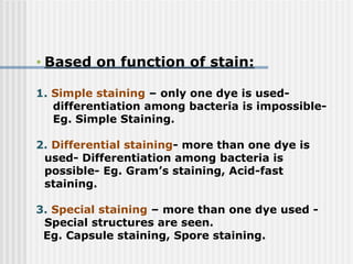
• Based onfunction of stain:
1. Simple staining – only one dye is used-
differentiation among bacteria is impossible-
Eg. Simple Staining.
2. Differential staining- more than one dye is
used- Differentiation among bacteria is
possible- Eg. Gram’s staining, Acid-fast
staining.
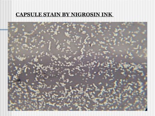
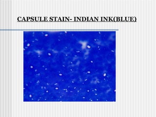
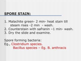
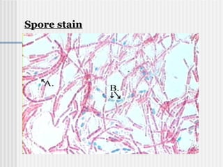
3. Special staining – more than one dye used -
Special structures are seen.
Eg. Capsule staining, Spore staining.
3.
Each staining methodshave own principles but
the following steps may be common:
Basic stain(+ve charge) –
To stain -ve charged molecules of bacteria
Mostly used because cell surface is –ve charge.
Acidic Stain(-ve charge)
To stain +ve charged molecules of bacteria.
Used to stain the bacterial capsules.
As cell surface is –ve charged- Basic dyes
mostly used.
Principle of staining:
4.
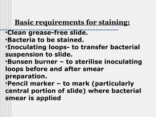
•Clean grease-free slide.
•Bacteriato be stained.
•Inoculating loops- to transfer bacterial
suspension to slide.
•Bunsen burner – to sterilise inoculating
loops before and after smear
preparation.
•Pencil marker – to mark (particularly
central portion of slide) where bacterial
smear is applied
Basic requirements for staining:
5.
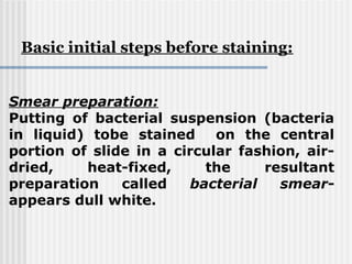
Smear preparation:
Putting ofbacterial suspension (bacteria
in liquid) tobe stained on the central
portion of slide in a circular fashion, air-
dried, heat-fixed, the resultant
preparation called bacterial smear-
appears dull white.
Basic initial steps before staining:
6.
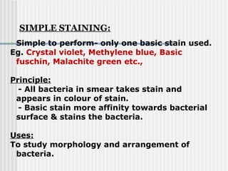
• Simple toperform- only one basic stain used.
Eg. Crystal violet, Methylene blue, Basic
fuschin, Malachite green etc.,
Principle:
- All bacteria in smear takes stain and
appears in colour of stain.
- Basic stain more affinity towards bacterial
surface & stains the bacteria.
Uses:
To study morphology and arrangement of
bacteria.
SIMPLE STAINING:
7.
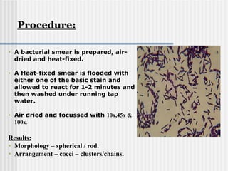
• A bacterialsmear is prepared, air-
dried and heat-fixed.
• A Heat-fixed smear is flooded with
either one of the basic stain and
allowed to react for 1-2 minutes and
then washed under running tap
water.
• Air dried and focussed with 10x,45x &
100x.
Results:
• Morphology – spherical / rod.
• Arrangement – cocci – clusters/chains.
Procedure:
8.
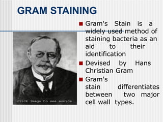
GRAM STAINING
■ Gram'sStain is a
widely used method of
staining bacteria as an
aid to their
identification
■ Devised by Hans
Christian Gram
■ Gram's
stain differentiates
between two major
cell wall types.
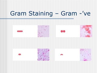
9.
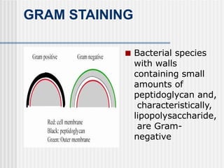
GRAM STAINING
■ Bacterialspecies
with walls
containing small
amounts of
peptidoglycan and,
characteristically,
lipopolysaccharide,
are Gram-
negative
10.
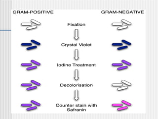
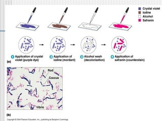
The process includesthe use of:
a primary stain (crystal violet)
a mordant (helper) iodine solution, a
decolorizer (95% ethanol),
a counterstain (safranin).
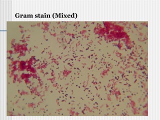
11.
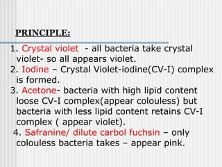
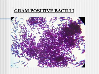
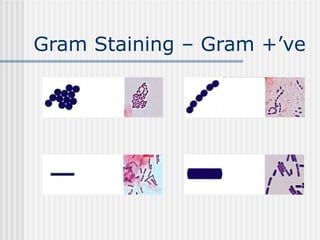
1. Crystal violet- all bacteria take crystal
violet- so all appears violet.
2. Iodine – Crystal Violet-iodine(CV-I) complex
is formed.
3. Acetone- bacteria with high lipid content
loose CV-I complex(appear colouless) but
bacteria with less lipid content retains CV-I
complex ( appear violet).
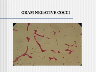
4. Safranine/ dilute carbol fuchsin – only
colouless bacteria takes – appear pink.
PRINCIPLE:
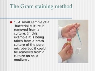
The Gram stainingmethod
■ 1. A small sample of a
bacterial culture is
removed from a
culture. In this
example it is being
taken from a broth
culture of the pure
microbe but it could
be removed from a
culture on solid
medium .
22.
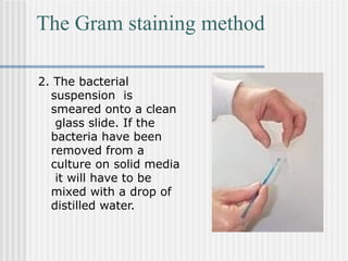
The Gram stainingmethod
2. The bacterial
suspension is
smeared onto a clean
glass slide. If the
bacteria have been
removed from a
culture on solid media
it will have to be
mixed with a drop of
distilled water.
23.
The Gram stainingmethod
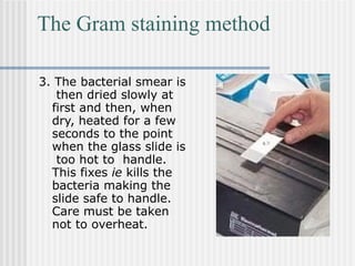
3. The bacterial smear is
then dried slowly at
first and then, when
dry, heated for a few
seconds to the point
when the glass slide is
too hot to handle.
This fixes ie kills the
bacteria making the
slide safe to handle.
Care must be taken
not to overheat.
24.
The Gram stainingmethod
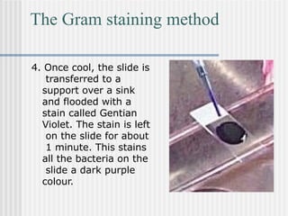
4. Once cool, the slide is
transferred to a
support over a sink
and flooded with a
stain called Gentian
Violet. The stain is left
on the slide for about
1 minute. This stains
all the bacteria on the
slide a dark purple
colour.
25.
The Gram stainingmethod
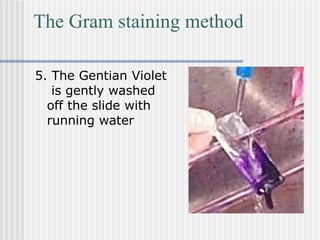
5. The Gentian Violet
is gently washed
off the slide with
running water
26.
The Gram stainingmethod
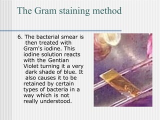
6. The bacterial smear is
then treated with
Gram's iodine. This
iodine solution reacts
with the Gentian
Violet turning it a very
dark shade of blue. It
also causes it to be
retained by certain
types of bacteria in a
way which is not
really understood.
27.
The Gram stainingmethod
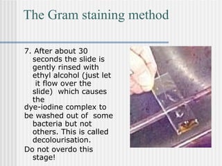
7. After about 30
seconds the slide is
gently rinsed with
ethyl alcohol (just let
it flow over the
slide) which causes
the
dye-iodine complex to
be washed out of some
bacteria but not
others. This is called
decolourisation.
Do not overdo this
stage!
28.
The Gram stainingmethod
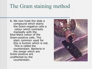
8. We now treat the slide a
compound which stains
the Gram-negative cells a
colour which contrasts
markedly with the
blue-black colour of the
Gram-positive cells. The
stain common used for
this is fuchsin which is red.
This is called the
counterstain. Bacteria in
the smear which are
Gram-positive are
unaffected by the
counterstain.
29.
The Gram stainingmethod
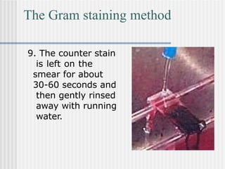
9. The counter stain
is left on the
smear for about
30-60 seconds and
then gently rinsed
away with running
water.
30.
The Gram stainingmethod
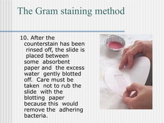
10. After the
counterstain has been
rinsed off, the slide is
placed between
some absorbent
paper and the excess
water gently blotted
off. Care must be
taken not to rub the
slide with the
blotting paper
because this would
remove the adhering
bacteria.
31.
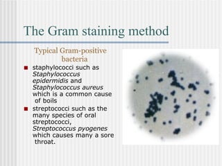
The Gram stainingmethod
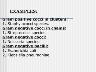
Typical Gram-positive
bacteria
■ staphylococci such as
Staphylococcus
epidermidis and
Staphylococcus aureus
which is a common cause
of boils
■ streptococci such as the
many species of oral
streptococci,
Streptococcus pyogenes
which causes many a sore
throat.
32.
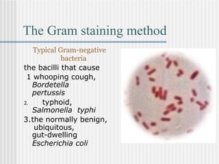
The Gram stainingmethod
Typical Gram-negative
bacteria
the bacilli that cause
1 whooping cough,
Bordetella
pertussis
2. typhoid,
Salmonella typhi
3.the normally benign,
ubiquitous,
gut-dwelling
Escherichia coli
33.
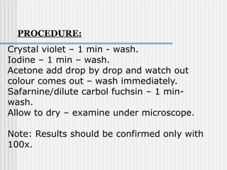
Crystal violet –1 min - wash.
Iodine – 1 min – wash.
Acetone add drop by drop and watch out
colour comes out – wash immediately.
Safarnine/dilute carbol fuchsin – 1 min-
wash.
Allow to dry – examine under microscope.
Note: Results should be confirmed only with
100x.
PROCEDURE:
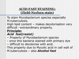
• To stainMycobacterium species especially
M.tuberculosis.
• High lipid content – makes decolorisation very
difficult –extraordinary property.
• Principle:
• Acid fast(resist)
• – Property of Mycobacterium species
• - once this bacteria stained with primary dye
• – difficult to decolorise with acid.
• This property due to Mycolic acid in cell wall of
• M.tuberculosis – also Alcohol fast
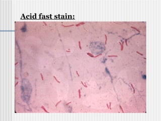
ACID-FAST STAINING:
(Ziehl-Neelsen stain)
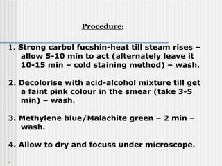
1. Strong carbolfucshin-heat till steam rises –
allow 5-10 min to act (alternately leave it
10-15 min – cold staining method) – wash.
2. Decolorise with acid-alcohol mixture till get
a faint pink colour in the smear (take 3-5
min) – wash.
3. Methylene blue/Malachite green – 2 min –
wash.
4. Allow to dry and focuss under microscope.
•
Procedure:
39.
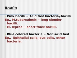
• Pink bacilli– Acid fast bacteria/bacilli
Eg., M.tuberculosis – long slender
bacilli.
• M. leprae – short thick bacilli.
• Blue colored bacteria – Non-acid fast
Eg., Epithelial cells, pus cells, other
bacteria.
Result:
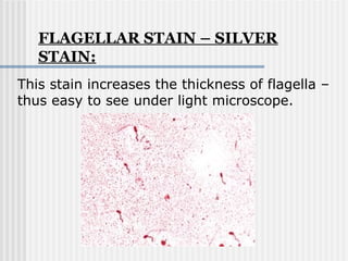
FLAGELLAR STAIN –SILVER
STAIN:
This stain increases the thickness of flagella –
thus easy to see under light microscope.
48.
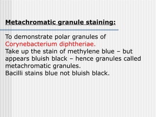
Metachromatic granule staining:
Todemonstrate polar granules of
Corynebacterium diphtheriae.
Take up the stain of methylene blue – but
appears bluish black – hence granules called
metachromatic granules.
Bacilli stains blue not bluish black.