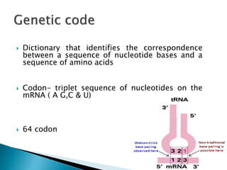
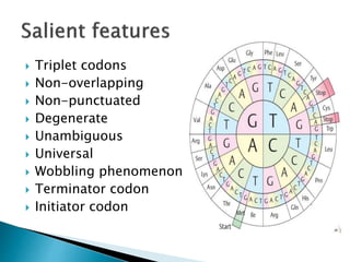
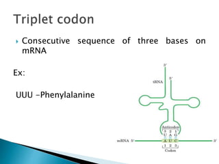
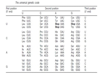
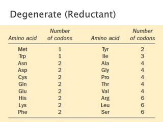
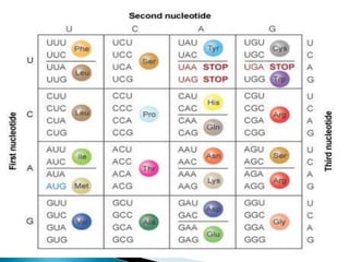
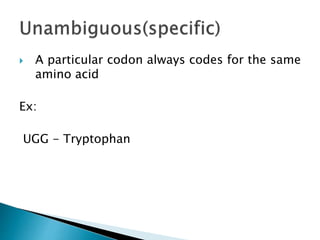
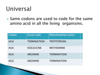
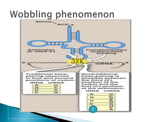
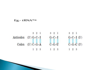
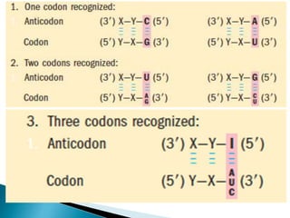
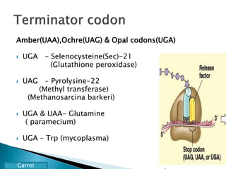
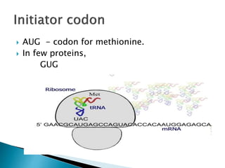
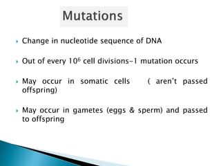
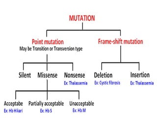
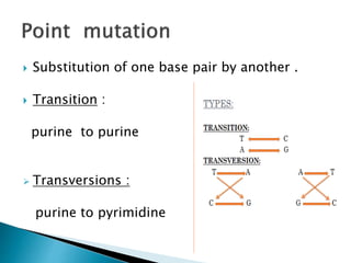
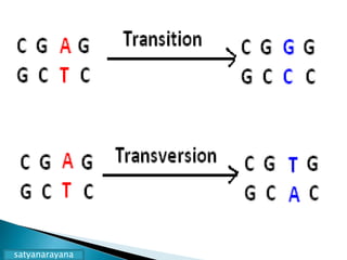
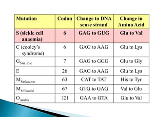
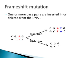
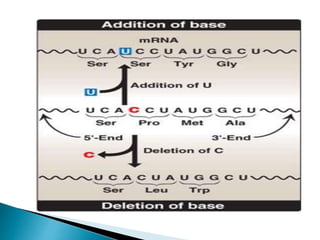
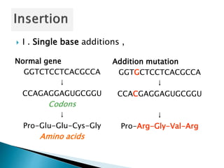
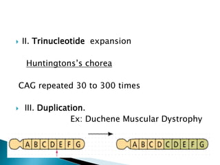
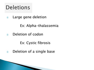
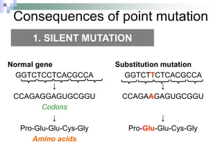
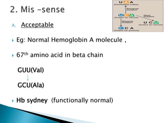
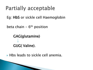
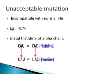
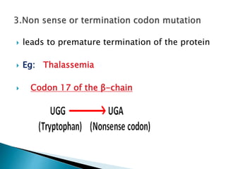
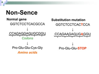
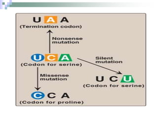
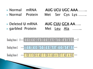
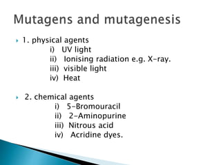
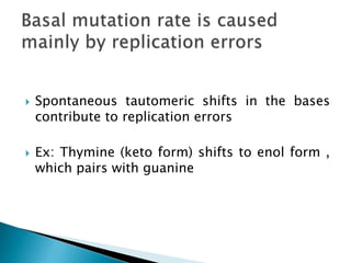
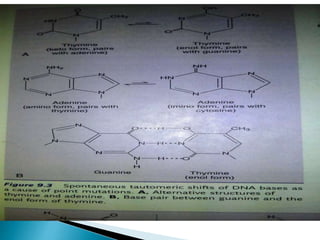
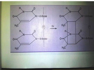
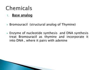
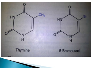
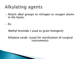
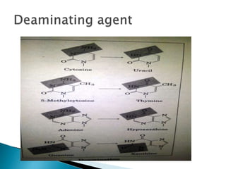
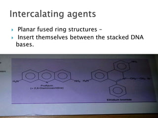
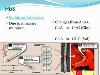
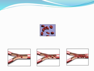
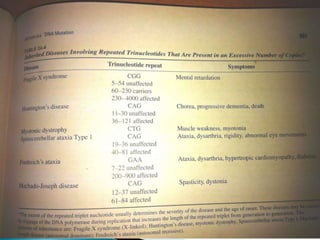
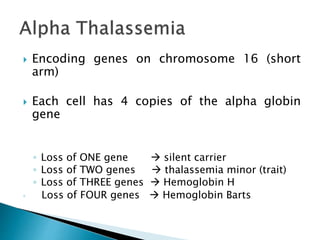
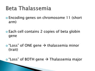
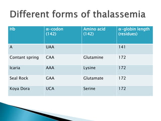
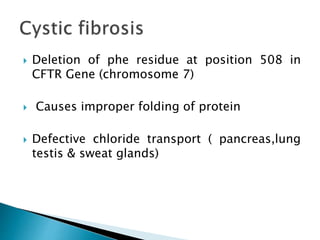
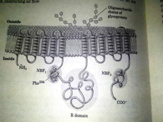
Moderator : Dr. M. S. Somannavar
Presenter : Jay prakash sah
 Genetic code
 Salient features
 Mutation
 Types of mutation
 Mutagens
 Clinical correlations
 Crick, Brenner et al. experiment - Demonstrate
that codons consist of three DNA bases
 Marshall Nirenberg and Heinrich J. Matthaei in
1961 reported the first three letter code words for
each amino acid
 H. Gobind Khorana- Genetic code dictionary and
previous results confirmed
 Dictionary that identifies the correspondence
between a sequence of nucleotide bases and a
sequence of amino acids
 Codon- triplet sequence of nucleotides on the
mRNA ( A G,C & U)
 64 codon
 Triplet codons
 Non-overlapping
 Non-punctuated
 Degenerate
 Unambiguous
 Universal
 Wobbling phenomenon
 Terminator codon
 Initiator codon
 Consecutive sequence of three bases on
mRNA
Ex:
UUU -Phenylalanine

AUACGAGUC
 A U A C G A G U C
1 2 3
overlapping code
A U A C G A G U C
 Consecutive or continuous
 A particular codon always codes for the same
amino acid
Ex:
UGG - Tryptophan
 Same codons are used to code for the same
amino acid in all the living organisms.
I
Amber(UAA),Ochre(UAG) & Opal codons(UGA)
 UGA - Selenocysteine(Sec)-21
(Glutathione peroxidase)
 UAG - Pyrolysine-22
(Methyl transferase)
(Methanosarcina barkeri)
 UGA & UAA- Glutamine
( paramecium)
 UGA – Trp (mycoplasma)
Garret
 AUG - codon for methionine.
 In few proteins,
GUG
 Change in nucleotide sequence of DNA
 Out of every 106 cell divisions-1 mutation occurs
 May occur in somatic cells ( aren’t passed
offspring)
 May occur in gametes (eggs & sperm) and passed
to offspring
 Substitution of one base pair by another .
 Transition :
purine to purine
 Transversions :
purine to pyrimidine
satyanarayana
Mutations: Substitutions
Substitution mutation
GGTCACCTCACGCCA
↓
CCAGUGGAGUGCGGU
↓
Pro-Arg-Glu-Cys-Gly
Normal gene
GGTCTCCTCACGCCA
↓
CCAGAGGAGUGCGGU
Codons
↓
Pro-Glu-Glu-Cys-Gly
Amino acids
Mutation Codon Change to DNA
sense strand
Change in
Amino Acid
S (sickle cell
anaemia)
6 GAG to GUG Glu to Val
C (cooley’s
syndrome)
6 GAG to AAG Glu to Lys
GSan Jose 7 GAG to GGG Glu to Gly
E 26 GAG to AAG Glu to Lys
MSaskatoon 63 CAT to TAT His to Tyr
MMilwauki 67 GTG to GAG Val to Glu
OArabia 121 GAA to GTA Glu to Val
 One or more base pairs are inserted in or
deleted from the DNA .
 I . Single base additions ,
Normal gene
GGTCTCCTCACGCCA
↓
CCAGAGGAGUGCGGU
Codons
↓
Pro-Glu-Glu-Cys-Gly
Amino acids
Addition mutation
GGTGCTCCTCACGCCA
↓
CCACGAGGAGUGCGGU
↓
Pro-Arg-Gly-Val-Arg
 II. Trinucleotide expansion
Huntingtons’s chorea
CAG repeated 30 to 300 times
 III. Duplication.
Ex: Duchene Muscular Dystrophy
i) Large gene deletion
Ex: Alpha-thalassemia
i) Deletion of codon
Ex: Cystic fibrosis
i) Deletion of a single base
Consequences of point mutation
Normal gene
GGTCTCCTCACGCCA
↓
CCAGAGGAGUGCGGU
Codons
↓
Pro-Glu-Glu-Cys-Gly
Amino acids
Substitution mutation
GGTCTTCTCACGCCA
↓
CCAGAAGAGUGCGGU
↓
Pro-Glu-Glu-Cys-Gly
1. SILENT MUTATION
A. Acceptable
 Eg: Normal Hemoglobin A molecule ,
 67th amino acid in beta chain
GUU(Val)
GCU(Ala)
 Hb sydney (functionally normal)
Eg: HbS or sickle cell Haemoglobin
beta chain - 6th position
GAG(glutamine)
GUG( Valine).
 Hbs leads to sickle cell anemia.
 Incompatible with normal life
 Eg : HbM
 Distal histidine of alpha chain.
 leads to premature termination of the protein
 Eg: Thalassemia
 Codon 17 of the β-chain
Non-Sence
Normal gene
GGTCTCCTCACGCCA
↓
CCAGAGGAGUGCGGU
Codons
↓
Pro-Glu-Glu-Cys-Gly
Amino acids
Substitution mutation
GGTCTCCTCACTCCA
↓
CCAGAAGAGUGAGGU
↓
Pro-Glu-Glu-STOP
 Insertion or deletion of base in a gene results
in an altered reading frame of the mRNA
 A ‘garbled’ protein - produced.
 Normal mRNA AUG UCU UGC AAA……..
 Normal Protein Met Ser Cys Lys …….
 Deleted U mRNA AUG CUU GCA AA…..
 garbled Protein Met Leu Ala ……..
 1. physical agents
i) UV light
ii) Ionising radiation e.g. X-ray.
iii) visible light
iv) Heat
 2. chemical agents
i) 5-Bromouracil
ii) 2-Aminopurine
iii) Nitrous acid
iv) Acridine dyes.
 Spontaneous tautomeric shifts in the bases
contribute to replication errors
 Ex: Thymine (keto form) shifts to enol form ,
which pairs with guanine
 Double strand DNA breaks
 It penetrates the whole body –
cause both somatic and Germ line mutations
 Mutagenic component of sunlight
 Can not penetrate beyond the outer layer of
the skin and - unable cause germ line
mutations.
 only causes sunburn and skin cancer mainly
through the formation of pyrimidine dimers
1. Base analog
 Bromouracil (structural analog of Thymine)
 Enzyme of nucleotide synthesis and DNA synthesis
treat Bromouracil as thymine and incorporate it
into DNA , where it pairs with adenine
 Attach alkyl groups to nitrogen or oxygen atoms
in the bases.
 Ex:
Methyl bromide ( used as grain fumigent)
Ethylene oxide (used for sterilization of surgical
instruments)
 Planar fused ring structures –
 Insert themselves between the stacked DNA
bases.
 Salmonella Typhimurium (his-) are selected
 Mutagenesis – indicated by his+ phenotype.
 The compound to be tested is mixed with
bacteria and introduced into histidine deficient
medium.
 Reverse mutation.
 Number of colonies -proportional to quantity of
mutagen.
Ethyl methanasulphonate
Spontaneous revertant
colonies
1.Single strand conformation polymorphism
(SSCP technique)
2.Heteroduplex Analysis
3.Conformation sensitive gel electrophoresis
4.Protein truncation test(PTT)
5.Denaturing HPLC
CLINICAL CORRELATION
HbS
 Sickle cell disease:
Due to missense
mutation.
Changes from A to U
GAA or GAG (Glu)
GUA or GUG (Val)
 Hemoglobin C disease:
Due to missense
mutation.
Changes from G to A
GAA or GAG (Glu)
AAA or AAG (Lys)
In Hb Mckees Rocks
 145 ( beta chain)
UAU or UAC (Tyrosine)
UAA or UAG(terminator codon )
 Shortening of the beta chain from its normal 146
residue to 144 residues
 cause overproduction of red blood cells
>36
>200
100-1000
 Encoding genes on chromosome 16 (short
arm)
 Each cell has 4 copies of the alpha globin
gene
◦ Loss of ONE gene  silent carrier
◦ Loss of TWO genes  thalassemia minor (trait)
◦ Loss of THREE genes  Hemoglobin H
◦ Loss of FOUR genes  Hemoglobin Barts
 Encoding genes on chromosome 11 (short
arm)
 Each cell contains 2 copies of beta globin
gene
 “Loss” of ONE gene  thalassemia minor
(trait)
 “Loss” of BOTH gene  Thalassemia major
Hb α-codon
(142)
Amino acid
(142)
α-globin length
(residues)
A UAA 141
Contant spring CAA Glutamine 172
Icaria AAA Lysine 172
Seal Rock GAA Glutamate 172
Koya Dora UCA Serine 172
 Deletion of phe residue at position 508 in
CFTR Gene (chromosome 7)
 Causes improper folding of protein
 Defective chloride transport ( pancreas,lung
testis & sweat glands)
 Harper’s Review of Biochemistry
 Lehniger’s principle of Biochemistry
 Lippincott’s Illustrated Review of Biochemistry
 Text Book of Biochemistry with clinical
correlations- Devlin TM
 Text Book of Biochemistry by Vasudevan
 Text book of biochemistry, satyanarayana
 Principle of biochemistry, William H. simmons.
zxcvbnmasfghhjlkjhgfddssaqwertyiopplkjhgfdsazxcvbnmmlkjhgdssaaaqwwerlkj

Genetic code & mutations

  • 1.
    Moderator : Dr.M. S. Somannavar Presenter : Jay prakash sah
  • 2.
     Genetic code Salient features  Mutation  Types of mutation  Mutagens  Clinical correlations
  • 3.
     Crick, Brenneret al. experiment - Demonstrate that codons consist of three DNA bases  Marshall Nirenberg and Heinrich J. Matthaei in 1961 reported the first three letter code words for each amino acid  H. Gobind Khorana- Genetic code dictionary and previous results confirmed
  • 4.
     Dictionary thatidentifies the correspondence between a sequence of nucleotide bases and a sequence of amino acids  Codon- triplet sequence of nucleotides on the mRNA ( A G,C & U)  64 codon
  • 5.
     Triplet codons Non-overlapping  Non-punctuated  Degenerate  Unambiguous  Universal  Wobbling phenomenon  Terminator codon  Initiator codon
  • 6.
     Consecutive sequenceof three bases on mRNA Ex: UUU -Phenylalanine
  • 8.
     AUACGAGUC  A UA C G A G U C 1 2 3 overlapping code A U A C G A G U C
  • 9.
  • 13.
     A particularcodon always codes for the same amino acid Ex: UGG - Tryptophan
  • 14.
     Same codonsare used to code for the same amino acid in all the living organisms.
  • 15.
  • 18.
    Amber(UAA),Ochre(UAG) & Opalcodons(UGA)  UGA - Selenocysteine(Sec)-21 (Glutathione peroxidase)  UAG - Pyrolysine-22 (Methyl transferase) (Methanosarcina barkeri)  UGA & UAA- Glutamine ( paramecium)  UGA – Trp (mycoplasma) Garret
  • 19.
     AUG -codon for methionine.  In few proteins, GUG
  • 20.
     Change innucleotide sequence of DNA  Out of every 106 cell divisions-1 mutation occurs  May occur in somatic cells ( aren’t passed offspring)  May occur in gametes (eggs & sperm) and passed to offspring
  • 22.
     Substitution ofone base pair by another .  Transition : purine to purine  Transversions : purine to pyrimidine
  • 23.
  • 24.
    Mutations: Substitutions Substitution mutation GGTCACCTCACGCCA ↓ CCAGUGGAGUGCGGU ↓ Pro-Arg-Glu-Cys-Gly Normalgene GGTCTCCTCACGCCA ↓ CCAGAGGAGUGCGGU Codons ↓ Pro-Glu-Glu-Cys-Gly Amino acids
  • 25.
    Mutation Codon Changeto DNA sense strand Change in Amino Acid S (sickle cell anaemia) 6 GAG to GUG Glu to Val C (cooley’s syndrome) 6 GAG to AAG Glu to Lys GSan Jose 7 GAG to GGG Glu to Gly E 26 GAG to AAG Glu to Lys MSaskatoon 63 CAT to TAT His to Tyr MMilwauki 67 GTG to GAG Val to Glu OArabia 121 GAA to GTA Glu to Val
  • 26.
     One ormore base pairs are inserted in or deleted from the DNA .
  • 28.
     I .Single base additions , Normal gene GGTCTCCTCACGCCA ↓ CCAGAGGAGUGCGGU Codons ↓ Pro-Glu-Glu-Cys-Gly Amino acids Addition mutation GGTGCTCCTCACGCCA ↓ CCACGAGGAGUGCGGU ↓ Pro-Arg-Gly-Val-Arg
  • 29.
     II. Trinucleotideexpansion Huntingtons’s chorea CAG repeated 30 to 300 times  III. Duplication. Ex: Duchene Muscular Dystrophy
  • 30.
    i) Large genedeletion Ex: Alpha-thalassemia i) Deletion of codon Ex: Cystic fibrosis i) Deletion of a single base
  • 31.
    Consequences of pointmutation Normal gene GGTCTCCTCACGCCA ↓ CCAGAGGAGUGCGGU Codons ↓ Pro-Glu-Glu-Cys-Gly Amino acids Substitution mutation GGTCTTCTCACGCCA ↓ CCAGAAGAGUGCGGU ↓ Pro-Glu-Glu-Cys-Gly 1. SILENT MUTATION
  • 32.
    A. Acceptable  Eg:Normal Hemoglobin A molecule ,  67th amino acid in beta chain GUU(Val) GCU(Ala)  Hb sydney (functionally normal)
  • 33.
    Eg: HbS orsickle cell Haemoglobin beta chain - 6th position GAG(glutamine) GUG( Valine).  Hbs leads to sickle cell anemia.
  • 34.
     Incompatible withnormal life  Eg : HbM  Distal histidine of alpha chain.
  • 35.
     leads topremature termination of the protein  Eg: Thalassemia  Codon 17 of the β-chain
  • 36.
  • 38.
     Insertion ordeletion of base in a gene results in an altered reading frame of the mRNA  A ‘garbled’ protein - produced.
  • 39.
     Normal mRNAAUG UCU UGC AAA……..  Normal Protein Met Ser Cys Lys …….  Deleted U mRNA AUG CUU GCA AA…..  garbled Protein Met Leu Ala ……..
  • 40.
     1. physicalagents i) UV light ii) Ionising radiation e.g. X-ray. iii) visible light iv) Heat  2. chemical agents i) 5-Bromouracil ii) 2-Aminopurine iii) Nitrous acid iv) Acridine dyes.
  • 41.
     Spontaneous tautomericshifts in the bases contribute to replication errors  Ex: Thymine (keto form) shifts to enol form , which pairs with guanine
  • 43.
     Double strandDNA breaks  It penetrates the whole body – cause both somatic and Germ line mutations
  • 44.
     Mutagenic componentof sunlight  Can not penetrate beyond the outer layer of the skin and - unable cause germ line mutations.  only causes sunburn and skin cancer mainly through the formation of pyrimidine dimers
  • 46.
    1. Base analog Bromouracil (structural analog of Thymine)  Enzyme of nucleotide synthesis and DNA synthesis treat Bromouracil as thymine and incorporate it into DNA , where it pairs with adenine
  • 48.
     Attach alkylgroups to nitrogen or oxygen atoms in the bases.  Ex: Methyl bromide ( used as grain fumigent) Ethylene oxide (used for sterilization of surgical instruments)
  • 51.
     Planar fusedring structures –  Insert themselves between the stacked DNA bases.
  • 52.
     Salmonella Typhimurium(his-) are selected  Mutagenesis – indicated by his+ phenotype.  The compound to be tested is mixed with bacteria and introduced into histidine deficient medium.  Reverse mutation.  Number of colonies -proportional to quantity of mutagen.
  • 53.
  • 54.
    1.Single strand conformationpolymorphism (SSCP technique) 2.Heteroduplex Analysis 3.Conformation sensitive gel electrophoresis 4.Protein truncation test(PTT) 5.Denaturing HPLC
  • 55.
  • 56.
    HbS  Sickle celldisease: Due to missense mutation. Changes from A to U GAA or GAG (Glu) GUA or GUG (Val)
  • 59.
     Hemoglobin Cdisease: Due to missense mutation. Changes from G to A GAA or GAG (Glu) AAA or AAG (Lys)
  • 60.
    In Hb MckeesRocks  145 ( beta chain) UAU or UAC (Tyrosine) UAA or UAG(terminator codon )  Shortening of the beta chain from its normal 146 residue to 144 residues  cause overproduction of red blood cells
  • 61.
  • 63.
     Encoding geneson chromosome 16 (short arm)  Each cell has 4 copies of the alpha globin gene ◦ Loss of ONE gene  silent carrier ◦ Loss of TWO genes  thalassemia minor (trait) ◦ Loss of THREE genes  Hemoglobin H ◦ Loss of FOUR genes  Hemoglobin Barts
  • 64.
     Encoding geneson chromosome 11 (short arm)  Each cell contains 2 copies of beta globin gene  “Loss” of ONE gene  thalassemia minor (trait)  “Loss” of BOTH gene  Thalassemia major
  • 65.
    Hb α-codon (142) Amino acid (142) α-globinlength (residues) A UAA 141 Contant spring CAA Glutamine 172 Icaria AAA Lysine 172 Seal Rock GAA Glutamate 172 Koya Dora UCA Serine 172
  • 66.
     Deletion ofphe residue at position 508 in CFTR Gene (chromosome 7)  Causes improper folding of protein  Defective chloride transport ( pancreas,lung testis & sweat glands)
  • 69.
     Harper’s Reviewof Biochemistry  Lehniger’s principle of Biochemistry  Lippincott’s Illustrated Review of Biochemistry  Text Book of Biochemistry with clinical correlations- Devlin TM  Text Book of Biochemistry by Vasudevan  Text book of biochemistry, satyanarayana  Principle of biochemistry, William H. simmons.
  • 70.

Editor's Notes

  • #5 Central dogma of molecular biology.
  • #9 Addition and deletion – cause frameshift mutation by altering reading frame,
  • #13 First two bases will be same only third base ll be deferent. Reduces the effect of mutation.
  • #14 , hence the genetic code is highly specific or unambiguous
  • #15 the, the same for “elephant or E.coli”. The genetic codon has been highly preserved during evolution.
  • #16 The pairing of codon and anticodon can wobble at the third letter. The reduced stringency between the third base of the codon and the complementary nucleotide in the anticodon is called wobbling Eg; Due to wobbling a single tRNA can recognize more than one codons for a single amino acid GGU,GGC & GGA are the codon for glycine ; all three will pair with the anticodon CCI (I= Inosinic acid) of glycine-tRNA,
  • #17 Eg glycine Single tRNA can recognise more than one codon
  • #18 31 tRNA for 61 amino acids
  • #19 non-sense codons” or ‘punctuator codon or terminator codons, They mark end of protein synthesis. UGA is a stop codon, but in special circumstances, it stands for seleno-cysteine (21st amino acid).
  • #21  Base substitutions in the coding sequence of genes are responsible for about 60% of disease causing mutation . 20-25% - due to insertion and deletion. less than 1% of single gene disorders
  • #23 Affect only one codon
  • #25 Substitutions will only affect a single codon Their effects may not be serious unless they affect an amino acid that is essential for the structure and function of the finished protein molecule (e.g. sickle cell anaemia)
  • #27 First codon in sequence establishes the reading frame.
  • #30 this leads to a polyglutamine repeat in the protein.
  • #32 Change base may code for same amino acid It is due to degeneracy.
  • #33 Change base may code for different amino acid
  • #34 ( transversion).A to U
  • #35 HbM results from histidine to tyrosine substitution { CAU to UAU} of the distal histidine residue of alpha chain . There is methemoglobinemia
  • #36 Change base become a termination codon. Due to premature termination so functional activity may be destroyed . codon 17 of the β-chain is changed from UGG to UGA and results in the conversion of a codon tryptophan to a nonsense codon One type of thalassemia
  • #37 Production of shortened protein Premature termination
  • #39 Insertion or deletion of base in a gene results in an altered reading frame of the mRNA { hence the name frameshift} A ‘garbled’ [completely irrelevant] protein , with altered amino acid sequence is produced.
  • #40 Deletion of one uracil changes , useless protein is produced . Frameshift mutation can also lead to thalassemia, premature chain termination and run on polypeptide
  • #41 Agent which will increase DNA damage. Induced mutation
  • #42 Thymine normally present is present in the keto form and pairs with adenine , very rarely ,however it shifts spontaneously to the enol form , which pairs with guanine If thymine in the template strand happens to be in the rare enol form at the moment of DNA replication, G instead of A is incorporated in the new strand.
  • #47 It is mutagenic because the enol form is more stable in bromouracil than in thymine , causing mutations through sponatneous tautomeric shifts.
  • #50 These agent turn the bases adenine ,guanine and cytosine into hypoxanthine , xanthine and uracil respectively, These base make aberrant base pairing and lead to errors during DNA replication.
  • #51 Cause error during replication
  • #52 causing frameshift mutations during DNA replications.
  • #53 Special strains of salmonella typhimurium ( bacteria causing typhoid are selected. They have the mutated histidine gene. They will grow only when histidine is provided in the culture medium. They are sensitive to mutagens because they are defective in excision repair system for correcting DNA damage. The compound to be tested is mixed with bacteria and introduced into histidine deficient medium. All bacteria will die,except those who have reverted back to wild type and acquire the capacity to synthesize histidine . This is called reverse mutation. The number of colonies will be proportional to quantity of mutagen.
  • #55 1.Sponataneous mutation detection 2.Scan for point mutation 3. Detect mutation upto 500bp size pcr fragments But other only 150-350b size High sentivity(99.9%) 4. For coding region of gene for mutation that result in premature termination 5. Detect single nucleotide substitution
  • #61 The UAU or UAC codon normally d esignating tyrosine in position 145 of beta chain has mutated to the terminator codon UAA or UAG, resulting in thick blood subject to abnormal clotting and bleeding
  • #67 Symptoms frameshift mutation Thick mucus in the lungs and digestive track Constant lung infections and impaired digestion Defect in cftr protein
  • #68 Cystic fibrosis transmemebrane conductance regulator-cftr 12-transmembrane helices 3 functionally significant domain R domain for site for phosphorylation.-camp dependent protein kinase Staph aureus& pseudomonas aeruginosa.