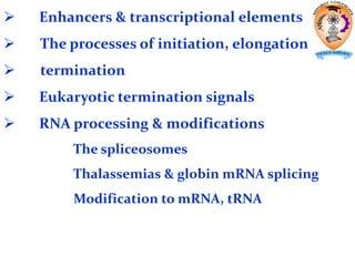
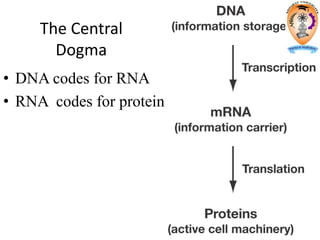
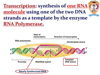
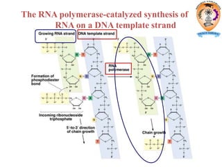
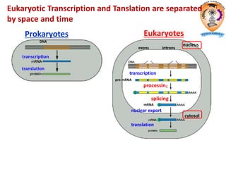
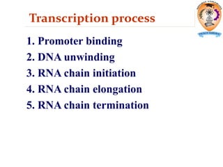
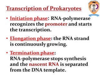
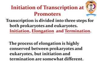
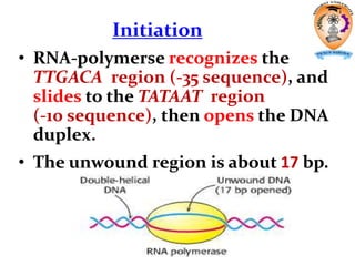
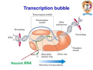
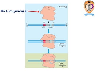
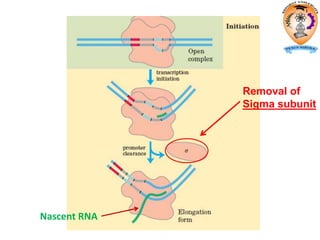
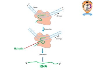
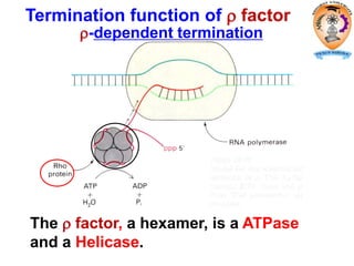
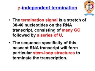
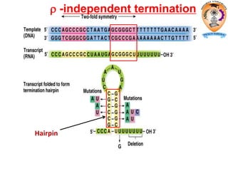
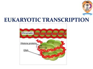
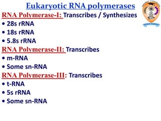
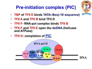
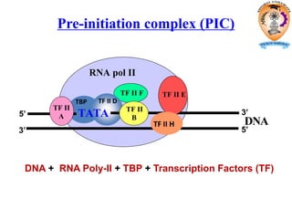
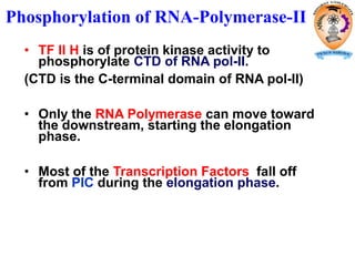
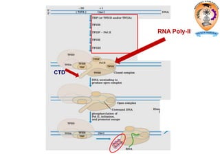
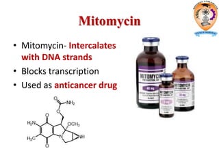
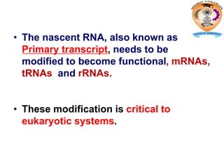
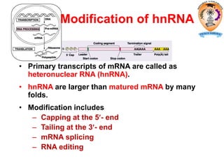
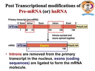
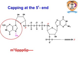
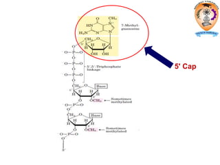
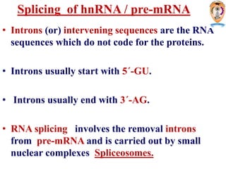
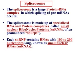
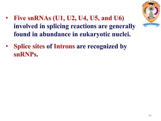
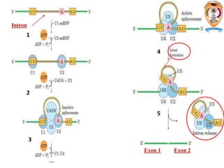
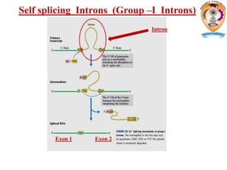
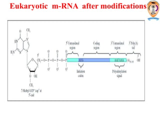
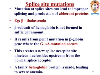
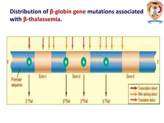
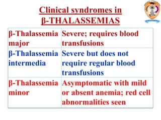
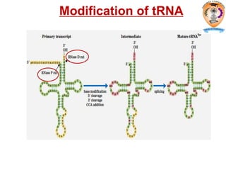
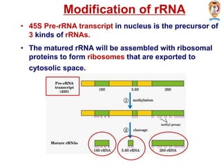
The document discusses transcription in prokaryotes and eukaryotes. In prokaryotes, RNA polymerase binds to promoter sequences and transcribes DNA into RNA through initiation, elongation, and termination. Transcription requires RNA polymerase and proceeds similarly in eukaryotes but involves multiple RNA polymerases and occurs in the nucleus. Eukaryotic transcription is more complex, utilizing regulatory sequences, transcription factors, and RNA processing to modify pre-mRNA into mature mRNA through splicing, capping, polyadenylation, and other modifications. Mutations can affect splicing and cause genetic disorders like beta-thalassemia.
it describes transcription with simple diagram and animation. its steps and inhibitors are described for both eukaryotes and prokaryotes. it will be easily understood by UG students . post transcriptional modification of all the RNA are also described with diagrams.
An Overview...
Definition of Translation.
Def. of Eukaryotes.
Translation: An Overview.
Components of Translation.
Some Enzymes .
Ribosome Role.
Mechanism of Translation.
Initiation.
Scanning Model of Initiation.
Initiation Factors.
Animation.
Elongation.
Chain Elongation: Translocation.
Animation.
Termination.
Animation....
It's not perfect still... what are your views friends?
Basics of Undergraduate/university fellows
Transcription is more complicated in eukaryotes than in prokaryotes because
eukaryotes possess three different classes of RNA polymerases and because of the
way in which transcripts are processed to their functional forms.
More proteins and transcription factors are involved in eukaryotic transcription.
RNA- A polymer of ribonucleotides, is a single stranded structure. There are three major types of RNA- m RNA,t RNA and r RNA. Besides that there are small nuclear,micro RNAs, small interfering and heterogeneous RNAs. Each of them has a specific structure and performs a specific function.
Microbial genetics is a subject area within microbiology and genetic engineering. This involves the study of the genotype of microbial species and also the expression system in the form of phenotypes
This presentation explains DNA transcription and RNA Processing.
It gives details about prokaryotic DNA transcription and eukaryotic DNA transcription. it also explains post-transcriptional modification both in prokaryotes and eukaryotes.
Prokaryotes are organisms that consist of a single prokaryotic cell. Eukaryotic cells are found in plants, animals, fungi, and protists. They range from 10–100 μm in diameter, and their DNA is contained within a membrane-bound nucleus.Prokaryotes do not have membrane-enclosed nuclei. Therefore, the processes of transcription, translation, and mRNA degradation can all occur simultaneously.
This Powerpoint consists of RNA synthesis (transcription) in prokaryotes and eukaryotes. This also explains about the post-transcriptional modifications in the mRNA. How the post transcriptionla modifications help in the gene expression.
Richard's aventures in two entangled wonderlandsRichard Gill
Since the loophole-free Bell experiments of 2020 and the Nobel prizes in physics of 2022, critics of Bell's work have retreated to the fortress of super-determinism. Now, super-determinism is a derogatory word - it just means "determinism". Palmer, Hance and Hossenfelder argue that quantum mechanics and determinism are not incompatible, using a sophisticated mathematical construction based on a subtle thinning of allowed states and measurements in quantum mechanics, such that what is left appears to make Bell's argument fail, without altering the empirical predictions of quantum mechanics. I think however that it is a smoke screen, and the slogan "lost in math" comes to my mind. I will discuss some other recent disproofs of Bell's theorem using the language of causality based on causal graphs. Causal thinking is also central to law and justice. I will mention surprising connections to my work on serial killer nurse cases, in particular the Dutch case of Lucia de Berk and the current UK case of Lucy Letby.
THE IMPORTANCE OF MARTIAN ATMOSPHERE SAMPLE RETURN.Sérgio Sacani
The return of a sample of near-surface atmosphere from Mars would facilitate answers to several first-order science questions surrounding the formation and evolution of the planet. One of the important aspects of terrestrial planet formation in general is the role that primary atmospheres played in influencing the chemistry and structure of the planets and their antecedents. Studies of the martian atmosphere can be used to investigate the role of a primary atmosphere in its history. Atmosphere samples would also inform our understanding of the near-surface chemistry of the planet, and ultimately the prospects for life. High-precision isotopic analyses of constituent gases are needed to address these questions, requiring that the analyses are made on returned samples rather than in situ.
Observation of Io’s Resurfacing via Plume Deposition Using Ground-based Adapt...Sérgio Sacani
Since volcanic activity was first discovered on Io from Voyager images in 1979, changes
on Io’s surface have been monitored from both spacecraft and ground-based telescopes.
Here, we present the highest spatial resolution images of Io ever obtained from a groundbased telescope. These images, acquired by the SHARK-VIS instrument on the Large
Binocular Telescope, show evidence of a major resurfacing event on Io’s trailing hemisphere. When compared to the most recent spacecraft images, the SHARK-VIS images
show that a plume deposit from a powerful eruption at Pillan Patera has covered part
of the long-lived Pele plume deposit. Although this type of resurfacing event may be common on Io, few have been detected due to the rarity of spacecraft visits and the previously low spatial resolution available from Earth-based telescopes. The SHARK-VIS instrument ushers in a new era of high resolution imaging of Io’s surface using adaptive
optics at visible wavelengths.
Multi-source connectivity as the driver of solar wind variability in the heli...Sérgio Sacani
The ambient solar wind that flls the heliosphere originates from multiple
sources in the solar corona and is highly structured. It is often described
as high-speed, relatively homogeneous, plasma streams from coronal
holes and slow-speed, highly variable, streams whose source regions are
under debate. A key goal of ESA/NASA’s Solar Orbiter mission is to identify
solar wind sources and understand what drives the complexity seen in the
heliosphere. By combining magnetic feld modelling and spectroscopic
techniques with high-resolution observations and measurements, we show
that the solar wind variability detected in situ by Solar Orbiter in March
2022 is driven by spatio-temporal changes in the magnetic connectivity to
multiple sources in the solar atmosphere. The magnetic feld footpoints
connected to the spacecraft moved from the boundaries of a coronal hole
to one active region (12961) and then across to another region (12957). This
is refected in the in situ measurements, which show the transition from fast
to highly Alfvénic then to slow solar wind that is disrupted by the arrival of
a coronal mass ejection. Our results describe solar wind variability at 0.5 au
but are applicable to near-Earth observatories.
10. RNA Polymerase
• The enzyme responsible for the RNA
synthesis is DNA-dependent RNA
polymerase.
–The prokaryotic RNA polymerase
is a multiple-subunit protein of
~480kD.
11. 1. Requires no primer for polymerization.
2. Requires DNA for activity and is most
active with a double-stranded DNA as
template.
3. 5’ 3’ synthesis.
4. Require Mg2+ for RNA synthesis activity.
5. lacks 3’ 5’ exonuclease activity, and the
error rate of nucleotides incorporation is
10-4 to 10-5.
6. Usually are multisubunit enzyme.
RNA Polymerase
(NMP)n + NTP (NMP)n+1 + PPi
12.
13. The holoenzyme of RNA-polymerase in E.coli
consists of 5 different subunits: 2
Subunit MW Function
36.5 KD
Determines the DNA to be
transcribed
150 KD Catalyzes polymerization
155.5 KD Binds & open DNA template
70 KD
Recognizes the promoter
for synthesis initiation
ω 11 KD Subunit packing
RNA Polymerase of E. Coli
17. Structure of bacterial prokaryotic
promoter region
TATA-Box / Pribnow box
• This is a stretch of 6
nucleotides ( 5'- TATAAT-3')
centered about 8-10
nucleotides to the left of the
transcription start site.
-35 Sequence
• A second consensus
nucleotide sequence
( 5'- TTGACA-3'), is centered
about 35 bases to the left of
the transcription start site.
Biochemistry For Medics- Lecture Notes 17
19. • Initiation phase: RNA-polymerase
recognizes the promoter and starts
the transcription.
• Elongation phase: the RNA strand
is continuously growing.
• Termination phase:
RNA-polymerase stops synthesis
and the nascent RNA is separated
from the DNA template.
Transcription of Prokaryotes
20. Initiation of Transcription at
Promoters
Transcription is divided into three steps for
both prokaryotes and eukaryotes.
Initiation, Elongation and Termination.
The process of elongation is highly
conserved between prokaryotes and
eukaryotes, but initiation and
termination are somewhat different.
21. Initiation
• RNA-polymerse recognizes the
TTGACA region (-35 sequence), and
slides to the TATAAT region
(-10 sequence), then opens the DNA
duplex.
• The unwound region is about 17 bp.
22. • The first nucleotide on RNA transcript
is always purine triphosphate. GTP is
more often than ATP.
• The pppGpN-OH structure remains on
the RNA transcript until the RNA
synthesis is completed.
• The three molecules form a
transcription initiation complex.
RNA-pol (2) - DNA - pppGpN- OH 3
23. • No primer is needed for RNA synthesis.
• The subunit falls off from the
RNA-polymerase once the first
3,5-phosphodiester bond is formed.
• The core enzyme moves along the DNA
template to enter the elongation phase.
24. Elongation
• The release of the subunit causes
the conformational change of the
core enzyme. The core enzyme slides
on the DNA template toward the 3
end.
• Free NTPs are added sequentially to
the 3 -OH of the nascent RNA strand.
(NMP)n + NTP (NMP)n+1 + PPi
RNA strand substrate
elongated
RNA strand
25. • RNA-polymerase, DNA segment of
~40nt and the nascent RNA form a
complex called the transcription
bubble.
• The 3 segment of the nascent RNA
hybridizes with the DNA template, and
its 5 end extends out the
transcription bubble as the synthesis
is processing.
31. Termination
• The RNA Polymerase stops moving on
the DNA template. The RNA transcript
falls off from the transcription complex.
• The termination occurs in either
-dependent or -independent manner.
32. The factor, a hexamer, is a ATPase
and a Helicase.
Termination function of factor
-dependent termination
33. -independent termination
• The termination signal is a stretch of
30-40 nucleotides on the RNA
transcript, consisting of many GC
followed by a series of U.
• The sequence specificity of this
nascent RNA transcript will form
particular stem-loop structures to
terminate the transcription.
36. Cleavage of this transcript produces 5S,
16S, and 23S rRNA molecules and a tRNA
molecule.
Spacer regions are shown in yellow.
Synthesis of rRNA and tRNA
39. It binds to the β-subunit of the RNA Polymerase
to block the initiation of transcription.
40. It binds with the β-subunit of prokaryotic
RNA Polymerase and thus inhibits the
Elongation phase of Transcription.
41. The tricyclic ring system (phenoxazone) of
Actinomycin D intercalates between adjacent
G-C base pairs, and the cyclic polypeptide
arms fill the nearby narrow groove and
inhibits Elongation phase of Transcription.
Actinomycin D
42. It inhibits the Elongation phase of Transcription
Cordycepin (3-deoxy Adenosine)
46. RNA POLYMERASE-II
•RNA polymerase II is central to eukaryotic
gene expression and has been studied
extensively.
•RNA polymerase II is a multi subunit
enzyme with 12 subunits.
•RNA polymerase II requires an array of
other proteins, called transcription factors
(TF II) in order to form the active
transcription complex.
48. α-Amanitin ( Fungal toxin from Amanita
phalloides) - cyclic octapeptide with
unussual amino acids.
Inhibitor of eukaryotic RNA polymerase
(mainly of type II )
49.
50. Eukaryotic Transcription
Promoters
Much more complex than those found in bacteria.
These are consensus sequences located at the
upstream regions of Coding strand.
Mutation of this region usually significantly lowers
the rate of transcription.
51. 1) TATA box ( Hogness Box)
Very similar to the prokaryotic TATA box,
except the sequence is slightly different
(TATAAA) and it is located in between
-25 to -30.
2) CAAT box
Located in between -70 to -80.
Always contains CCAAT.
3) GC box
Usually has the sequence GGGCGG
and is typically found at -110.
52. ENHANCERS :
Enhancers elements are the
sequences located in a variety of
regions of a gene both upstream
and downstream of the
transcription start site and even
within the transcribed portions of
some genes.
Enhancers increases the
transcription rate by several folds.
54. • RNA-pol II does not bind to the
promoter sequences directly.
• RNA-pol II associates with six
transcription factors.
• TFII A, TFII B, TFII D, TFII E, TFII F
and TFII H
Transcription factors
55.
56. Pre-initiation complex (PIC)
RNA pol II
TF II F
TBP TF IID
TATA
DNA
TF II
A
TF II
B
TF II E
TF II H
• TBP of TFII D binds TATA–Box(-10 sequence)
• TFII A and TFII B bind TFII D
• TFII F- RNA-pol complex binds TFII B
• TFII F and TFII E open the dsDNA (helicase
and ATPase)
• TFII H: completion of PIC
57. Pre-initiation complex (PIC)
RNA pol II
TF II F
TBP TF II D
TATA
DNA
TF II
A
TF II
B
TF II E
TF II H
DNA + RNA Poly-II + TBP + Transcription Factors (TF)
5’ 3’
5’3’
58. • TF II H is of protein kinase activity to
phosphorylate CTD of RNA pol-II.
(CTD is the C-terminal domain of RNA pol-II)
• Only the RNA Polymerase can move toward
the downstream, starting the elongation
phase.
• Most of the Transcription Factors fall off
from PIC during the elongation phase.
Phosphorylation of RNA-Polymerase-II
60. • When the RNA Polymerase transcribes the
terminator region of the DNA, the polymerase
releases the mRNA
• The termination sequence is AATAAA followed
by GT repeats.
Termination
61. Elongation
TFIIF remains associated with RNA Pol-II
throughout elongation.
The activity of the RNA poly-II is greatly
enhanced by proteins called Elongation factors
71. • The nascent RNA, also known as
Primary transcript, needs to be
modified to become functional, mRNAs,
tRNAs and rRNAs.
• These modification is critical to
eukaryotic systems.
72.
73. • Primary transcripts of mRNA are called as
heteronuclear RNA (hnRNA).
• hnRNA are larger than matured mRNA by many
folds.
• Modification includes
– Capping at the 5- end
– Tailing at the 3- end
– mRNA splicing
– RNA editing
Modification of hnRNA
74. Post Transcriptional modifications of
Pre-mRNA (or) hnRNA
• Introns are removed from the primary
transcript in the nucleus, exons (coding
sequences) are ligated to form the mRNA
molecule.
Exons
78. • The 5- cap structure is found on hnRNA
too. The capping process occurs in
nuclei.
• The cap structure of mRNA will be
recognized by the cap-binding protein
required for translation.
• The capping occurs prior to the splicing.
80. The matured mRNAs are much shorter than
the DNA templates.
DNA
mRNA
mRNA splicing
81. A~G no-coding region 1~7 coding region
L 1 2 3 4 5 6 7
7 700 bp
The structural genes are composed of Coding
(Exons) and Non-coding (Introns)regions
that are alternatively separated.
Split gene
EA B C D F G
82. • Introns (or) intervening sequences are the RNA
sequences which do not code for the proteins.
• Introns usually start with 5´-GU.
• Introns usually end with 3´-AG.
• RNA splicing involves the removal introns
from pre-mRNA and is carried out by small
nuclear complexes Spliceosomes.
Splicing of hnRNA / pre-mRNA
83. Spliceosome
• The spliceosome is a large Protein-RNA
complex in which splicing of pre-mRNAs
occurs.
• The spliceosome is made up of specialized
RNA and Protein complexes called small
nuclear RiboNucleoProteins (snRNPs, often
pronounced “snurps”).
• Each snRNP contains RNAs with 100 to 200
nucleotides long, known as small nuclear
RNAs (snRNAs).
84. 84
• Five snRNAs (U1, U2, U4, U5, and U6)
involved in splicing reactions are generally
found in abundance in eukaryotic nuclei.
• Splice sites of Introns are recognized by
snRNPs.
89. 89
Splice site mutations
• Mutation at splice sites can lead to improper
splicing and production of abberant proteins
• Eg: - thalassemia
• β-subunit of hemoglobin is not formed in
sufficient amount.
• It results from point mutation in -globin
gene where the GA mutation occurs.
• This creates a new splice acceptor site
nineteen nucleotides upstream from the
normal splice acceptor
• A faulty beta-globin protein is made, leading
to severe anemia.
93. Clinical syndromes in
β-THALASSEMIAS
β-Thalassemia
major
Severe; requires blood
transfusions
β-Thalassemia
intermedia
Severe but does not
require regular blood
transfusions
β-Thalassemia
minor
Asymptomatic with mild
or absent anemia; red cell
abnormalities seen
95. 95
Endo- and exonucleases to generate
ends of tRNA
• Endonuclease RNase P cleaves to generate the
5´ end.
• Exonuclease RNase D trims 3′ to 5′, leaving the
mature 3´ end.
99. Modification of rRNA
• 45S Pre-rRNA transcript in nucleus is the precursor of
3 kinds of rRNAs.
• The matured rRNA will be assembled with ribosomal
proteins to form ribosomes that are exported to
cytosolic space.