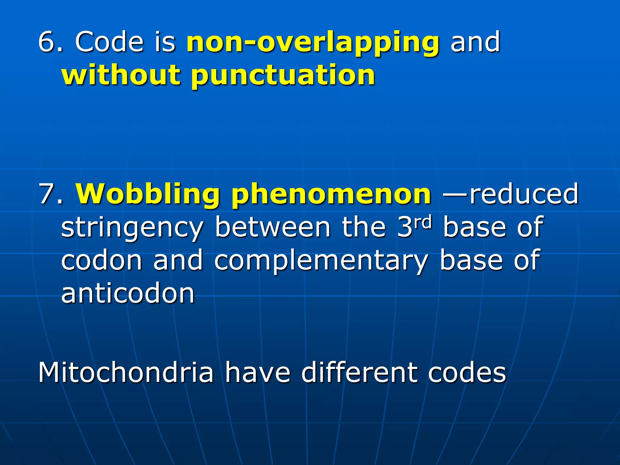
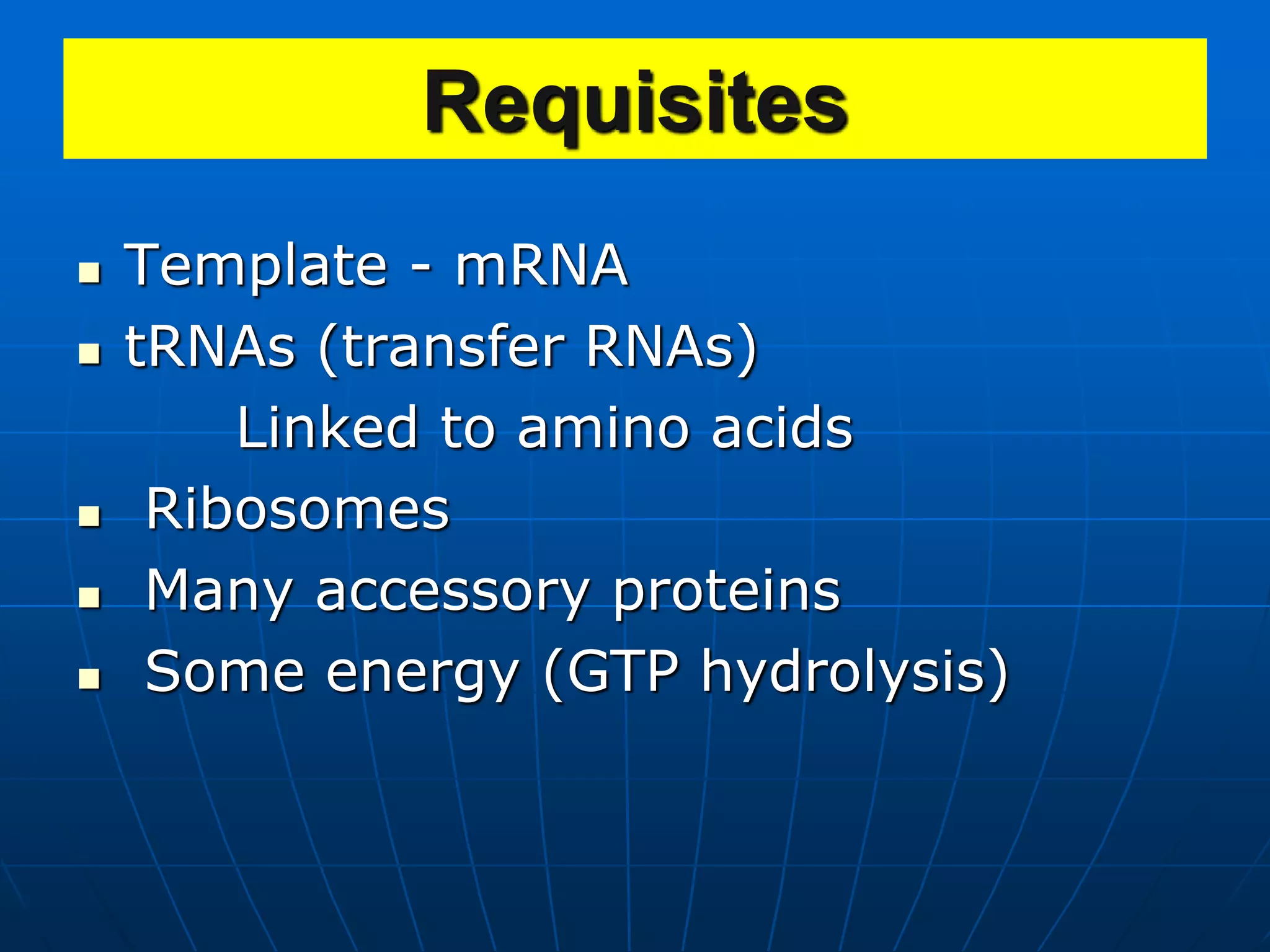
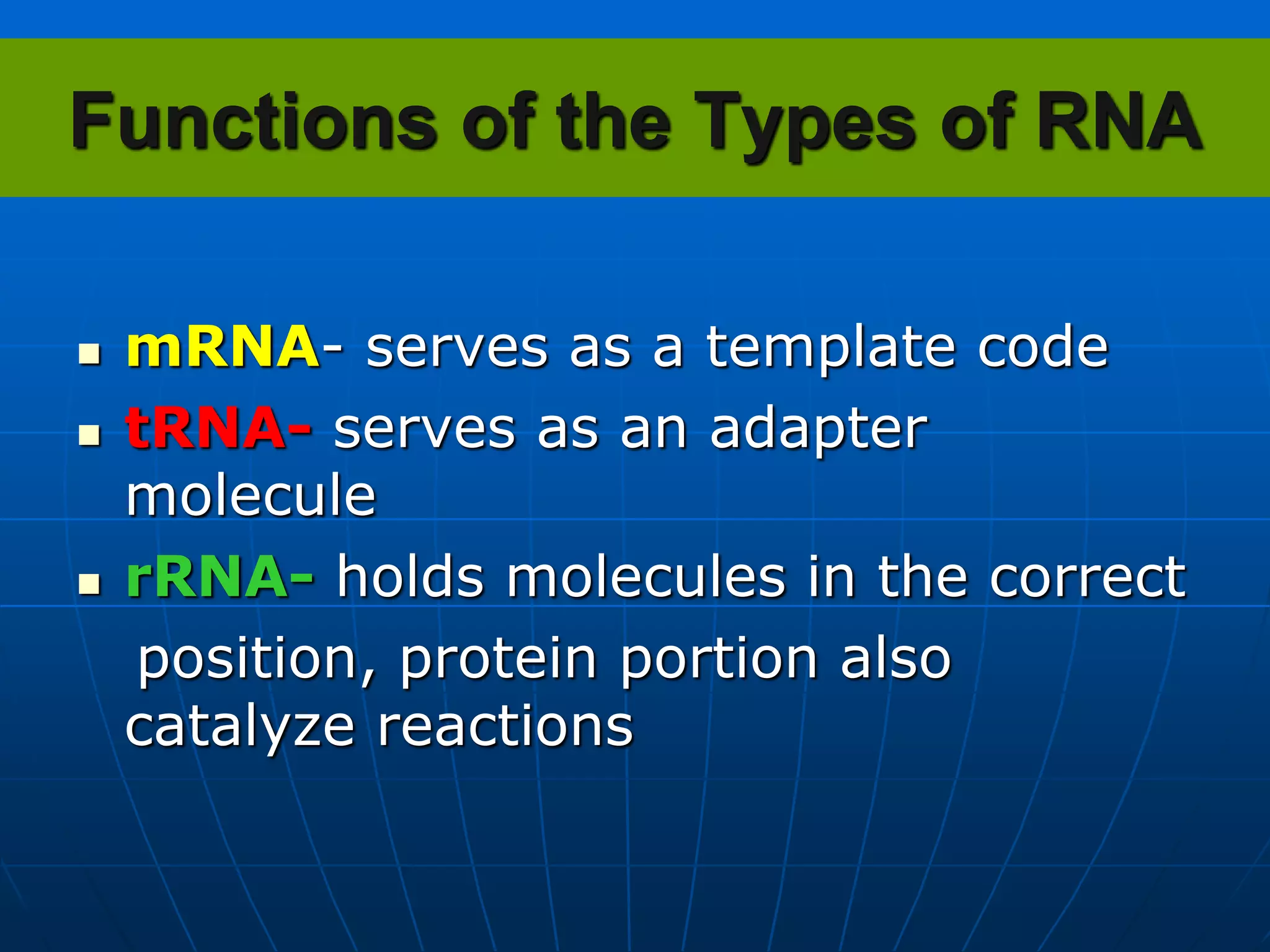
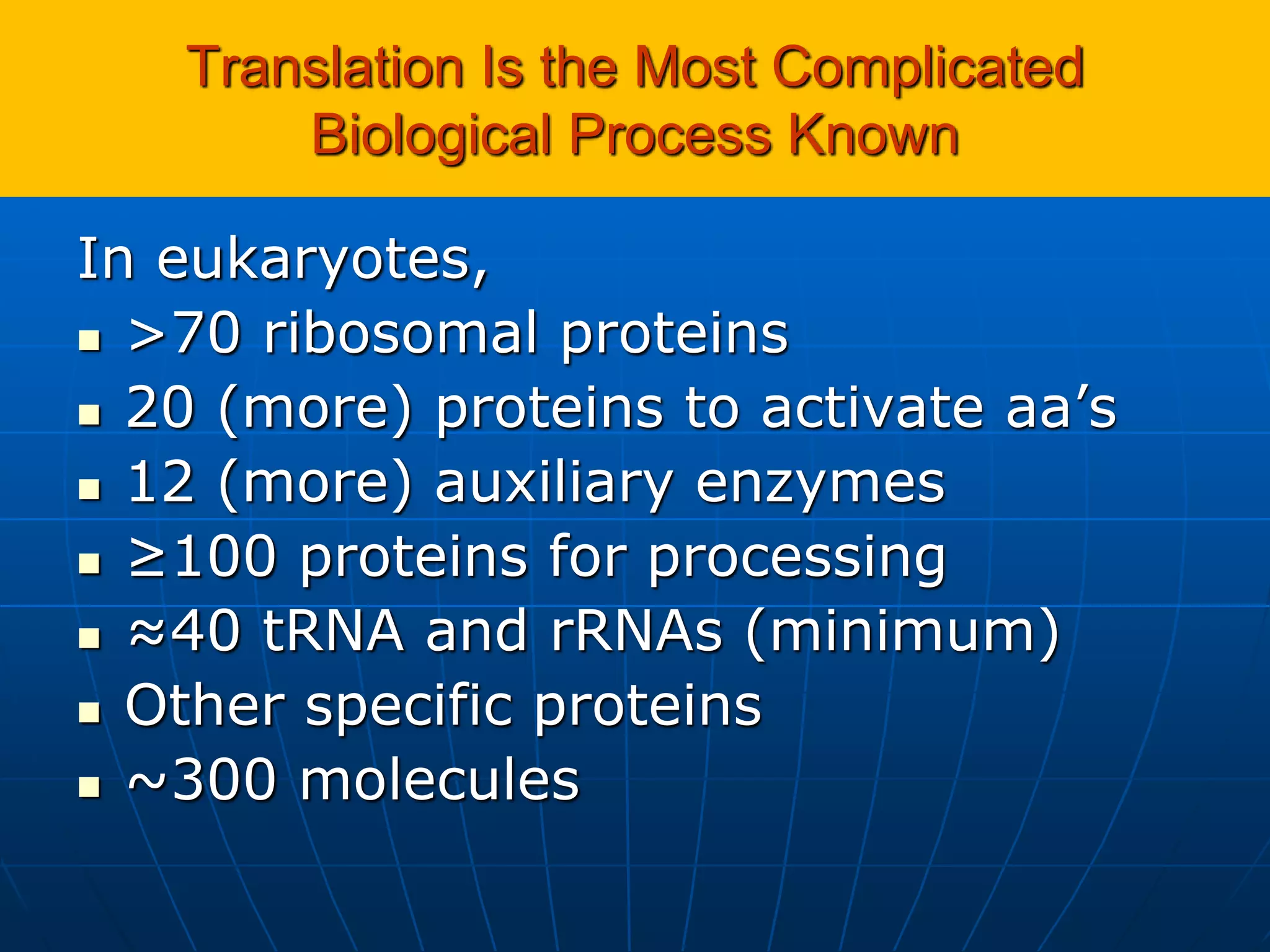
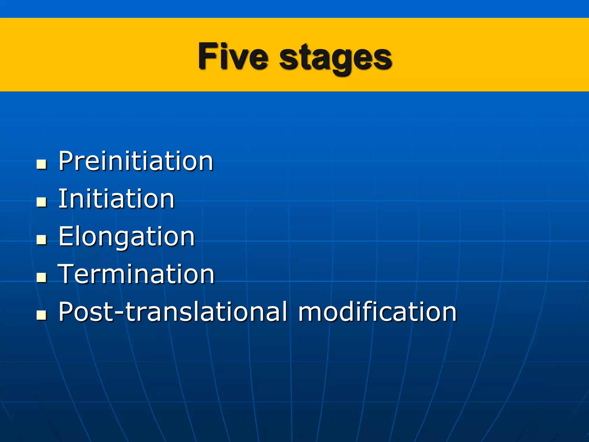
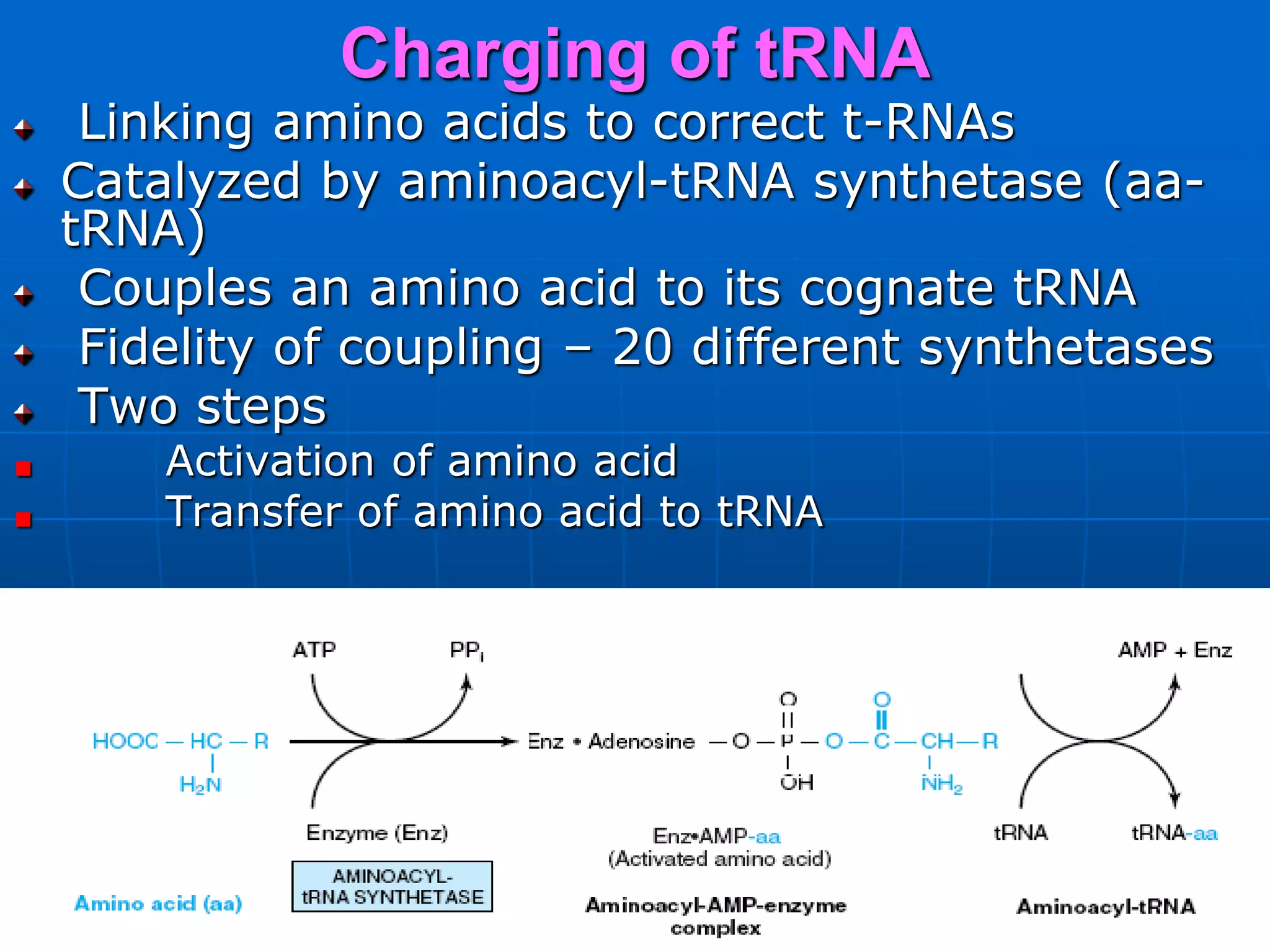
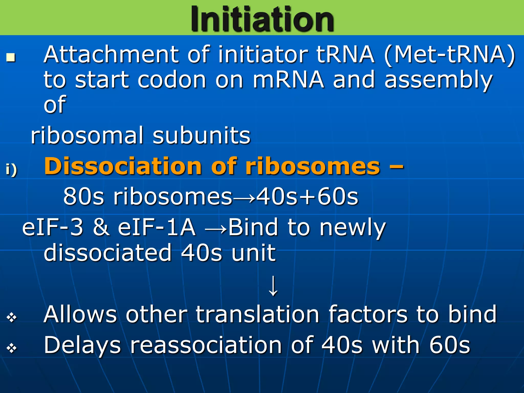
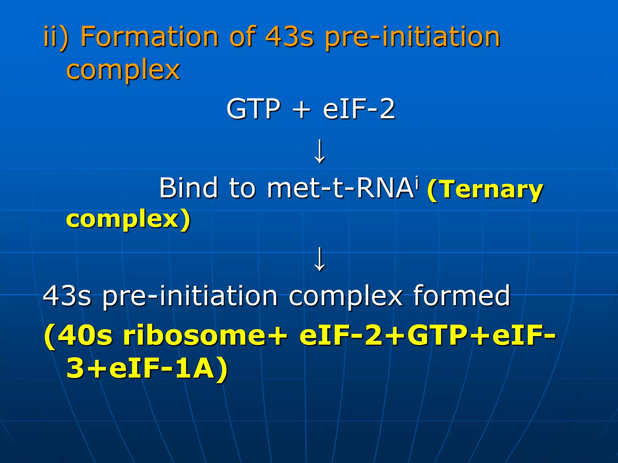
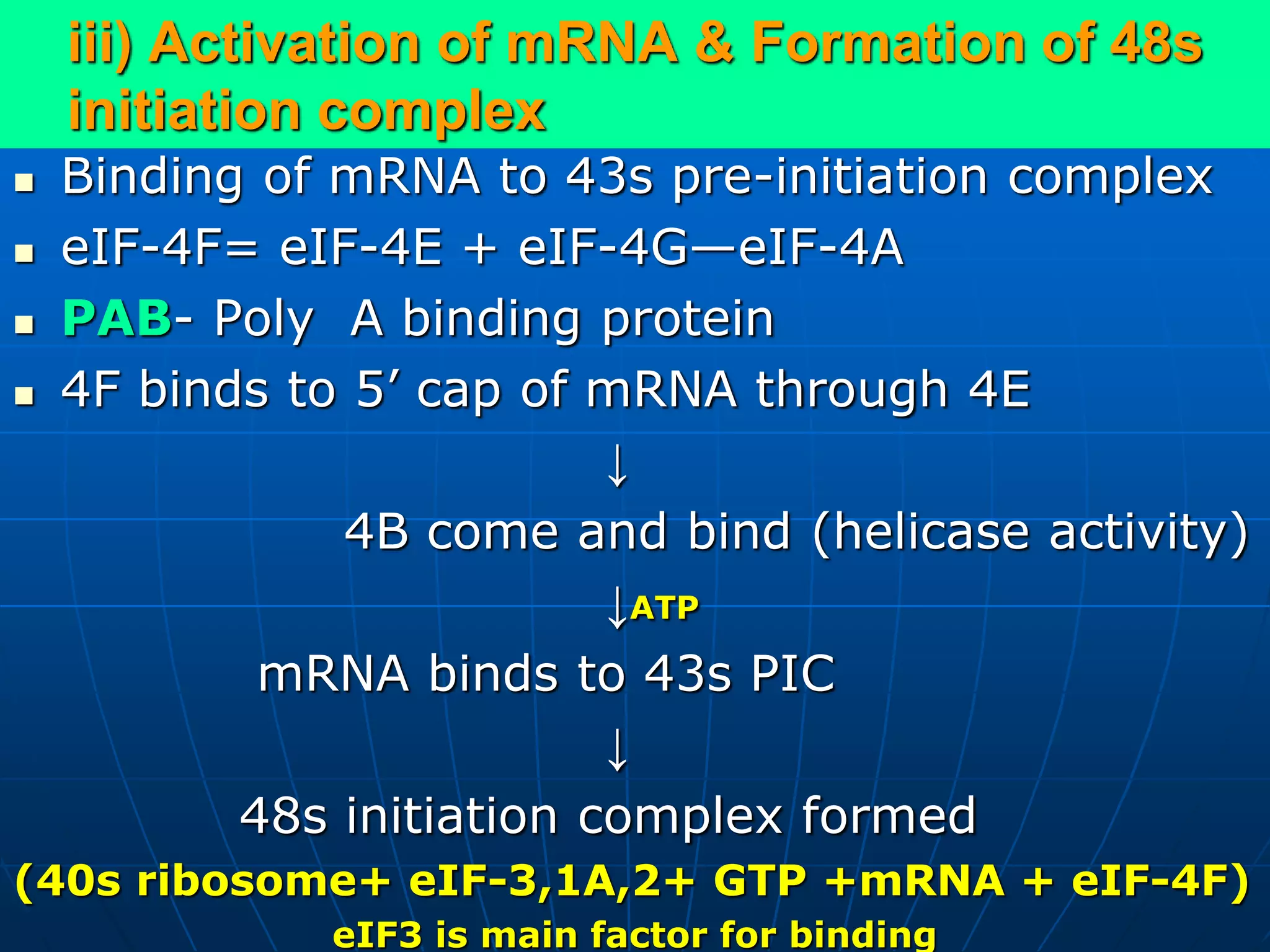
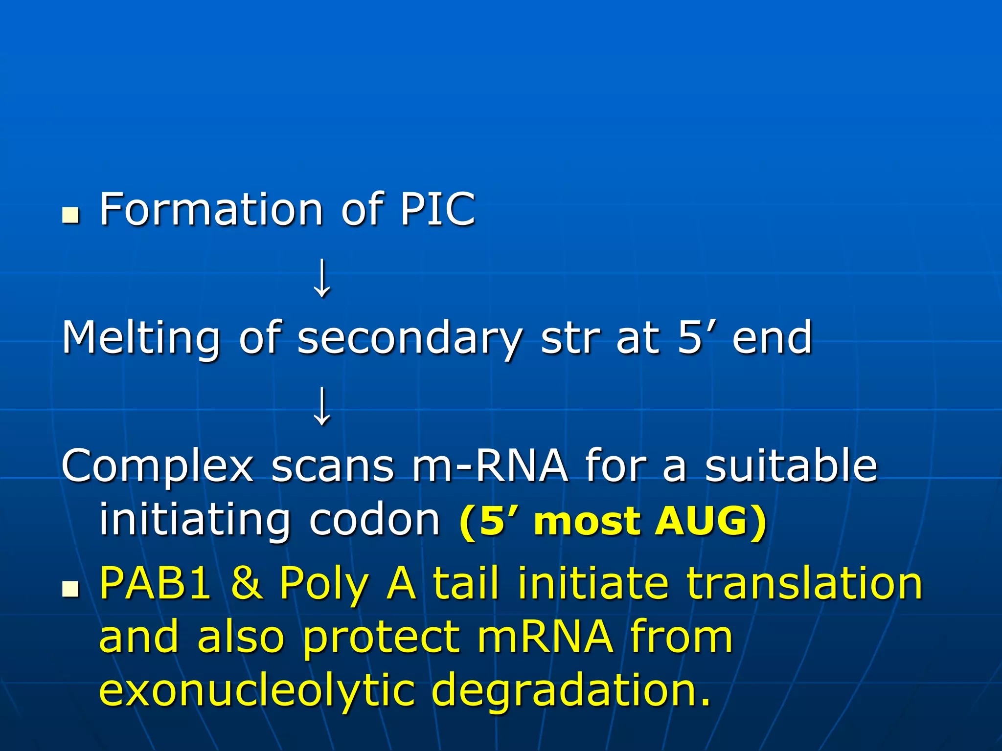
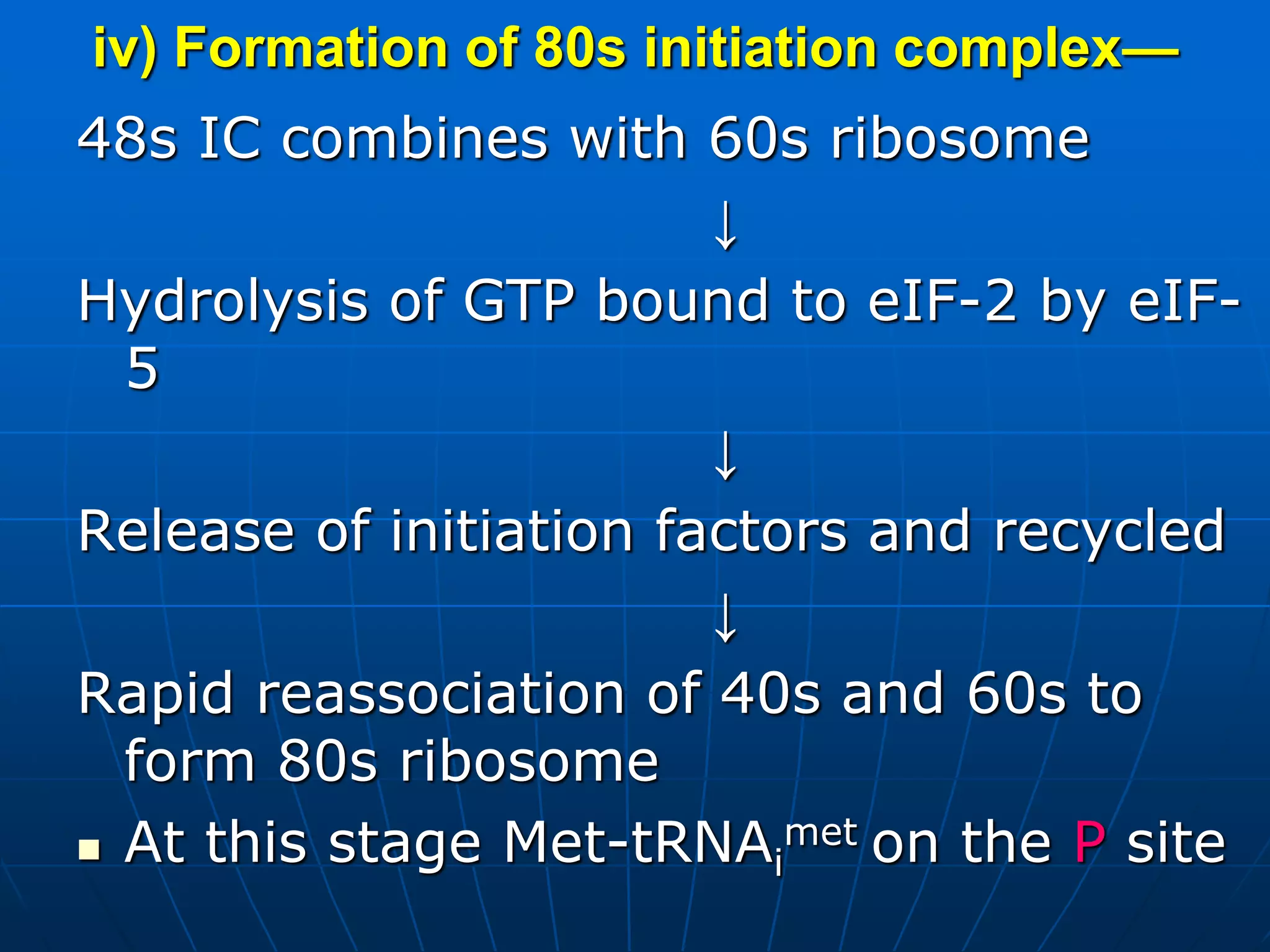
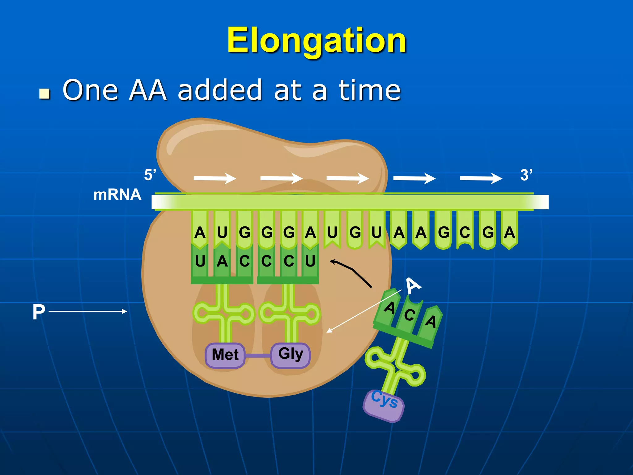
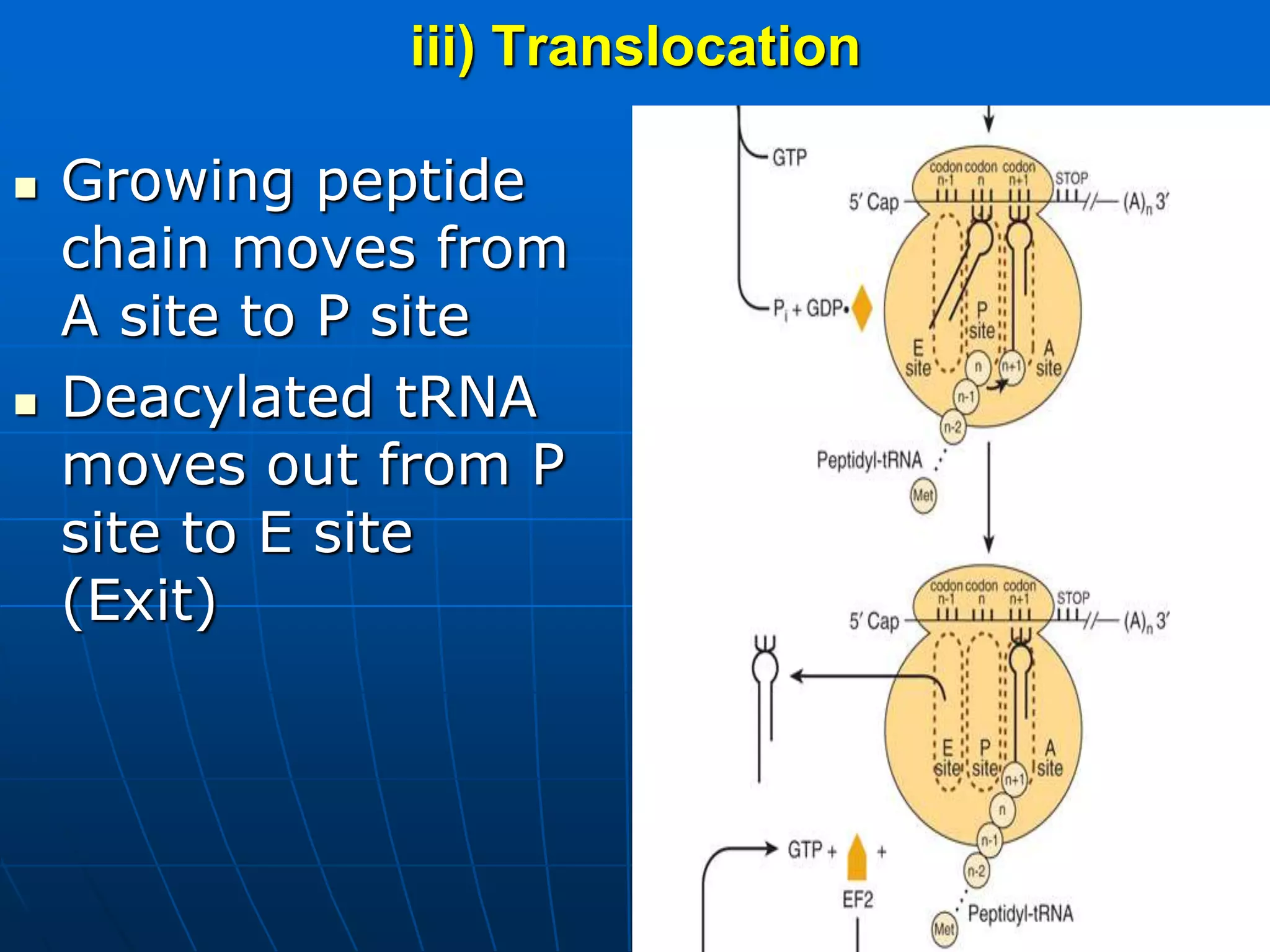
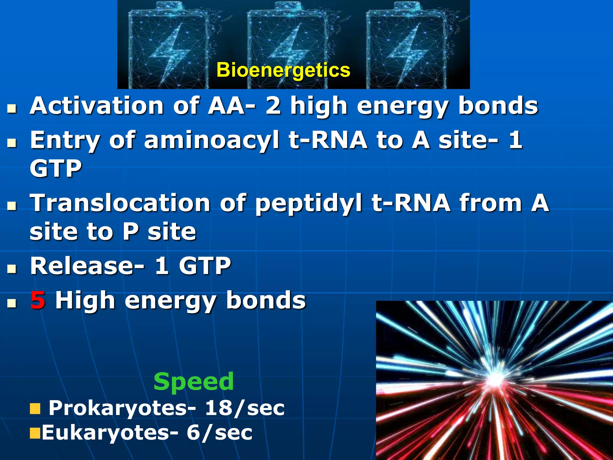
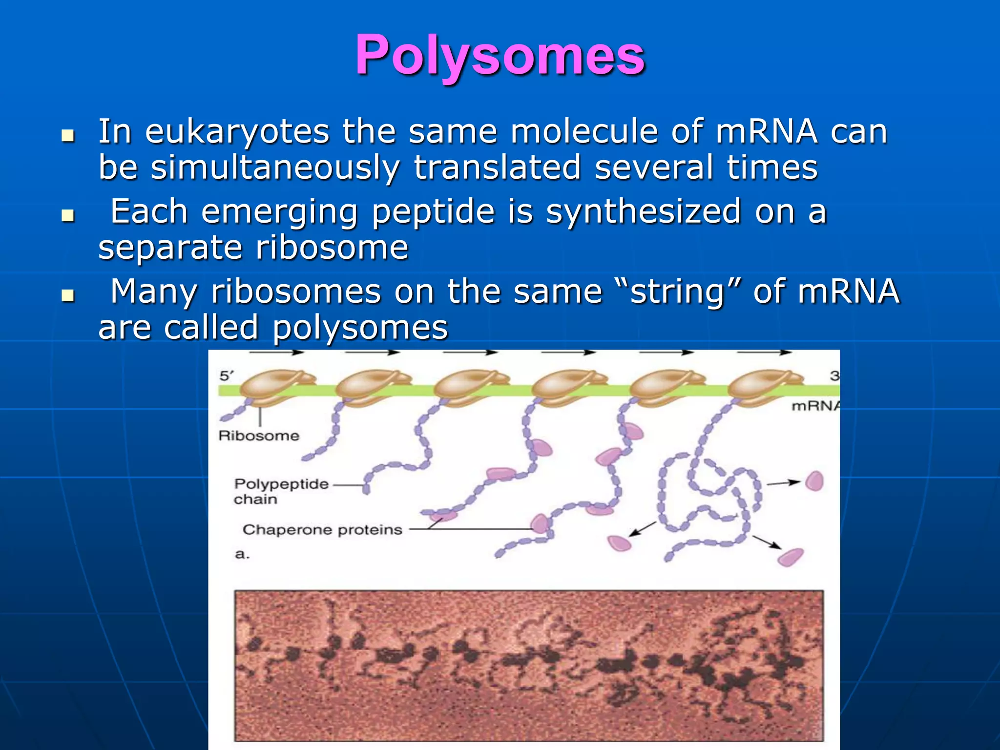
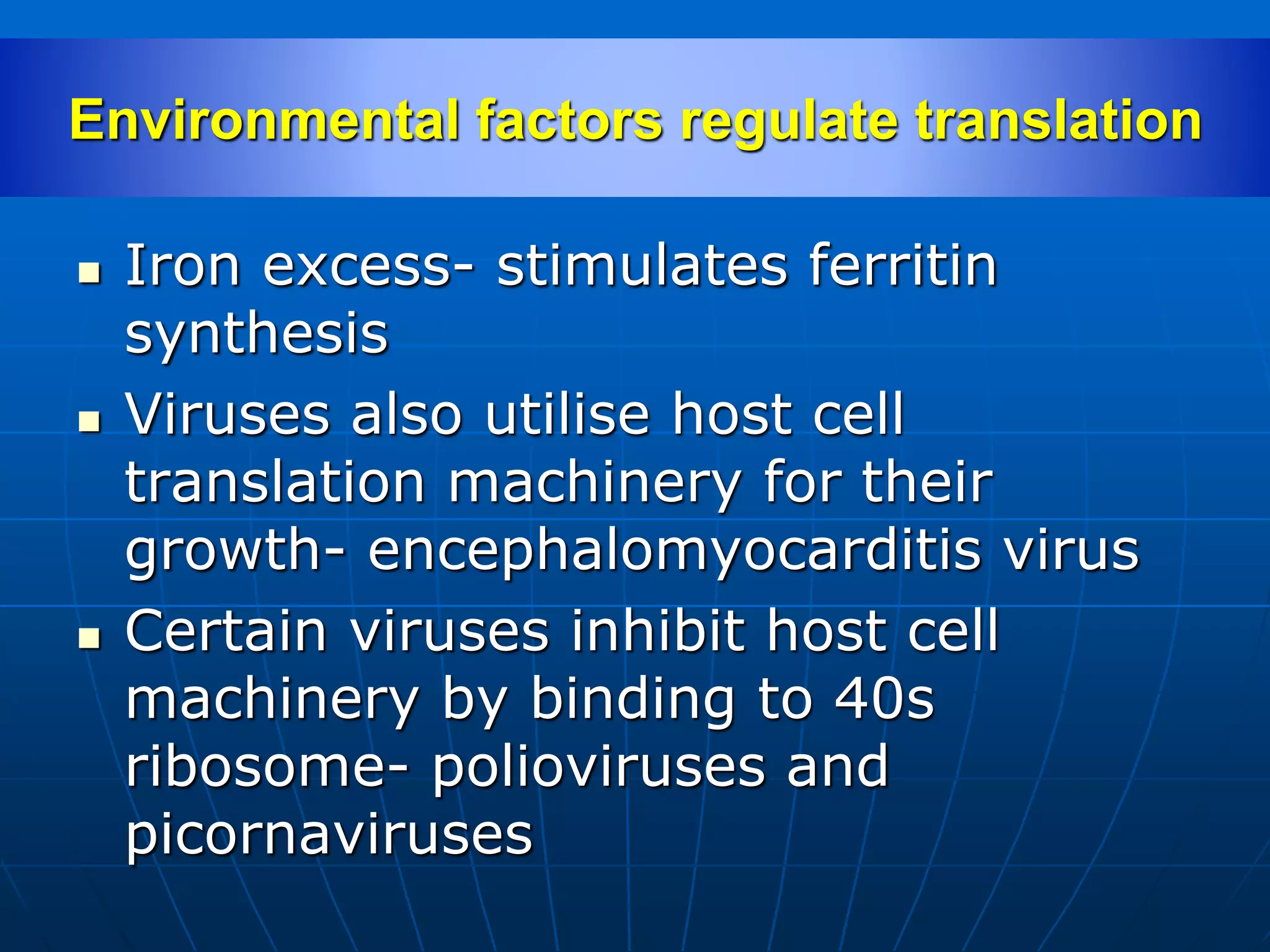
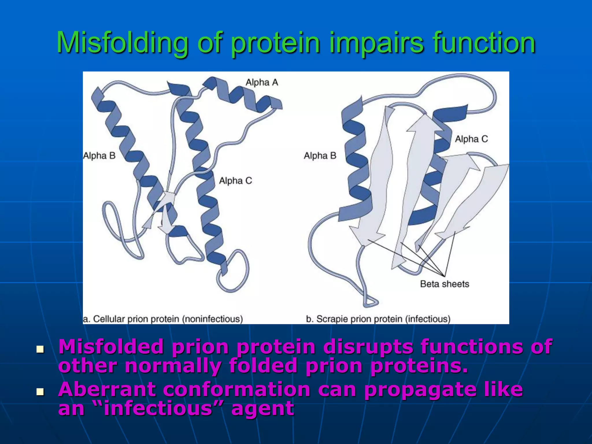
Prof (Dr.) V.P.Acharya
GENETIC CODE
Definition: System of nucleotide
sequences of mRNA that designates
particular amino acid sequences in
the process of translation.
 Genetic code is the relation between
the sequence bases in DNA and the
sequence of amino acids in protein
 Code words for amino acids--Codons
Indian-American scientist
He showed the order
of nucleotides in nucleic
acids, which carry
the genetic code of the
cell and control the
cell’s synthesis of
proteins.
Shared NP with Nirenberg
and Holley
Characteristics
1.64 possible codons
4 nucleotides– A,G,C,U
Triplet base codons
43=64
2. UNIVERSAL
Few exceptions– AUA codes for
Met in mitochondria
AUA– Ile in cytoplasm
UGA– stop codon in Cytoplasm
UGA– Trp in Mt
Bacteria– GUG,UUG, AUU,CUG
initiating codons
3. Stop codon and Initiating codon
AUG– Start codon
UAA,UGA and UAG– stop/ non-sense
codon
Exception– 21st AA Selenocystine--
UGA
4.Genetic code is degenerate
5. It is unambiguous
6. Code is non-overlapping and
without punctuation
7. Wobbling phenomenon —reduced
stringency between the 3rd base of
codon and complementary base of
anticodon
Mitochondria have different codes
TRANSLATION
Translation (An Overview)
 Translation is defined as protein synthesis.
 Occurs on ribosomes—mRNA → Potein
 mRNA is translated in the 5’ to 3’
direction.
 Highly regulated
 Very fast– 20AA/sec
 Amino acids are brought to the ribosome
bound to a specific tRNA molecule.
 The mRNA and tRNA are responsible for
the correct recognition of each amino acid
in the growing polypeptide
Requisites
 Template - mRNA
 tRNAs (transfer RNAs)
Linked to amino acids
 Ribosomes
 Many accessory proteins
 Some energy (GTP hydrolysis)
Functions of the Types of RNA
 mRNA- serves as a template code
 tRNA- serves as an adapter
molecule
 rRNA- holds molecules in the correct
position, protein portion also
catalyze reactions
Translation Is the Most Complicated
Biological Process Known
In eukaryotes,
 >70 ribosomal proteins
 20 (more) proteins to activate aa’s
 12 (more) auxiliary enzymes
 ≥100 proteins for processing
 ≈40 tRNA and rRNAs (minimum)
 Other specific proteins
 ~300 molecules
Five stages
 Preinitiation
 Initiation
 Elongation
 Termination
 Post-translational modification
Pre-initiation/Charging of AA
Aminoacyl t-RNA
Charging of tRNA
Linking amino acids to correct t-RNAs
Catalyzed by aminoacyl-tRNA synthetase (aa-
tRNA)
Couples an amino acid to its cognate tRNA
Fidelity of coupling – 20 different synthetases
Two steps
Activation of amino acid
Transfer of amino acid to tRNA
Initiation
 Attachment of initiator tRNA (Met-tRNA)
to start codon on mRNA and assembly
of
ribosomal subunits
i) Dissociation of ribosomes –
80s ribosomes→40s+60s
eIF-3 & eIF-1A →Bind to newly
dissociated 40s unit
↓
 Allows other translation factors to bind
 Delays reassociation of 40s with 60s
ii) Formation of 43s pre-initiation
complex
GTP + eIF-2
↓
Bind to met-t-RNAi (Ternary
complex)
↓
43s pre-initiation complex formed
(40s ribosome+ eIF-2+GTP+eIF-
3+eIF-1A)
eIF-2 is a control point
 α, β, γ subunits
 eIF 2α – phosphorylated by 4 different protein
kinases- HCR, PKR, PERK, and GCN2
Cell under stress (AA/Glucose starvation,
viral infection, mis-folded protein, serum
deprivation, hyperosmolality, heat shock )
↓
Kinase activated
↓
Translation inactivated
 PKR- actvated in viral infection
 eIF2α binds tightly to and deactivates GTP-
GDP recycling
iii) Activation of mRNA & Formation of 48s
initiation complex
 Binding of mRNA to 43s pre-initiation complex
 eIF-4F= eIF-4E + eIF-4G—eIF-4A
 PAB- Poly A binding protein
 4F binds to 5’ cap of mRNA through 4E
↓
4B come and bind (helicase activity)
↓ATP
mRNA binds to 43s PIC
↓
48s initiation complex formed
(40s ribosome+ eIF-3,1A,2+ GTP +mRNA + eIF-4F)
eIF3 is main factor for binding
 Usually to 5’ most AUG is chosen but it is
decided by Kozak consensus sequences-
surrounding AUG codon
 Prokaryotes– Shine-Dalgarno
sequence--a sequence of nucleotide
bases on m-RNA that facilitates m-RNA
binding to the pre-initiation complex
Eukaryotes– Kozak consensus sequence
 A purine present at -3 and +4 positions
of the initiating codon
 Formation of PIC
↓
Melting of secondary str at 5’ end
↓
Complex scans m-RNA for a suitable
initiating codon (5’ most AUG)
 PAB1 & Poly A tail initiate translation
and also protect mRNA from
exonucleolytic degradation.
iv) Formation of 80s initiation complex—
48s IC combines with 60s ribosome
↓
Hydrolysis of GTP bound to eIF-2 by eIF-
5
↓
Release of initiation factors and recycled
↓
Rapid reassociation of 40s and 60s to
form 80s ribosome
 At this stage Met-tRNAi
met on the P site
Regulation of initiation
 eIF-4F– rate of translation
 4E– recognises the cap of mRNA
 4G– Scaffolding protein
-- binds to helicase complex that
helps in unwinding the RNA
 eIF-2– one of the key regulators
 PAB regulates the initiation of
translation
Phosphorylated 4E binds more avidly to
mRNA cap structure
 1. Phosphorylation of 4E (Ser
209 or Thr 210)is controlled by
insulin and mitogenic factors
 Components of MAP kinase,
mTOR, PI3K, RAS, s6 kinase
pathways- involved in
regulation
 2nd path of regulation- Binding
of binding proteins keep 4E
inactivated
 Insulin & GF- cause
phosphorylation of BP and its
release from 4E
Insulin and GF enhance
post-transcriptional protein
synthesis in liver, muscle
and adipose tissues
Elongation
 One AA added at a time
CU A
Met
mRNA
5’ 3’
C C U
Gly
U U U CG G G G GGA A A A A
P
i) Binding of aminoacyl t-RNA to the A-
site
A site empty
Binding of proper AA-t-RNA requires
codon recognition
EF-1A (Elongation factor)+ GTP+
Aminoacyl t-RNA
↓
Ternary complex
↓
Enters A site
↓ EF-1A,GDP+Pi
Aminoacyl-tRNA
ii) Peptide bond formation
 Peptidyl transferase– catalyses
peptide bond formation
 Found on 28s rRNA
 Ribozyme
 No energy required
iii) Translocation
 Growing peptide
chain moves from
A site to P site
 Deacylated tRNA
moves out from P
site to E site
(Exit)
 EF-2 factor binds
↓
Displaces peptidyl tRNA from A site to
P site
↓
Energy from GTP; mRNA moves by
one codon
 Process of peptide synthesis occurs
until a termination codon is reached
Termination
Termination
Stop codon appears on A site
No tRNA available
Releasing factor (RF-1) recognises its
presence in A site
RF-1+RF-3+GTP→ Hydrolysis of bond
between peptide and tRNA occupying
P site (a water molecule is added)
↓
Protein, tRNA, 60s & 40s
ribosomes, GDP released
 Activation of AA- 2 high energy bonds
 Entry of aminoacyl t-RNA to A site- 1
GTP
 Translocation of peptidyl t-RNA from A
site to P site
 Release- 1 GTP
 5 High energy bonds
Bioenergetics
Speed
Prokaryotes- 18/sec
Eukaryotes- 6/sec
Polysomes
 In eukaryotes the same molecule of mRNA can
be simultaneously translated several times
 Each emerging peptide is synthesized on a
separate ribosome
 Many ribosomes on the same “string” of mRNA
are called polysomes
P bodies
 Non-translating mRNAs can
form Ribonucleoprotein
particles (mRNPs)-
accumulate in Cytoplasmic
organelles as P bodies
 P bodies- sites for
translation repression and
mRNA decay
 35 distinct proteins are found
in P bodies like mRNA
decapping enzyem, RNA
helicases, exonucleases etc
 Few mRNAs are retrieved at
the time of need
Environmental factors regulate translation
 Iron excess- stimulates ferritin
synthesis
 Viruses also utilise host cell
translation machinery for their
growth- encephalomyocarditis virus
 Certain viruses inhibit host cell
machinery by binding to 40s
ribosome- polioviruses and
picornaviruses
Four levels of protein structure
Chaperones and protein folding
 Heat shock proteins
Hsp70
Chaperonin (Hsp60)
 Prevent aggregation
 Put all the hydrophobic ends inside
 Misfolding causes diseases e.g.
Cystic fibrosis, Prion diseases
Misfolding of protein impairs function
 Misfolded prion protein disrupts functions of
other normally folded prion proteins.
 Aberrant conformation can propagate like
an “infectious” agent
POST-TRANSLATIONAL
MODIFICATION
Post-translational modification of Polypeptide
Protein folding
Proteolytic cleavage Intein splicing
Covalent modification
Different structures
Insulin, Zymogens
Inteins removed & Exteins spliced
a. Phosphorylation
b. Hydroxylation
c. Glycosylation
d. Carboxylation
e. Ubiquitylation
Inhibitors of Translation
A. Reversible inhibitor
a.Tetracyclin– Binds to 30s ribosome
↓
Inhibit attachment of aminoacyl tRNA to the
A site
b. Chloramphenicol– Inhibits peptidyl
transferase
c. Erythromycin & Clindamycin– Prevent
translocation
B. Irreversible inhibitor
Streptomycin and
Aminoglycosides—Bind to 30s
ribosomal sub-unit
Low conc.- Misreading of protein
↓
Useless bacterial proteins
Pharma. Conc.- Inhibit IC synthesis
↓
Protein synthesis inhibited
C. Inhibitors in mammals
1. Puromycin– Structural analogue of
tyrosinyl t-RNA
2. Cycloheximide– Inhibits peptidyl
transferase
3. Diphtheria toxin—Inactivation of
EF-2 by attachment of ADP to EF-2
4. Ricin– Inactivates 28s rRNA
Protein Targeting/Sorting of proteins
For more ppt onMedicalBiochemistrypleasevisit
www.vpacharya.com

Genetic code and translation

  • 1.
  • 2.
    GENETIC CODE Definition: Systemof nucleotide sequences of mRNA that designates particular amino acid sequences in the process of translation.  Genetic code is the relation between the sequence bases in DNA and the sequence of amino acids in protein  Code words for amino acids--Codons
  • 3.
    Indian-American scientist He showedthe order of nucleotides in nucleic acids, which carry the genetic code of the cell and control the cell’s synthesis of proteins. Shared NP with Nirenberg and Holley
  • 5.
    Characteristics 1.64 possible codons 4nucleotides– A,G,C,U Triplet base codons 43=64
  • 6.
    2. UNIVERSAL Few exceptions–AUA codes for Met in mitochondria AUA– Ile in cytoplasm UGA– stop codon in Cytoplasm UGA– Trp in Mt Bacteria– GUG,UUG, AUU,CUG initiating codons
  • 7.
    3. Stop codonand Initiating codon AUG– Start codon UAA,UGA and UAG– stop/ non-sense codon Exception– 21st AA Selenocystine-- UGA 4.Genetic code is degenerate 5. It is unambiguous
  • 9.
    6. Code isnon-overlapping and without punctuation 7. Wobbling phenomenon —reduced stringency between the 3rd base of codon and complementary base of anticodon Mitochondria have different codes
  • 10.
  • 11.
    Translation (An Overview) Translation is defined as protein synthesis.  Occurs on ribosomes—mRNA → Potein  mRNA is translated in the 5’ to 3’ direction.  Highly regulated  Very fast– 20AA/sec  Amino acids are brought to the ribosome bound to a specific tRNA molecule.  The mRNA and tRNA are responsible for the correct recognition of each amino acid in the growing polypeptide
  • 13.
    Requisites  Template -mRNA  tRNAs (transfer RNAs) Linked to amino acids  Ribosomes  Many accessory proteins  Some energy (GTP hydrolysis)
  • 14.
    Functions of theTypes of RNA  mRNA- serves as a template code  tRNA- serves as an adapter molecule  rRNA- holds molecules in the correct position, protein portion also catalyze reactions
  • 15.
    Translation Is theMost Complicated Biological Process Known In eukaryotes,  >70 ribosomal proteins  20 (more) proteins to activate aa’s  12 (more) auxiliary enzymes  ≥100 proteins for processing  ≈40 tRNA and rRNAs (minimum)  Other specific proteins  ~300 molecules
  • 16.
    Five stages  Preinitiation Initiation  Elongation  Termination  Post-translational modification
  • 17.
  • 18.
    Charging of tRNA Linkingamino acids to correct t-RNAs Catalyzed by aminoacyl-tRNA synthetase (aa- tRNA) Couples an amino acid to its cognate tRNA Fidelity of coupling – 20 different synthetases Two steps Activation of amino acid Transfer of amino acid to tRNA
  • 20.
    Initiation  Attachment ofinitiator tRNA (Met-tRNA) to start codon on mRNA and assembly of ribosomal subunits i) Dissociation of ribosomes – 80s ribosomes→40s+60s eIF-3 & eIF-1A →Bind to newly dissociated 40s unit ↓  Allows other translation factors to bind  Delays reassociation of 40s with 60s
  • 22.
    ii) Formation of43s pre-initiation complex GTP + eIF-2 ↓ Bind to met-t-RNAi (Ternary complex) ↓ 43s pre-initiation complex formed (40s ribosome+ eIF-2+GTP+eIF- 3+eIF-1A)
  • 23.
    eIF-2 is acontrol point  α, β, γ subunits  eIF 2α – phosphorylated by 4 different protein kinases- HCR, PKR, PERK, and GCN2 Cell under stress (AA/Glucose starvation, viral infection, mis-folded protein, serum deprivation, hyperosmolality, heat shock ) ↓ Kinase activated ↓ Translation inactivated
  • 24.
     PKR- actvatedin viral infection  eIF2α binds tightly to and deactivates GTP- GDP recycling
  • 26.
    iii) Activation ofmRNA & Formation of 48s initiation complex  Binding of mRNA to 43s pre-initiation complex  eIF-4F= eIF-4E + eIF-4G—eIF-4A  PAB- Poly A binding protein  4F binds to 5’ cap of mRNA through 4E ↓ 4B come and bind (helicase activity) ↓ATP mRNA binds to 43s PIC ↓ 48s initiation complex formed (40s ribosome+ eIF-3,1A,2+ GTP +mRNA + eIF-4F) eIF3 is main factor for binding
  • 28.
     Usually to5’ most AUG is chosen but it is decided by Kozak consensus sequences- surrounding AUG codon  Prokaryotes– Shine-Dalgarno sequence--a sequence of nucleotide bases on m-RNA that facilitates m-RNA binding to the pre-initiation complex Eukaryotes– Kozak consensus sequence  A purine present at -3 and +4 positions of the initiating codon
  • 29.
     Formation ofPIC ↓ Melting of secondary str at 5’ end ↓ Complex scans m-RNA for a suitable initiating codon (5’ most AUG)  PAB1 & Poly A tail initiate translation and also protect mRNA from exonucleolytic degradation.
  • 30.
    iv) Formation of80s initiation complex— 48s IC combines with 60s ribosome ↓ Hydrolysis of GTP bound to eIF-2 by eIF- 5 ↓ Release of initiation factors and recycled ↓ Rapid reassociation of 40s and 60s to form 80s ribosome  At this stage Met-tRNAi met on the P site
  • 31.
    Regulation of initiation eIF-4F– rate of translation  4E– recognises the cap of mRNA  4G– Scaffolding protein -- binds to helicase complex that helps in unwinding the RNA  eIF-2– one of the key regulators  PAB regulates the initiation of translation
  • 32.
    Phosphorylated 4E bindsmore avidly to mRNA cap structure  1. Phosphorylation of 4E (Ser 209 or Thr 210)is controlled by insulin and mitogenic factors  Components of MAP kinase, mTOR, PI3K, RAS, s6 kinase pathways- involved in regulation  2nd path of regulation- Binding of binding proteins keep 4E inactivated  Insulin & GF- cause phosphorylation of BP and its release from 4E Insulin and GF enhance post-transcriptional protein synthesis in liver, muscle and adipose tissues
  • 34.
    Elongation  One AAadded at a time CU A Met mRNA 5’ 3’ C C U Gly U U U CG G G G GGA A A A A P
  • 36.
    i) Binding ofaminoacyl t-RNA to the A- site A site empty Binding of proper AA-t-RNA requires codon recognition EF-1A (Elongation factor)+ GTP+ Aminoacyl t-RNA ↓ Ternary complex ↓ Enters A site ↓ EF-1A,GDP+Pi Aminoacyl-tRNA
  • 38.
    ii) Peptide bondformation  Peptidyl transferase– catalyses peptide bond formation  Found on 28s rRNA  Ribozyme  No energy required
  • 39.
    iii) Translocation  Growingpeptide chain moves from A site to P site  Deacylated tRNA moves out from P site to E site (Exit)
  • 40.
     EF-2 factorbinds ↓ Displaces peptidyl tRNA from A site to P site ↓ Energy from GTP; mRNA moves by one codon  Process of peptide synthesis occurs until a termination codon is reached
  • 41.
  • 42.
  • 43.
    Stop codon appearson A site No tRNA available Releasing factor (RF-1) recognises its presence in A site RF-1+RF-3+GTP→ Hydrolysis of bond between peptide and tRNA occupying P site (a water molecule is added) ↓ Protein, tRNA, 60s & 40s ribosomes, GDP released
  • 44.
     Activation ofAA- 2 high energy bonds  Entry of aminoacyl t-RNA to A site- 1 GTP  Translocation of peptidyl t-RNA from A site to P site  Release- 1 GTP  5 High energy bonds Bioenergetics Speed Prokaryotes- 18/sec Eukaryotes- 6/sec
  • 45.
    Polysomes  In eukaryotesthe same molecule of mRNA can be simultaneously translated several times  Each emerging peptide is synthesized on a separate ribosome  Many ribosomes on the same “string” of mRNA are called polysomes
  • 46.
    P bodies  Non-translatingmRNAs can form Ribonucleoprotein particles (mRNPs)- accumulate in Cytoplasmic organelles as P bodies  P bodies- sites for translation repression and mRNA decay  35 distinct proteins are found in P bodies like mRNA decapping enzyem, RNA helicases, exonucleases etc  Few mRNAs are retrieved at the time of need
  • 47.
    Environmental factors regulatetranslation  Iron excess- stimulates ferritin synthesis  Viruses also utilise host cell translation machinery for their growth- encephalomyocarditis virus  Certain viruses inhibit host cell machinery by binding to 40s ribosome- polioviruses and picornaviruses
  • 48.
    Four levels ofprotein structure
  • 49.
    Chaperones and proteinfolding  Heat shock proteins Hsp70 Chaperonin (Hsp60)  Prevent aggregation  Put all the hydrophobic ends inside  Misfolding causes diseases e.g. Cystic fibrosis, Prion diseases
  • 50.
    Misfolding of proteinimpairs function  Misfolded prion protein disrupts functions of other normally folded prion proteins.  Aberrant conformation can propagate like an “infectious” agent
  • 51.
  • 52.
    Post-translational modification ofPolypeptide Protein folding Proteolytic cleavage Intein splicing Covalent modification Different structures Insulin, Zymogens Inteins removed & Exteins spliced a. Phosphorylation b. Hydroxylation c. Glycosylation d. Carboxylation e. Ubiquitylation
  • 53.
    Inhibitors of Translation A.Reversible inhibitor a.Tetracyclin– Binds to 30s ribosome ↓ Inhibit attachment of aminoacyl tRNA to the A site b. Chloramphenicol– Inhibits peptidyl transferase c. Erythromycin & Clindamycin– Prevent translocation
  • 54.
    B. Irreversible inhibitor Streptomycinand Aminoglycosides—Bind to 30s ribosomal sub-unit Low conc.- Misreading of protein ↓ Useless bacterial proteins Pharma. Conc.- Inhibit IC synthesis ↓ Protein synthesis inhibited
  • 55.
    C. Inhibitors inmammals 1. Puromycin– Structural analogue of tyrosinyl t-RNA 2. Cycloheximide– Inhibits peptidyl transferase 3. Diphtheria toxin—Inactivation of EF-2 by attachment of ADP to EF-2 4. Ricin– Inactivates 28s rRNA
  • 57.
  • 58.
    For more pptonMedicalBiochemistrypleasevisit www.vpacharya.com