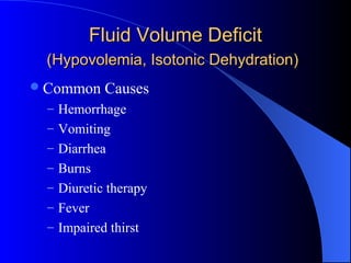
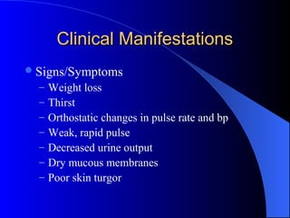
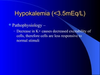
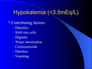
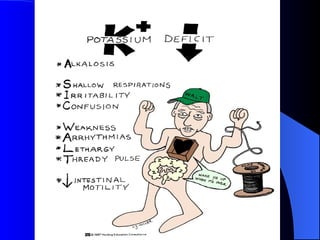
Fluid & Electrolyte ImbalanceFluid & Electrolyte Imbalance
Fluid ImbalanceFluid Imbalance
Fluid Volume DeficitFluid Volume Deficit
(Hypovolemia, Isotonic Dehydration)(Hypovolemia, Isotonic Dehydration)
Common Causes
– Hemorrhage
– Vomiting
– Diarrhea
– Burns
– Diuretic therapy
– Fever
– Impaired thirst
Clinical ManifestationsClinical Manifestations
Signs/Symptoms
– Weight loss
– Thirst
– Orthostatic changes in pulse rate and bp
– Weak, rapid pulse
– Decreased urine output
– Dry mucous membranes
– Poor skin turgor
Treatment/Interventions (FVD)Treatment/Interventions (FVD)
Fluid Management
– Diet therapy – Mild to moderate dehydration.
Correct with oral fluid replacement.
– Oral rehydration therapy – Solutions containing
glucose and electrolytes. E.g., Pedialyte,
Rehydralyte.
– IV therapy – Type of fluid ordered depends on
the type of dehydration and the clients
cardiovascular status.
Nursing ImplicationsNursing Implications
Monitor postural heart rate and bp
when getting patients out of bed
Fluid Volume ExcessFluid Volume Excess
Common Causes:
– Congestive Heart Failure
– Early renal failure
– IV therapy
– Excessive sodium ingestion
Clinical ManifestationsClinical Manifestations
Signs/Symptoms
– Increased BP
– Bounding pulse
– Venous distention
– Pulmonary edema
 Dyspnea
 Orthopnea (diff. breathing when supine)
 crackles
Treatment/InterventionsTreatment/Interventions
Drug therapy
– Diuretics may be ordered if renal failure is not
the cause.
Restriction of sodium and saline intake
Electrolyte ImbalanceElectrolyte Imbalance
Hypokalemia (<3.5mEq/L)Hypokalemia (<3.5mEq/L)
Pathophysiology –
– Decrease in K+ causes decreased excitability of
cells, therefore cells are less responsive to
normal stimuli
Hypokalemia (<3.5mEq/L)Hypokalemia (<3.5mEq/L)
Contributing factors:
– Diuretics
– Shift into cells
– Digitalis
– Water intoxication
– Corticosteroids
– Diarrhea
– Vomiting
Hypokalemia (<3.5mEq/L)Hypokalemia (<3.5mEq/L)
Interventions
– Assess and identify those at risk
– Encourage potassium-rich foods
– K+ replacement (IV or PO)
– Monitor lab values
– D/c potassium-wasting diuretics
– Treat underlying cause
Hyperkalemia (>5.0mEq/L)Hyperkalemia (>5.0mEq/L)
Pathophysiology – An inc. in K+ causes
increased excitability of cells.
Hyperkalemia (>5.0mEq/L)Hyperkalemia (>5.0mEq/L)
Contributing factors:
– Increase in K+ intake
– Renal failure
– K+ sparing diuretics
– Shift of K+ out of the cells
Hyperkalemia (>5.0mEq/L)Hyperkalemia (>5.0mEq/L)
Interventions
– Need to restore normal K+ balance:
– Eliminate K+ administration
– Inc. K+ excretion
 Lasix
 Kayexalate (Polystyrene sulfonate)
– Infuse glucose and insulin
– Cardiac Monitoring
Hyponatremia (<135mEq/L)Hyponatremia (<135mEq/L)
Contributing Factors
– Excessive diaphoresis
– Wound Drainage
– NPO
– CHF
– Low salt diet
– Renal Disease
– Diuretics
Hyponatremia (<135mEq/L)Hyponatremia (<135mEq/L)
Assessment findings:
– Neuro - Generalized skeletal muscle weakness.
Headache / personality changes.
– Resp.- Shallow respirations
– CV - Cardiac changes depend on fluid volume
– GI – Increased GI motility, Nausea, Diarrhea
(explosive)
– GU - Increased urine output
Hyponatremia (<135mEq/L)Hyponatremia (<135mEq/L)
Interventions/Treatment
– Restore Na levels to normal and prevent further
decreases in Na.
– Drug Therapy –
 - IV therapy to restore both fluid and Na. If severe
may see 2-3% saline.
 – Administer osmotic diuretic (Mannitol) to excrete
the water rather than the sodium.
– Increase oral sodium intake and restrict oral
fluid intake.
Hypernatremia (>145mEq/L)Hypernatremia (>145mEq/L)
Contributing Factors
– Hyperaldosteronism
– Renal failure
– Corticosteroids
– Increase in oral Na intake
– Na containing IV fluids
– Decreased urine output with increased urine
concentration
Hypernatremia (>145mEq/L)Hypernatremia (>145mEq/L)
Contributing factors (cont’d):
– Diarrhea
– Dehydration
– Fever
– Hyperventilation
Hypernatremia (>145mEq/L)Hypernatremia (>145mEq/L)
Assessment findings:
– Neuro - Spontaneous muscle twitches.
Irregular contractions. Skeletal muscle wkness.
Diminished deep tendon reflexes
– Resp. – Pulmonary edema
– CV – Diminished CO. HR and BP depend on
vascular volume.
Hypernatremia (>145mEq/L)Hypernatremia (>145mEq/L)
GU – Dec. urine output. Inc. specific
gravity
Skin – Dry, flaky skin. Edema r/t fluid
volume changes.
Hypernatremia (>145mEq/L)Hypernatremia (>145mEq/L)
Interventions/Treatment
– Drug therapy
 .45% NSS. If caused by both Na and fluid loss,
will administer NaCL. If inadequate renal excretion
of sodium, will administer diuretics.
– Diet therapy
 Mild – Ensure water intake
Hypocalcemia (<9.0mg/dL)Hypocalcemia (<9.0mg/dL)
Contributing factors:
– Dec. oral intake
– Lactose intolerance
– Dec. Vitamin D intake
– End stage renal disease
– Diarrhea
Hypocalcemia (<9.0mg/dL)Hypocalcemia (<9.0mg/dL)
Contributing factors (cont’d):
Acute pancreatitis
Hyperphosphatemia
Immobility
Removal or destruction of parathyroid
gland
Hypocalcemia (<9.0mg/dL)Hypocalcemia (<9.0mg/dL)
Assessment findings:
– Neuro –Irritable muscle twitches.
 Positive Trousseau’s sign.
 Positive Chvostek’s sign.
– Resp. – Resp. failure d/t muscle tetany.
– CV – Dec. HR., dec. BP, diminished
peripheral pulses
– GI – Inc. motility. Inc. BS. Diarrhea
Positive Trousseau’s SignPositive Trousseau’s Sign
Positive Chvostek’s SignPositive Chvostek’s Sign
Hypocalcemia (<9.0mg/dL)Hypocalcemia (<9.0mg/dL)
Interventions/Treatment
– Drug Therapy
 Calcium supplements
 Vitamin D
– Diet Therapy
 High calcium diet
– Prevention of Injury
 Seizure precautions
Hypercalcemia (>10.5mg/dL)Hypercalcemia (>10.5mg/dL)
Contributing factors:
– Excessive calcium intake
– Excessive vitamin D intake
– Renal failure
– Hyperparathyroidism
– Malignancy
– Hyperthyroidism
Hypercalcemia (>10.5mg/dL)Hypercalcemia (>10.5mg/dL)
Assessment findings:
– Neuro – Disorientation, lethargy, coma, profound
muscle weakness
– Resp. – Ineffective resp. movement
– CV - Inc. HR, Inc. BP. , Bounding peripheral pulses,
Positive Homan’s sign.
Late Phase – Bradycardia, Cardiac arrest
– GI – Dec. motility. Dec. BS. Constipation
– GU – Inc. urine output. Formation of renal calculi
Hypercalcemia (>10.5mg/dL)Hypercalcemia (>10.5mg/dL)
Interventions/Treatment
– Eliminate calcium administration
– Drug Therapy
– Isotonic NaCL (Inc. the excretion of Ca)
– Diuretics
– Calcium reabsorption inhibitors (Phosphorus)
– Cardiac Monitoring
Hypomagnesemia (<1.4mEq/L)Hypomagnesemia (<1.4mEq/L)
Contributing factors:
– Malnutrition
– Starvation
– Diuretics
– Aminoglycoside antibiotics
– Hyperglycemia
– Insulin administration
Hypomagnesemia (<1.4mEq/L)Hypomagnesemia (<1.4mEq/L)
Assessment findings:
*Neuro - Positive Trousseau’s sign.
Positive Chvostek’s sign. Hyperreflexia.
Seizures
*CV – ECG changes. Dysrhythmias. HTN
*Resp. – Shallow resp.
*GI – Dec. motility. Anorexia. Nausea
Hypomagnesemia (<1.4mEq/L)Hypomagnesemia (<1.4mEq/L)
Interventions:
– Eliminate contributing drugs
– IV MgSO4
– Assess DTR’s hourly with MgSO4
– Diet Therapy
Hypermagnesemia (>2.0mEq/L)Hypermagnesemia (>2.0mEq/L)
Contributing factors:
– Increased Mag intake
– Decreased renal excretion
Hypermagnesemia (>2.0mEq/L)Hypermagnesemia (>2.0mEq/L)
Assessment findings:
Neuro – Reduced or weak DTR’s. Weak
voluntary muscle contractions. Drowsy to
the point of lethargy
CV – Bradycardia, peripheral
vasodilatation, hypotension. ECG changes.
Hypermagnesemia (>2.0mg/dL)Hypermagnesemia (>2.0mg/dL)
Interventions
– Eliminate contributing drugs
– Administer diuretic
– Calcium gluconate reverses cardiac effects
– Diet restrictions

Fluid & Electrolyte Imbalance