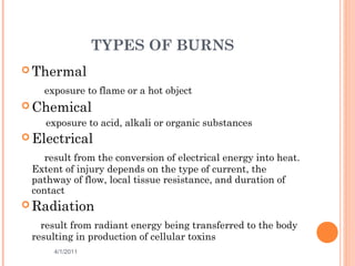
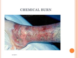
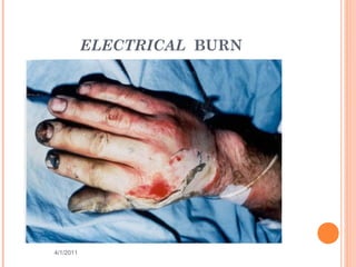
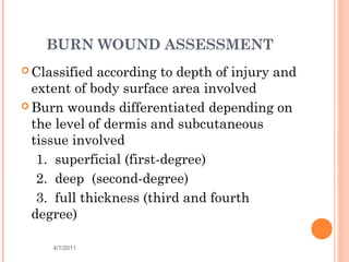
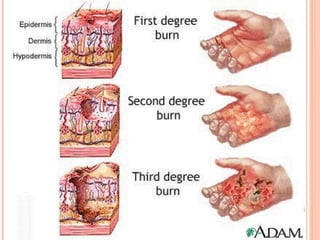
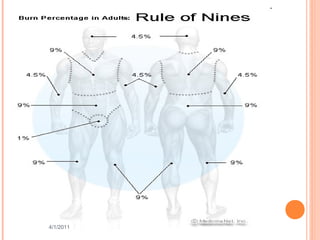
BURN INJURIES & ITS
MANAGEMENT
Dr Ibraheem Bashayreh, RN, PhD
4/1/2011
1
BURNS
Wounds caused by exposure to:
1. Excessive heat
2. Chemicals
3. Fire/steam
4. Radiation
5. Electricity
4/1/2011 2
BURNS
 Results in 10-20 thousand deaths annually
 Survival best at ages 15-45
 Children, elderly, and diabetics
 Survival best burns cover less than 20% of TBA
4/1/2011 3
TYPES OF BURNS
 Thermal
exposure to flame or a hot object
 Chemical
exposure to acid, alkali or organic substances
 Electrical
result from the conversion of electrical energy into heat.
Extent of injury depends on the type of current, the
pathway of flow, local tissue resistance, and duration of
contact
 Radiation
result from radiant energy being transferred to the body
resulting in production of cellular toxins
4/1/2011 4
CHEMICAL BURN
4/1/2011 5
ELECTRICAL BURN
4/1/2011 6
BURN WOUND ASSESSMENT
 Classified according to depth of injury and
extent of body surface area involved
 Burn wounds differentiated depending on
the level of dermis and subcutaneous
tissue involved
1. superficial (first-degree)
2. deep (second-degree)
3. full thickness (third and fourth
degree)
4/1/2011 7
4/1/2011
8
SUPERFICIAL BURNS
(FIRST DEGREE)
 Epidermal tissue only affected
 Erythema, blanching on pressure, mild swelling
no vesicles or blister initially
 Not serious unless large areas involved
 i.e. sunburn
4/1/2011 9
4/1/2011
10
4/1/2011
11
4/1/2011
12
DEEP (SECOND DEGREE)
*Involves the epidermis and deep layer of the
dermis
Fluid-filled vesicles –red, shiny, wet, severe pain
Hospitalization required if over 25% of body
surface involved
i.e. tar burn, flame
4/1/2011 13
4/1/2011
14
4/1/2011
15
4/1/2011
16
FULL THICKNESS
(THIRD/FOURTH DEGREE)
 Destruction of all skin layers
 Requires immediate hospitalization
 Dry, waxy white, leathery, or hard skin, no pain
 Exposure to flames, electricity or chemicals can
cause 3rd
degree burns
4/1/2011 17
4/1/2011
18
4/1/2011
19
CALCULATION OF BURNED BODY
SURFACE AREA
Calculation of Burned
Body Surface Area
4/1/2011 20
TOTAL BODY SURFACE AREA
(TBSA)
 Superficial burns are not involved in the
calculation
 Lund and Browder Chart is the most accurate
because it adjusts for age
 Rule of nines divides the body – adequate for
initial assessment for adult burns
4/1/2011 21
LUND BROWDER CHART USED FOR
DETERMINING BSA
4/1/2011 22Evans, 18.1, 2007)
RULES OF NINES
 Head & Neck = 9%
 Each upper extremity (Arms) = 9%
 Each lower extremity (Legs) = 18%
 Anterior trunk= 18%
 Posterior trunk = 18%
 Genitalia (perineum) = 1%
4/1/2011 23
4/1/2011 24
VASCULAR CHANGES
RESULTING FROM BURN
INJURIES
 Circulatory disruption occurs at the burn
site immediately after a burn injury
 Blood flow decreases or cease due to
occluded blood vessels
 Damaged macrophages within the tissues
release chemicals that cause constriction
of vessel
 Blood vessel thrombosis may occur
causing necrosis
 Macrophage: A type of white blood that ingests (takes in) foreign
material. Macrophages are key players in the immune response to foreign
invaders such as infectious microorganisms.
4/1/2011 25
FLUID SHIFT
 Occurs after initial vasoconstriction, then
dilation
 Blood vessels dilate and leak fluid into
the interstitial space
 Known as third spacing or capillary leak
syndrome
 Causes decreased blood volume and blood
pressure
 Occurs within the first 12 hours after the
burn and can continue to up to 36 hours
4/1/2011 26
FLUID IMBALANCES
 Occur as a result of fluid shift and cell
damage
 Hypovolemia
 Metabolic acidosis
 Hyperkalemia
 Hyponatremia
 Hemoconcentration (elevated blood
osmolarity, hematocrit/hemoglobin) due to
dehydration
4/1/2011 27
FLUID REMOBILIZATION
 Occurs after 24 hours
 Capillary leak stops
 See diuretic stage where edema fluid
shifts from the interstitial spaces into the
vascular space
 Blood volume increases leading to
increased renal blood flow and diuresis
 Body weight returns to normal
 See Hypokalemia
4/1/2011 28
CURLING’S ULCER
 Acute ulcerative gastro duodenal disease
 Occur within 24 hours after burn
 Due to reduced GI blood flow and mucosal
damage
 Treat clients with H2 blockers, mucoprotectants,
and early enteral nutrition
 Watch for sudden drop in hemoglobin
4/1/2011 29
PHASES OF BURN INJURIES
 Emergent (24-48 hrs)
 Acute
 Rehabilitative
4/1/2011 30
EMERGENT PHASE
*Immediate problem is fluid loss, edema,
reduced blood flow (fluid and electrolyte
shifts)
 Goals:
1. secure airway
2. support circulation by fluid
replacement
3. keep the client comfortable with
analgesics
4. prevent infection through wound care
5. maintain body temperature
6. provide emotional support
4/1/2011 31
EMERGENT PHASE
 Knowledge of circumstances surrounding the
burn injury
 Obtain client’s pre-burn weight (dry weight) to
calculate fluid rates
 Calculations based on weight obtained after fluid
replacement is started are not accurate because
of water-induced weight gain
 Height is important in determining body surface
area (BSA) which is used to calculate nutritional
needs
 Know client’s health history because the
physiologic stress seen with a burn can make a
latent disease process develop symptoms
4/1/2011 32
CLINICAL MANIFESTATIONS IN THE
EMERGENT PHASE
 Clients with major burn injuries and with inhalation injury
are at risk for respiratory problems
 Inhalation injuries are present in 20% to 50% of the clients
admitted to burn centers
 Assess the respiratory system by inspecting the mouth, nose,
and pharynx
 Burns of the lips, face, ears, neck, eyelids, eyebrows, and
eyelashes are strong indicators that an inhalation injury may
be present
 Change in respiratory pattern may indicate a pulmonary
injury.
 The client may: become progressively hoarse, develop a brassy
cough, drool or have difficulty swallowing, produce expiratory
sounds that include audible wheezes, crowing, and stridor
 Upper airway edema and inhalation injury are most common
in the trachea and mainstem bronchi
 Auscultate these areas for wheezes
 If wheezes disappear, this indicates impending airway
obstruction and demands immediate intubation4/1/2011 33
CLINICAL MANIFESTATIONS
 Cardiovascular will begin immediately
which can include shock (Shock is a
common cause of death in the emergent
phase in clients with serious injuries)
 Obtain a baseline EKG
 Monitor for edema, measure central and
peripheral pulses, blood pressure,
capillary refill and pulse oximetry
4/1/2011 34
CLINICAL MANIFESTATIONS
 Changes in renal function are related to
decreased renal blood flow
 Urine is usually highly concentrated and
has a high specific gravity
 Urine output is decreased during the first
24 hours of the emergent phase
 Fluid resuscitation is provided at the rate
needed to maintain adult urine output at
30 to 50- mL/hr.
 Measure BUN, creat and NA levels
4/1/2011 35
CLINICAL MANIFESTATIONS
 Sympathetic stimulation during the
emergent phase causes reduced GI
motility and paralytic ileus
 Auscultate the abdomen to assess bowel
sounds which may be reduced
 Monitor for n/v and abdominal distention
 Clients with burns of 25% TBSA or who
are intubated generally require a NG tube
inserted to prevent aspiration and
removal of gastric secretions
4/1/2011 36
SKIN ASSESSMENT
 Assess the skin to determine the size and
depth of burn injury
 The size of the injury is first estimated in
comparison to the total body surface area
(TBSA). For example, a burn that
involves 40% of the TBSA is a 40% burn
 Use the rule of nines for clients whose
weights are in normal proportion to their
heights
4/1/2011 37
IV FLUID THERAPY
 Infusion of IV fluids is needed to maintain sufficient
blood volume for normal CO
 Clients with burns involving 15% to 20% of the TBSA
require IV fluid
 Purpose is to prevent shock by maintaining adequate
circulating blood fluid volume
 Severe burn requires large fluid loads in a short time
to maintain blood flow to vital organs
 Fluid replacement formulas are calculated from the
time of injury and not from the time of arrival at the
hospital
 Diuretics should not be given to increase urine output.
Change the amount and rate of fluid administration.
Diuretics do not increase CO; they actually decrease
circulating volume and CO by pulling fluid from the
circulating blood volume to enhance diuresis
4/1/2011 38
COMMON FLUIDS
 Protenate or 5% albumin in isotonic saline (1/2
given in first 8 hr; ½ given in next 16 hr)
 LR (Lactate Ringer) without dextrose (1/2 given
in first 8 hr; ½ given in next 16 hr)
 Crystalloid (hypertonic saline) adjust to maintain
urine output at 30 mL/hr
 Crystalloid only (lactated ringers)
4/1/2011 39
NURSING DIAGNOSIS IN THE
EMERGENT PHASE
 Decreased CO
 Deficient fluid volume r/t active fluid volume loss
 Ineffective Tissue perfusion
 Ineffective breathing pattern
4/1/2011 40
ACUTE PHASE OF BURN INJURY
• Lasts until wound closure is complete
• Care is directed toward continued assessment and
maintenance of the cardiovascular and respiratory
system
• Pneumonia is a concern which can result in respiratory
failure requiring mechanical ventilation
• Infection (Topical antibiotics – Silvadene)
• Tetanus toxoid
• Weight daily without dressings or splints and compare
to pre-burn weight
• A 2% loss of body weight indicates a mild deficit
• A 10% or greater weight loss requires modification of
calorie intake
• Monitor for signs of infection
4/1/2011 41
LOCAL AND SYSTEMIC SIGNS
OF INFECTION- GRAM
NEGATIVE BACTERIA
 Pseudomonas, Proteus
 May led to septic shock
 Conversion of a partial-thickness injury to a full-thickness
injury
 Ulceration of health skin at the burn site
 Erythematous, nodular lesions in uninvolved skin
 Excessive burn wound drainage
 Odor
 Sloughing of grafts
 Altered level of consciousness
 Changes in vital signs
 Oliguria
 GI dysfunction such as diarrhea, vomiting
 Metabolic acidosis
4/1/2011 42
LAB VALUES
 Na – hyponatremia or Hypernatremia
 K – Hyperkalemia or Hypokalemia
 WBC – 10,000-20,000
4/1/2011 43
NURSING DIAGNOSIS IN THE
ACUTE PHASE
 Impaired skin integrity
 Risk for infection
 Imbalanced nutrition
 Impaired physical mobility
 Disturbed body image
4/1/2011 44
PLANNING AND
IMPLEMENTATION
 Nonsurgical management: removal of exudates
and necrotic tissue, cleaning the area,
stimulating granulation and revascularization
and applying dressings. Debridement may be
needed
4/1/2011 45
DRESSING THE BURN WOUND
 After burn wounds are cleaned and debrided,
topical antibiotics are reapplied to prevent
infection
 Standard wound dressings are multiple layers of
gauze applied over the topical agents on the burn
wound
4/1/2011 46
REHABILITATIVE PHASE OF
BURN INJURY
 Started at the time of admission
 Technically begins with wound closure
and ends when the client returns to the
highest possible level of functioning
 Provide psychosocial support
 Assess home environment, financial
resources, medical equipment, prosthetic
rehab
 Health teaching should include symptoms
of infection, drugs regimens, f/u
appointments, comfort measures to reduce
pruritis
4/1/2011 47
DIET
 Initially NPO
 Begin oral fluids after bowel sounds return
 Do not give ice chips or free water lead to
electrolyte imbalance
 High protein, high calorie
4/1/2011 48
GOALS
 Prevent complications (contractures)
 Vital signs hourly
 Assess respiratory function
 Tetanus booster
 Anti-infective
 Analgesics
 No aspirin
 Strict surgical asepsis
 Turn q2h to prevent contractures
 Emotional support
4/1/2011 49
DEBRIDEMENT
 Done with forceps and curved scissor or through
hydrotherapy (application of water for treatment)
 Only loose eschar removed
 Blisters are left alone to serve as a protector –
controversial
4/1/2011 50
SKIN GRAFTS
 Done during the acute phase
 Used for full-thickness and deep partial-
thickness wounds
4/1/2011 51
POST CARE OF SKIN GRAFTS
 Maintain dressing
 Use aseptic technique
 Graft should look pink if it has taken after 5 days
 Skeletal traction may be used to prevent
contractures
 Elastic bandages may be applied for 6 mo to 1
year to prevent hypertrophic scarring
4/1/2011 52
THE END
QUESTIONS
4/1/2011
53

Burn Injuries and Its Management

  • 1.
    BURN INJURIES &ITS MANAGEMENT Dr Ibraheem Bashayreh, RN, PhD 4/1/2011 1
  • 2.
    BURNS Wounds caused byexposure to: 1. Excessive heat 2. Chemicals 3. Fire/steam 4. Radiation 5. Electricity 4/1/2011 2
  • 3.
    BURNS  Results in10-20 thousand deaths annually  Survival best at ages 15-45  Children, elderly, and diabetics  Survival best burns cover less than 20% of TBA 4/1/2011 3
  • 4.
    TYPES OF BURNS Thermal exposure to flame or a hot object  Chemical exposure to acid, alkali or organic substances  Electrical result from the conversion of electrical energy into heat. Extent of injury depends on the type of current, the pathway of flow, local tissue resistance, and duration of contact  Radiation result from radiant energy being transferred to the body resulting in production of cellular toxins 4/1/2011 4
  • 5.
  • 6.
  • 7.
    BURN WOUND ASSESSMENT Classified according to depth of injury and extent of body surface area involved  Burn wounds differentiated depending on the level of dermis and subcutaneous tissue involved 1. superficial (first-degree) 2. deep (second-degree) 3. full thickness (third and fourth degree) 4/1/2011 7
  • 8.
  • 9.
    SUPERFICIAL BURNS (FIRST DEGREE) Epidermal tissue only affected  Erythema, blanching on pressure, mild swelling no vesicles or blister initially  Not serious unless large areas involved  i.e. sunburn 4/1/2011 9
  • 10.
  • 11.
  • 12.
  • 13.
    DEEP (SECOND DEGREE) *Involvesthe epidermis and deep layer of the dermis Fluid-filled vesicles –red, shiny, wet, severe pain Hospitalization required if over 25% of body surface involved i.e. tar burn, flame 4/1/2011 13
  • 14.
  • 15.
  • 16.
  • 17.
    FULL THICKNESS (THIRD/FOURTH DEGREE) Destruction of all skin layers  Requires immediate hospitalization  Dry, waxy white, leathery, or hard skin, no pain  Exposure to flames, electricity or chemicals can cause 3rd degree burns 4/1/2011 17
  • 18.
  • 19.
  • 20.
    CALCULATION OF BURNEDBODY SURFACE AREA Calculation of Burned Body Surface Area 4/1/2011 20
  • 21.
    TOTAL BODY SURFACEAREA (TBSA)  Superficial burns are not involved in the calculation  Lund and Browder Chart is the most accurate because it adjusts for age  Rule of nines divides the body – adequate for initial assessment for adult burns 4/1/2011 21
  • 22.
    LUND BROWDER CHARTUSED FOR DETERMINING BSA 4/1/2011 22Evans, 18.1, 2007)
  • 23.
    RULES OF NINES Head & Neck = 9%  Each upper extremity (Arms) = 9%  Each lower extremity (Legs) = 18%  Anterior trunk= 18%  Posterior trunk = 18%  Genitalia (perineum) = 1% 4/1/2011 23
  • 24.
  • 25.
    VASCULAR CHANGES RESULTING FROMBURN INJURIES  Circulatory disruption occurs at the burn site immediately after a burn injury  Blood flow decreases or cease due to occluded blood vessels  Damaged macrophages within the tissues release chemicals that cause constriction of vessel  Blood vessel thrombosis may occur causing necrosis  Macrophage: A type of white blood that ingests (takes in) foreign material. Macrophages are key players in the immune response to foreign invaders such as infectious microorganisms. 4/1/2011 25
  • 26.
    FLUID SHIFT  Occursafter initial vasoconstriction, then dilation  Blood vessels dilate and leak fluid into the interstitial space  Known as third spacing or capillary leak syndrome  Causes decreased blood volume and blood pressure  Occurs within the first 12 hours after the burn and can continue to up to 36 hours 4/1/2011 26
  • 27.
    FLUID IMBALANCES  Occuras a result of fluid shift and cell damage  Hypovolemia  Metabolic acidosis  Hyperkalemia  Hyponatremia  Hemoconcentration (elevated blood osmolarity, hematocrit/hemoglobin) due to dehydration 4/1/2011 27
  • 28.
    FLUID REMOBILIZATION  Occursafter 24 hours  Capillary leak stops  See diuretic stage where edema fluid shifts from the interstitial spaces into the vascular space  Blood volume increases leading to increased renal blood flow and diuresis  Body weight returns to normal  See Hypokalemia 4/1/2011 28
  • 29.
    CURLING’S ULCER  Acuteulcerative gastro duodenal disease  Occur within 24 hours after burn  Due to reduced GI blood flow and mucosal damage  Treat clients with H2 blockers, mucoprotectants, and early enteral nutrition  Watch for sudden drop in hemoglobin 4/1/2011 29
  • 30.
    PHASES OF BURNINJURIES  Emergent (24-48 hrs)  Acute  Rehabilitative 4/1/2011 30
  • 31.
    EMERGENT PHASE *Immediate problemis fluid loss, edema, reduced blood flow (fluid and electrolyte shifts)  Goals: 1. secure airway 2. support circulation by fluid replacement 3. keep the client comfortable with analgesics 4. prevent infection through wound care 5. maintain body temperature 6. provide emotional support 4/1/2011 31
  • 32.
    EMERGENT PHASE  Knowledgeof circumstances surrounding the burn injury  Obtain client’s pre-burn weight (dry weight) to calculate fluid rates  Calculations based on weight obtained after fluid replacement is started are not accurate because of water-induced weight gain  Height is important in determining body surface area (BSA) which is used to calculate nutritional needs  Know client’s health history because the physiologic stress seen with a burn can make a latent disease process develop symptoms 4/1/2011 32
  • 33.
    CLINICAL MANIFESTATIONS INTHE EMERGENT PHASE  Clients with major burn injuries and with inhalation injury are at risk for respiratory problems  Inhalation injuries are present in 20% to 50% of the clients admitted to burn centers  Assess the respiratory system by inspecting the mouth, nose, and pharynx  Burns of the lips, face, ears, neck, eyelids, eyebrows, and eyelashes are strong indicators that an inhalation injury may be present  Change in respiratory pattern may indicate a pulmonary injury.  The client may: become progressively hoarse, develop a brassy cough, drool or have difficulty swallowing, produce expiratory sounds that include audible wheezes, crowing, and stridor  Upper airway edema and inhalation injury are most common in the trachea and mainstem bronchi  Auscultate these areas for wheezes  If wheezes disappear, this indicates impending airway obstruction and demands immediate intubation4/1/2011 33
  • 34.
    CLINICAL MANIFESTATIONS  Cardiovascularwill begin immediately which can include shock (Shock is a common cause of death in the emergent phase in clients with serious injuries)  Obtain a baseline EKG  Monitor for edema, measure central and peripheral pulses, blood pressure, capillary refill and pulse oximetry 4/1/2011 34
  • 35.
    CLINICAL MANIFESTATIONS  Changesin renal function are related to decreased renal blood flow  Urine is usually highly concentrated and has a high specific gravity  Urine output is decreased during the first 24 hours of the emergent phase  Fluid resuscitation is provided at the rate needed to maintain adult urine output at 30 to 50- mL/hr.  Measure BUN, creat and NA levels 4/1/2011 35
  • 36.
    CLINICAL MANIFESTATIONS  Sympatheticstimulation during the emergent phase causes reduced GI motility and paralytic ileus  Auscultate the abdomen to assess bowel sounds which may be reduced  Monitor for n/v and abdominal distention  Clients with burns of 25% TBSA or who are intubated generally require a NG tube inserted to prevent aspiration and removal of gastric secretions 4/1/2011 36
  • 37.
    SKIN ASSESSMENT  Assessthe skin to determine the size and depth of burn injury  The size of the injury is first estimated in comparison to the total body surface area (TBSA). For example, a burn that involves 40% of the TBSA is a 40% burn  Use the rule of nines for clients whose weights are in normal proportion to their heights 4/1/2011 37
  • 38.
    IV FLUID THERAPY Infusion of IV fluids is needed to maintain sufficient blood volume for normal CO  Clients with burns involving 15% to 20% of the TBSA require IV fluid  Purpose is to prevent shock by maintaining adequate circulating blood fluid volume  Severe burn requires large fluid loads in a short time to maintain blood flow to vital organs  Fluid replacement formulas are calculated from the time of injury and not from the time of arrival at the hospital  Diuretics should not be given to increase urine output. Change the amount and rate of fluid administration. Diuretics do not increase CO; they actually decrease circulating volume and CO by pulling fluid from the circulating blood volume to enhance diuresis 4/1/2011 38
  • 39.
    COMMON FLUIDS  Protenateor 5% albumin in isotonic saline (1/2 given in first 8 hr; ½ given in next 16 hr)  LR (Lactate Ringer) without dextrose (1/2 given in first 8 hr; ½ given in next 16 hr)  Crystalloid (hypertonic saline) adjust to maintain urine output at 30 mL/hr  Crystalloid only (lactated ringers) 4/1/2011 39
  • 40.
    NURSING DIAGNOSIS INTHE EMERGENT PHASE  Decreased CO  Deficient fluid volume r/t active fluid volume loss  Ineffective Tissue perfusion  Ineffective breathing pattern 4/1/2011 40
  • 41.
    ACUTE PHASE OFBURN INJURY • Lasts until wound closure is complete • Care is directed toward continued assessment and maintenance of the cardiovascular and respiratory system • Pneumonia is a concern which can result in respiratory failure requiring mechanical ventilation • Infection (Topical antibiotics – Silvadene) • Tetanus toxoid • Weight daily without dressings or splints and compare to pre-burn weight • A 2% loss of body weight indicates a mild deficit • A 10% or greater weight loss requires modification of calorie intake • Monitor for signs of infection 4/1/2011 41
  • 42.
    LOCAL AND SYSTEMICSIGNS OF INFECTION- GRAM NEGATIVE BACTERIA  Pseudomonas, Proteus  May led to septic shock  Conversion of a partial-thickness injury to a full-thickness injury  Ulceration of health skin at the burn site  Erythematous, nodular lesions in uninvolved skin  Excessive burn wound drainage  Odor  Sloughing of grafts  Altered level of consciousness  Changes in vital signs  Oliguria  GI dysfunction such as diarrhea, vomiting  Metabolic acidosis 4/1/2011 42
  • 43.
    LAB VALUES  Na– hyponatremia or Hypernatremia  K – Hyperkalemia or Hypokalemia  WBC – 10,000-20,000 4/1/2011 43
  • 44.
    NURSING DIAGNOSIS INTHE ACUTE PHASE  Impaired skin integrity  Risk for infection  Imbalanced nutrition  Impaired physical mobility  Disturbed body image 4/1/2011 44
  • 45.
    PLANNING AND IMPLEMENTATION  Nonsurgicalmanagement: removal of exudates and necrotic tissue, cleaning the area, stimulating granulation and revascularization and applying dressings. Debridement may be needed 4/1/2011 45
  • 46.
    DRESSING THE BURNWOUND  After burn wounds are cleaned and debrided, topical antibiotics are reapplied to prevent infection  Standard wound dressings are multiple layers of gauze applied over the topical agents on the burn wound 4/1/2011 46
  • 47.
    REHABILITATIVE PHASE OF BURNINJURY  Started at the time of admission  Technically begins with wound closure and ends when the client returns to the highest possible level of functioning  Provide psychosocial support  Assess home environment, financial resources, medical equipment, prosthetic rehab  Health teaching should include symptoms of infection, drugs regimens, f/u appointments, comfort measures to reduce pruritis 4/1/2011 47
  • 48.
    DIET  Initially NPO Begin oral fluids after bowel sounds return  Do not give ice chips or free water lead to electrolyte imbalance  High protein, high calorie 4/1/2011 48
  • 49.
    GOALS  Prevent complications(contractures)  Vital signs hourly  Assess respiratory function  Tetanus booster  Anti-infective  Analgesics  No aspirin  Strict surgical asepsis  Turn q2h to prevent contractures  Emotional support 4/1/2011 49
  • 50.
    DEBRIDEMENT  Done withforceps and curved scissor or through hydrotherapy (application of water for treatment)  Only loose eschar removed  Blisters are left alone to serve as a protector – controversial 4/1/2011 50
  • 51.
    SKIN GRAFTS  Doneduring the acute phase  Used for full-thickness and deep partial- thickness wounds 4/1/2011 51
  • 52.
    POST CARE OFSKIN GRAFTS  Maintain dressing  Use aseptic technique  Graft should look pink if it has taken after 5 days  Skeletal traction may be used to prevent contractures  Elastic bandages may be applied for 6 mo to 1 year to prevent hypertrophic scarring 4/1/2011 52
  • 53.