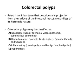
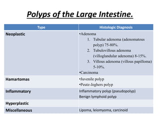
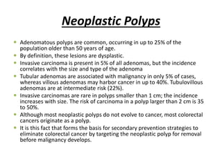
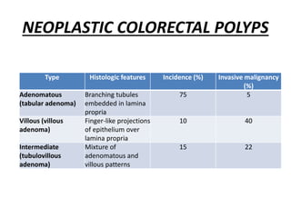
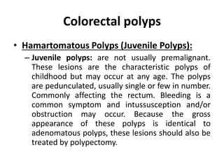
Colorectal polyps
• Polyp is a clinical term that describes any projection
from the surface of the intestinal mucosa regardless of
its histologic nature.
• Colorectal polyps may be classified as
A) Neoplastic (tubular adenoma, villous adenoma,
tubulovillous adenomas).
B) Hamartomatous (juvenile, Peutz-Jeghers, Cronkite-Canada
and Cowden).
C) Inflammatory (pseudopolyps and benign lymphoid polyp)
D) Hyperplastic.
Polyps of the Colon & Rectum
 Colorectal polyps are masses of tissue that project into
the lumen.
sessile or pedunculated,
benign or malignant,
mucosal, submucosal, or muscular lesions.
 "Polyp" is a morphologic term, and no histologic
diagnosis is implied.
 Polyposis is a term reserved for the presence of many
polyps in the large bowel.
 Estimates of the incidence of colonic and rectal polyps
in the general population range from 9% to 60%.
Polyps of the Colon & Rectum
• Approximately 50% of polyps occur in the
sigmoid or rectum.
• About 50% of patients with adenoma have more
than one lesion, and 15% have more than two
lesions.
• Approximately 25% of patients who have five or
more adenomatous polyps have a synchronous
colon cancer at the initial colonoscopy.
• An increased incidence of adenomas in breast
cancer patients has been reported.
Polyps of the Large Intestine.
Type Histologic Diagnosis
Neoplastic •Adenoma
1. Tubular adenoma (adenomatous
polyp) 75-80%.
2. Tubulovillous adenoma
(villoglandular adenoma) 8-15%.
3. Villous adenoma (villous papilloma)
5-10%.
•Carcinoma
Hamartomas •Juvenile polyp
•Peutz-Jeghers polyp
Inflammatory Inflammatory polyp (pseudopolyp)
Benign lymphoid polyp
Hyperplastic
Miscellaneous Lipoma, leiomyoma, carcinoid
Neoplastic Polyps
 Adenomatous polyps are common, occurring in up to 25% of the
population older than 50 years of age.
 By definition, these lesions are dysplastic.
 Invasive carcinoma is present in 5% of all adenomas, but the incidence
correlates with the size and type of the adenoma
 Tubular adenomas are associated with malignancy in only 5% of cases,
whereas villous adenomas may harbor cancer in up to 40%. Tubulovillous
adenomas are at intermediate risk (22%).
 Invasive carcinomas are rare in polyps smaller than 1 cm; the incidence
increases with size. The risk of carcinoma in a polyp larger than 2 cm is 35
to 50%.
 Although most neoplastic polyps do not evolve to cancer, most colorectal
cancers originate as a polyp.
 It is this fact that forms the basis for secondary prevention strategies to
eliminate colorectal cancer by targeting the neoplastic polyp for removal
before malignancy develops.
NEOPLASTIC COLORECTAL POLYPS
Type Histologic features Incidence (%) Invasive malignancy
(%)
Adenomatous
(tabular adenoma)
Branching tubules
embedded in lamina
propria
75 5
Villous (villous
adenoma)
Finger-like projections
of epithelium over
lamina propria
10 40
Intermediate
(tubulovillous
adenoma)
Mixture of
adenomatous and
villous patterns
15 22
Colorectal polyps
• Hamartomatous Polyps (Juvenile Polyps):
– Juvenile polyps: are not usually premalignant.
These lesions are the characteristic polyps of
childhood but may occur at any age. The polyps
are pedunculated, usually single or few in number.
Commonly affecting the rectum. Bleeding is a
common symptom and intussusception and/or
obstruction may occur. Because the gross
appearance of these polyps is identical to
adenomatous polyps, these lesions should also be
treated by polypectomy.
Colorectal polyps
• Hamartomatous Polyps (Juvenile Polyps):
– Peutz-Jeghers syndrome is characterized by polyposis of
the small intestine, and to a lesser extent, polyposis of the
colon and rectum. Characteristic melanin spots are often
noted on the buccal mucosa and lips of these patients. The
polyps of Peutz-Jeghers syndrome are generally considered
to be hamartomas and are not thought to be at significant
risk for malignant degeneration. However, carcinoma may
occasionally develop. Because the entire length of the
gastrointestinal tract may be affected, surgery is reserved
for symptoms such as obstruction or bleeding or for
patients in whom polyps develop adenomatous features.
Colorectal polyps
• FAMILIAL ADENOMATOUS POLYPOSIS ( FAP)(Familial Polyposis
Coli, FPC)
– Aetiology:
• This rare autosomal dominant condition.
• Clinically, patients develop hundreds to thousands of adenomatous polyps
shortly after puberty. The lifetime risk of colorectal cancer in FAP patients
approaches 100% by age 50 years.
– Pathology:
• Polyps usually start to appear at puberty, affecting the sigmoid colon and
rectum initially and by the age of 20 years the entire colon and rectum are
affected by immense numbers of polyps ( at least 100), ranging from 1 mm to
several cms.
• Polyps may be sessile or pedunculated.
• Three histological types are recognized; the tubular, tubulo-villous, and villous.
• Left untreated, carcinoma develops in 100% of the affected patients by the
fifth decade.
Colorectal polyps
• Clinical picture:
– The disease usually manifests itself soon after puberty
by attacks of lower abdominal pain, diarrhoea,
tenesmus with the passage of blood and mucus in the
stools. Loss of weight, anaemia and general debility
are common.
– Rectal examination reveals the adenomas.
• Investigations:
– Barium enema shows multiple rounded filling defects
throughout the colon and the rectum.
– Sigmoidoscopy or colonoscopy and biopsy prove the
nature of the disease.
Macroscopic appearance of the colon in classical familial
adenomatous polyposis. The specimen shows dense carpeting of the
colonic mucosa with thousands of adenomas.
Colorectal polyps
• Treatment:
– Once the diagnosis of FAP has been made and polyps
are developing, treatment is surgical.
– Three operative procedures (options) can be
considered:
1. total proctocolectomy: with either an end ileostomy or
continent (Kock's) ileostomy
2. Restorative proctocolectomy : with ileal pouch–anal
anastomosis with or without a temporary ileostomy
3. Total abdominal colectomy with ileorectal anastomosis;
the rectum is preserved, and polypi in the rectum are
followed endoscopically and removed periodically.
Carcinoma of the colon
• Incidence: Colorectal carcinoma is the most common malignancy of the
gastrointestinal tract. The incidence is similar in men and women.
• Risk Factors:
(1) Aging: Aging is the most important risk factor for colorectal cancer, with incidence rising
steadily after the age of 50 years. More than 90% of cases diagnosed are in people older
than age 50 years.
(2) Hereditary Risk Factors: Approximately 80% of colorectal cancers occur sporadically,
while 20% arise in patients with a known family history of colorectal cancer.
(3) Environmental and Dietary Factors: Colorectal carcinoma occurs more commonly in
populations that consume diets high in animal fat and low in fiber.
(4) Inflammatory bowel disease: Patients with long-standing ulcerative colitis are at
increased risk for the development of colorectal cancer. Crohn's pancolitis have similar
risk.
(5) Other Risk Factors:
Cigarette smoking reportedly increases the risk of colorectal cancer .
Patients with ureterosigmoidostomy are also at increased risk .
Also in women, gallstones (and the consequent cholecystectomy) are associated with colorectal
cancer, especially in the right colon.
Risk Factors Associated with Colon Cancer
Risk Factor Comment
Geographic variation Highest risk in Western countries and lowest risk in
developing countries
Age Risk increase sharply after the fifth decade
Diet Increased with total and animal fat diets
Physical inactivity Increased with obesity and sedentary life style
Adenoma Risk dependent on type and size
FAP penetrance in gene carriers 100%
HNPCC penetrance in gene carriers 80%
Hamartomatous syndromes Risk increased with Peutz-Jeghers syndrome and juvenile
polyposis but not isolated juvenile polyps
Previous history of colon cancer Increased risk for recurrent cancer
Ulcerative colitis 10–20% after 20 years
Radiation Associated with a mucinous histology and poor prognosis
Ureterosigmoidostomy 100–500 times increased risk at or adjacent to the uretero-
colonic anastomosis
Carcinoma of the colon
• Pathology:
– Site:
• The sigmoid colon is the commonest site (50%) probably
because its contents are solid, stagnant and irritant, and it is
often the seat of precancerous lesions (i.e., FAP and UC)
• Caecum and ascending colon: 25%
• Transverse colon: 20%.
• Descending colon: 5%.
• Multiple synchronous colonic cancers (i.e., two or more
carcinomas occurring simultaneously) are found in 5% of
patients. Metachronous cancer is a new primary lesion in a
patient who has had a previous resection for cancer.
Carcinoma of the colon
• Gross picture:
– Cauliflower growth: This occurs in the right colon.
– Scirrhous growth (annular and tubular): This occurs in
the left colon.
– Malignant ulcer: This occurs commonly in the caecum.
• Microscopic picture
– Histologically the tumour is an adenocarcinoma that
arises from the columnar epithelium. It may be well,
moderately, or poorly differentiated.
– Some tumours have a colloid structure, occur in
younger patients, and have a poor prognosis.
Carcinoma of the colon
• Spread:
– Direct spread:
• Carcinoma grows circumferentially and may completely encircle the
bowel before it is diagnosed; this is especially true in the left colon,
which has a smaller caliber than the right. It takes about 1 year for a
tumor to encircle three-fourths of the circumference of the bowel.
• Longitudinal submucosal extension occurs with invasion of the
intramural lymphatic network, but it rarely goes beyond 2 cm from the
edge of the tumor unless there is concomitant spread to lymph nodes.
• As the lesion extends radially, it penetrates the outer layers of the
bowel wall, and it may invade adjacent structures: the liver, the
greater curvature of the stomach, the duodenum, the small bowel,
the pancreas, the spleen, the bladder, the vagina, the kidneys and
ureters, and the abdominal wall. Subacute perforation with
inflammatory attachment of bowel to an adjacent viscous may be
indistinguishable from actual invasion on gross examination.
Carcinoma of the colon
• Lymphatic spread:
– Cancer spreads from the colon to the following lymph
node groups, in sequence:
1. Epicolic nodes on the bowel wall.
2. Paracolic nodes between the marginal artery and the bowel.
3. Intermediate nodes on the main vessels.
4. Principal nodes alongside the superior and inferior mesenteric
vessels.
– From the principal nodes further spread occurs to the
parpa-aortic lymph nodes, then to cysterna chyli and
thoracic duct. The left supraclavicular lymph nodes may be
involved by retrograde flow from the thoracic duct
(Virchow’s glands) giving positive Troisier’s sign.
Carcinoma of the colon
• Blood spread:
– Venous invasion may allow tumor cells be carried via
the portal venous system to the liver. Tumor
embolization also occurs through lumbar and
vertebral veins to the lungs and elsewhere.
Metastases to ovaries are mostly hematogenous; they
are found in 1–10% of women with colorectal cancer.
An attempt is made to avoid producing hematogenous
metastases during operation by minimizing
manipulation of the tumor prior to ligation of the
blood supply.
Carcinoma of the colon
• Transperitoneal spread:
– "Seeding" may occur when the tumor has extended
through the serosa and tumor cells enter the peritoneal
cavity, producing local implants or generalized abdominal
carcinomatosis. Large metastatic deposits in the pelvic cul-
de-sac are palpable as a hard shelf (Blumer's shelf).
• Intraluminal spread:
– Malignant cells shed from the surface of the tumor can be
swept with the fecal current. Implantation more distally on
intact mucosa occurs rarely, if ever, but viable exfoliated
cells presumably can be trapped in an anastomotic suture
or staple line during operation.
Carcinoma of the colon
• Complications:
1. Intestinal obstruction:
1. chronic obstruction is common especially in left colon cancer (20%). This
tendency is attributed to:
1. The smaller lumen of the left colon.
2. Stool tends to be more solid.
3. Carcinoma tends to be of the stenosing type.
2. Acute obstruction may occur due to faecal impaction or intussusception (in
carcinoma at the ileocaecal valve and sigmoid colon).
2. Haemorrhage: Chronic bleeding is the rule and causes iron
deficiency anaemia. Massive bleeding is rare.
3. Perforation: is rare and is usually gradual with the formation of a
pericolic abscess and faecal fistula.
4. Malignant ascites: results from liver secondaries, lymph nodes
compressing the portal vein and Transperitoneal spread.
Carcinoma of the colon
• Staging:
– Colorectal cancer staging is based upon tumor depth
and the presence or absence of nodal or distant
metastases.
– Old staging systems, such as the Dukes' Classification
and its Astler-Coller modification, have been largely
replaced by the TNM staging system (Table=).
– The preoperative evaluation usually identifies stage IV
disease. In colon cancer, differentiating stages I, II, and
III depends upon examination of the resected
specimen. In rectal cancer, endorectal ultrasound may
predict the stage .
Carcinoma of the colon
• Tumor Stage (T) Definition
– Tx Cannot be assessed
– T0 No evidence of cancer
– Tis Carcinoma in situ
– T1 Tumor invades submucosa
– T2 Tumor invades muscularis propria
– T3 Tumor invades through muscularis propria into subserosa or into nonperitonealized pericolic tissues
– T4 Tumor directly invades other organs or tissues or perforates the visceral peritoneum of specimen
• Nodal Stage (N)
– NX Regional lymph nodes cannot be assessed
– N0 No lymph node metastasis
– N1 Metastasis to one to three pericolic or perirectal lymph nodes
– N2 Metastasis to four or more pericolic or perirectal lymph nodes
– N3 Metastasis to any lymph node along a major named vascular trunk
• Distant Metastasis (M)
– MX Presence of distant metastasis cannot be assessed
– M0 No distant metastasis
– M1 Distant metastasis present
Stage Grouping
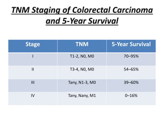
STAGE T N M DUKES[§]
0 Tis N0 M0
I T1 N0 M0 A
T2 N0 M0 A
IIA T3 N0 M0 B
IIB T4 N0 M0 B
IIIA T1-T2 N1 M0 C
IIIB T3-T4 N1 M0 C
IIIC Any T N2 M0 C
IV Any T Any N M1
TNM Staging of Colorectal Carcinoma
and 5-Year Survival
Stage TNM 5-Year Survival
I T1-2, N0, M0 70–95%
II T3-4, N0, M0 54–65%
III Tany, N1-3, M0 39–60%
IV Tany, Nany, M1 0–16%
Carcinoma of the colon
• Clinical features:
– Right colon cancer is more common in females,
while left colon cancer is more common in males.
• Clinical features depend upon the location of
the tumour, its size, and the presence of
metastases.
Rigid Sigmoidoscopy
Carcinoma of the colon
• A. Right colon cancer:
• (Irritative manifestations)
– The usual presentation is vague with anaemia, weakness and loss of
weight (Anaemia, Anorexia and Asthenia).
– The patient may present with recurrent attacks of pain in the right iliac
fossa.
– A hard mass may be present in the right side of the abdomen. It is
differentiated from appendicular mass by the long duration and
absence of toxaemia and tenderness. A malignant mass is hard, ill-
defined, irregular and fixed.
– The patient does not present by intestinal obstruction as
• The lesion is usually of the cauliflower variety.
• The contents are liquid.
• The lumen of the colon is wide.
– Obstruction occurs, rarely, if the lesion obstructs the ileocaecal valve.
Carcinoma of the colon
• B. Left colon cancer:
• (Obstructive manifestations)
1. The usual presentation is change of bowel habits, usually as progressive
constipation, but there may be diarrhoea or attacks of constipation
alternating with diarrhoea. These patients are usually diagnosed and treated
as having colitis. Spurious diarrhoea (early morning slime) may be present.
2. Large bowel obstruction: The patient may present as acute, subacute or
chronic large bowel obstruction. There is constipation, severe abdominal
distension but vomiting is late. Carcinoma of the sigmoid colon is a common
cause of intestinal obstruction in an elderly patient.
3. Bleeding per rectum: Carcinoma of the left colon is a common cause of fresh
bleeding per rectum but it is not a common cause of massive bleeding
(compare with diverticular disease and angiodysplasia).
4. Mass in the left side of abdomen: As the lesion is usually of the infiltrating
scirrhous type, it rarely presents by a mass. If a mass is palpable, it is usually
due to faecal impaction above the tumour.
Carcinoma of the colon
• Investigations
1. Blood picture may reveal microcytic hypochromic anaemia. Sigmoidoscopy is essential in all
patients with altered bowel habit or rectal bleeding. Biopsy is obtained from suspicious le-
sions. Total colonoscopy is important to exclude a second higher tumour.
2. Sigmoidoscopy is essential in all patients with altered bowel habit or rectal bleeding. Biopsy
is obtained from suspicious lesions. Total colonoscopy is important to exclude a second
higher tumour.
3. Barium enema. The tumour appears as a fixed irregular stricture or filling defect. Annular
strictures of the left colon show a characteristic "apple core appearance". Even with a
palpable rectal cancer, contrast radiography is useful to exclude a second higher tumour.
4. To detect spread liver function tests, abdominal ultrasound (or CT scan), and chest X-ray are
done. If the tumour is expected to be close to the ureter, an intravenous urogram (IVU) is
essential.
5. Carcinoembryonic antigen (CEA) is a tumour marker whose serum level is high in colorectal
cancer but is not specific. It is of prognostic rather than diagnostic value. The level drops
after a successful radical surgery. If it shows a rise in the follow up period, this signifies
recurrence.
6. For patients presenting with acute intestinal obstruction plain X-ray of the abdomen, blood
picture and electrolytes are needed .
Apple core appearance
A double-contrast barium enema showing a carcinoma in the
sigmoid colon (The classic apple core defect) .
Carcinoma of the colon
• Treatment
• General principles
– Surgery is the main line of treatment. Radical resection is the
only curative measure. For inoperable tumours resection also
offers the best palliation.
– Treatment depends on whether the tumour presents by acute
obstruction or not and whether the tumour is operable or not.
– Criteria of inoperability
1. Unfit patient.
2. Unresectable liver metastases.
3. Para-aortic lymph node involvement.
4. Peritoneal nodules
5. Irresectable primary tumour.
Carcinoma of the colon
• A. Treatment of colon cancer without acute
intestinal obstruction:
– Operable cases: elective radical resection. The
operation aims at cure and entails removal of the
tumour bearing segment together with its lymphatic
drainage area, in one mass; and then to restore bowel
continuity. Since the lymph nodes are so close to the
main blood vessels arising from the superior and
inferior mesenteric, their clearance requires ligation
and division of these vessels at their origin, and conse-
quently the whole part of the colon supplied by the
removed vessels should be resected.
Polyp Snaring
Principles of Surgical Management of
Colo-rectal Ca
 As a basic principle, any colorectal cancer is an indication for surgery unless
widespread tumor dissemination or general contraindications from the patient's
overall health status are present.
 The general goal for surgical management is either to achieve cure from the tumor
and extension of survival or disease-free survival
 Local tumor control generally is the primary treatment objective to prevent local
tumor complications, i.e., obstruction, perforation, bleeding, and pain. Even in the
presence of distant metastases in the liver or lung, resection of the primary tumor
remains a reasonable priority.
 Since solitary or a limited number of metastases in the liver or lung often may be
treated surgically by partial organ resection or metastasectomy with a cure rate of
up to 35%, their presence should not necessarily alter the surgical approach at the
primary site to do a curative resection.
 In contrast to rectal cancer, neoadjuvant treatment (i.e., preoperative
chemoradiation) is not indicated in the overwhelming majority of colonic cases
unless a locally very advanced lesion is treated with chemotherapy in anticipation
of an otherwise unresectable mass. Adjuvant (i.e., postoperative) treatment will be
discussed in a later section.
Preparation for Surgery
 Transfusion: Even though many colonic operations can be performed
without a blood transfusion, it is recommended to have the patient's blood
typed and crossed-matched, with a minimum of 2 units of blood available
at the beginning of the surgery.
 Bowel Cleansing: the products used generally are based on either
polyethylene glycol (e.g., GoLytely) or sodium phosphate (Fleet Phospho-
soda), the latter of which is contraindicated in patients with renal failure.
 Antibiotic Prophylaxis: Intravenous broad-spectrum antibiotics often
contain a combination of intravenous second- or third-generation
cephalosporin (cefoxitin or ceftriaxone) with metronidazole.
 Thromboembolic Prophylaxis
 Urinary Catheters/Stents
 Nasogastric Tube: is not necessary on a routine basis for patients
undergoing resection of the colon or rectum unless they present with a
complete or partial bowel obstruction.166
 Preoperative Marking of Ostomy Site
Carcinoma of the colon
a) Tumours of the caecum or the ascending colon: require a right hemicolectomy:
The terminal 10 inches of the ileum, the caecum, the ascending colon, the hepatic flexure, and the right
third of the transverse colon (to the right of the middle colic vessels) are resected. The operation is
completed by performing an ileotransverse anastomosis.
b) Tumours of the hepatic flexure: are treated by extended right hemicolectomy, where in addition to
the above, the middle colic vessels are included. The resection extends to the junction of the right
two thirds and the left third of the transverse colon.
c) Tumours of the transverse colon: are treated by transverse colectomy where the middle colic vessels
are divided flush at their origin and the transverse colon is resected with the hepatic and splenic
flexures, the transverse mesocolon and the omentum A colocolonic anastomosis is then performed.
d) Tumours of the splenic flexure or the descending colon: are treated by left hemicolectomy: the
resection includes the left half of the transverse colon (to the left of the middle colic vessels), the
splenic flexure and descending colon .
e) Tumours of the distal transverse colon and splenic flexure: are treated by extended left
hemicolectomy, where the left hemicolectomy is extended proximally to include the right branches
of the middle colic vessels.
g) Tumours arising in a long redundant sigmoid colon may be treated by sigmoid colectomy, where the
sigmoid vessels are divided at their origin from the inferior mesenteric artery and the resection
includes the sigmoid colon and mesocolon and sigmoid vessels.
Carcinoma of the colon
Extent of resection for carcinoma of the colon. A. Caecal and right colonic
cancer. B. Hepatic flexure cancer. C. Transverse colon cancer. D. Splenic
flexure cancer.
Carcinoma of the colon
Extent of resection for carcinoma of the colon . E. Descending
colon cancer. F. Sigmoid colon cancer.
Carcinoma of the colon
– Palliative operations for Inoperable cases : (whether
inoperability is diagnosed before or during operation)
• Whenever possible, palliative resection of the colon cancer
is preferred. Here there is no need for wide resection of the
bowel nor its lymphatics and vessels. The operation is
destined to obviate the risk of obstruction and bleeding.
• Tumours of the right colon require a side to side ileo-
transverse anastomosis.
• Tumours elsewhere in the colon require a proximal
colostomy.
– After palliative surgery, radiotherapy, chemotherapy (5-
flurouracil) and immunotherapy may be given to the patient.
Carcinoma of the colon
• B. Treatment of colon cancer with acute intestinal obstruction:
– Urgent surgery is required after adequate resuscitation.
– The best is colon resection (radical or palliative depending on operability).
– If the lesion is in the right colon, primary resection anastomosis is feasible .If
the tumour is in the left colon, the proximal end of the colon is brought to the
surface as end colostomy. The distal end is either closed as a Hartmann s
pouch or brought to the surface as a mucous fistula.
– N>B: Nowadays obstructed left colon cancer can be treated by resection and
primary anastomosis because it has a better prognosis as it allows earlier
removal of the tumour. The proximal colon should be decompressed and
cleaned by on-table lavage before anastomosis is done.
– Irresectable tumours of the right colon are treated by ileotransverse
anastomosis. Irresectable tumours of the left colon are treated by palliative
transverse or pelvic colostomy.
– For the critically ill, a proximal colostomy is all that can be done. After relief of
obstruction, most colonic carcinomas can be resected at a second-look
operation.
THANK YOU

Polyposis & Cancer Colon

  • 4.
    Colorectal polyps • Polypis a clinical term that describes any projection from the surface of the intestinal mucosa regardless of its histologic nature. • Colorectal polyps may be classified as A) Neoplastic (tubular adenoma, villous adenoma, tubulovillous adenomas). B) Hamartomatous (juvenile, Peutz-Jeghers, Cronkite-Canada and Cowden). C) Inflammatory (pseudopolyps and benign lymphoid polyp) D) Hyperplastic.
  • 5.
    Polyps of theColon & Rectum  Colorectal polyps are masses of tissue that project into the lumen. sessile or pedunculated, benign or malignant, mucosal, submucosal, or muscular lesions.  "Polyp" is a morphologic term, and no histologic diagnosis is implied.  Polyposis is a term reserved for the presence of many polyps in the large bowel.  Estimates of the incidence of colonic and rectal polyps in the general population range from 9% to 60%.
  • 6.
    Polyps of theColon & Rectum • Approximately 50% of polyps occur in the sigmoid or rectum. • About 50% of patients with adenoma have more than one lesion, and 15% have more than two lesions. • Approximately 25% of patients who have five or more adenomatous polyps have a synchronous colon cancer at the initial colonoscopy. • An increased incidence of adenomas in breast cancer patients has been reported.
  • 7.
    Polyps of theColon & Rectum • Approximately 50% of polyps occur in the sigmoid or rectum. • About 50% of patients with adenoma have more than one lesion, and 15% have more than two lesions. • Approximately 25% of patients who have five or more adenomatous polyps have a synchronous colon cancer at the initial colonoscopy. • An increased incidence of adenomas in breast cancer patients has been reported.
  • 8.
    Polyps of theLarge Intestine. Type Histologic Diagnosis Neoplastic •Adenoma 1. Tubular adenoma (adenomatous polyp) 75-80%. 2. Tubulovillous adenoma (villoglandular adenoma) 8-15%. 3. Villous adenoma (villous papilloma) 5-10%. •Carcinoma Hamartomas •Juvenile polyp •Peutz-Jeghers polyp Inflammatory Inflammatory polyp (pseudopolyp) Benign lymphoid polyp Hyperplastic Miscellaneous Lipoma, leiomyoma, carcinoid
  • 9.
    Neoplastic Polyps  Adenomatouspolyps are common, occurring in up to 25% of the population older than 50 years of age.  By definition, these lesions are dysplastic.  Invasive carcinoma is present in 5% of all adenomas, but the incidence correlates with the size and type of the adenoma  Tubular adenomas are associated with malignancy in only 5% of cases, whereas villous adenomas may harbor cancer in up to 40%. Tubulovillous adenomas are at intermediate risk (22%).  Invasive carcinomas are rare in polyps smaller than 1 cm; the incidence increases with size. The risk of carcinoma in a polyp larger than 2 cm is 35 to 50%.  Although most neoplastic polyps do not evolve to cancer, most colorectal cancers originate as a polyp.  It is this fact that forms the basis for secondary prevention strategies to eliminate colorectal cancer by targeting the neoplastic polyp for removal before malignancy develops.
  • 10.
    NEOPLASTIC COLORECTAL POLYPS TypeHistologic features Incidence (%) Invasive malignancy (%) Adenomatous (tabular adenoma) Branching tubules embedded in lamina propria 75 5 Villous (villous adenoma) Finger-like projections of epithelium over lamina propria 10 40 Intermediate (tubulovillous adenoma) Mixture of adenomatous and villous patterns 15 22
  • 11.
    Colorectal polyps • HamartomatousPolyps (Juvenile Polyps): – Juvenile polyps: are not usually premalignant. These lesions are the characteristic polyps of childhood but may occur at any age. The polyps are pedunculated, usually single or few in number. Commonly affecting the rectum. Bleeding is a common symptom and intussusception and/or obstruction may occur. Because the gross appearance of these polyps is identical to adenomatous polyps, these lesions should also be treated by polypectomy.
  • 12.
    Colorectal polyps • HamartomatousPolyps (Juvenile Polyps): – Peutz-Jeghers syndrome is characterized by polyposis of the small intestine, and to a lesser extent, polyposis of the colon and rectum. Characteristic melanin spots are often noted on the buccal mucosa and lips of these patients. The polyps of Peutz-Jeghers syndrome are generally considered to be hamartomas and are not thought to be at significant risk for malignant degeneration. However, carcinoma may occasionally develop. Because the entire length of the gastrointestinal tract may be affected, surgery is reserved for symptoms such as obstruction or bleeding or for patients in whom polyps develop adenomatous features.
  • 13.
    Colorectal polyps • FAMILIALADENOMATOUS POLYPOSIS ( FAP)(Familial Polyposis Coli, FPC) – Aetiology: • This rare autosomal dominant condition. • Clinically, patients develop hundreds to thousands of adenomatous polyps shortly after puberty. The lifetime risk of colorectal cancer in FAP patients approaches 100% by age 50 years. – Pathology: • Polyps usually start to appear at puberty, affecting the sigmoid colon and rectum initially and by the age of 20 years the entire colon and rectum are affected by immense numbers of polyps ( at least 100), ranging from 1 mm to several cms. • Polyps may be sessile or pedunculated. • Three histological types are recognized; the tubular, tubulo-villous, and villous. • Left untreated, carcinoma develops in 100% of the affected patients by the fifth decade.
  • 14.
    Colorectal polyps • Clinicalpicture: – The disease usually manifests itself soon after puberty by attacks of lower abdominal pain, diarrhoea, tenesmus with the passage of blood and mucus in the stools. Loss of weight, anaemia and general debility are common. – Rectal examination reveals the adenomas. • Investigations: – Barium enema shows multiple rounded filling defects throughout the colon and the rectum. – Sigmoidoscopy or colonoscopy and biopsy prove the nature of the disease.
  • 15.
    Macroscopic appearance ofthe colon in classical familial adenomatous polyposis. The specimen shows dense carpeting of the colonic mucosa with thousands of adenomas.
  • 16.
    Colorectal polyps • Treatment: –Once the diagnosis of FAP has been made and polyps are developing, treatment is surgical. – Three operative procedures (options) can be considered: 1. total proctocolectomy: with either an end ileostomy or continent (Kock's) ileostomy 2. Restorative proctocolectomy : with ileal pouch–anal anastomosis with or without a temporary ileostomy 3. Total abdominal colectomy with ileorectal anastomosis; the rectum is preserved, and polypi in the rectum are followed endoscopically and removed periodically.
  • 17.
    Carcinoma of thecolon • Incidence: Colorectal carcinoma is the most common malignancy of the gastrointestinal tract. The incidence is similar in men and women. • Risk Factors: (1) Aging: Aging is the most important risk factor for colorectal cancer, with incidence rising steadily after the age of 50 years. More than 90% of cases diagnosed are in people older than age 50 years. (2) Hereditary Risk Factors: Approximately 80% of colorectal cancers occur sporadically, while 20% arise in patients with a known family history of colorectal cancer. (3) Environmental and Dietary Factors: Colorectal carcinoma occurs more commonly in populations that consume diets high in animal fat and low in fiber. (4) Inflammatory bowel disease: Patients with long-standing ulcerative colitis are at increased risk for the development of colorectal cancer. Crohn's pancolitis have similar risk. (5) Other Risk Factors: Cigarette smoking reportedly increases the risk of colorectal cancer . Patients with ureterosigmoidostomy are also at increased risk . Also in women, gallstones (and the consequent cholecystectomy) are associated with colorectal cancer, especially in the right colon.
  • 18.
    Risk Factors Associatedwith Colon Cancer Risk Factor Comment Geographic variation Highest risk in Western countries and lowest risk in developing countries Age Risk increase sharply after the fifth decade Diet Increased with total and animal fat diets Physical inactivity Increased with obesity and sedentary life style Adenoma Risk dependent on type and size FAP penetrance in gene carriers 100% HNPCC penetrance in gene carriers 80% Hamartomatous syndromes Risk increased with Peutz-Jeghers syndrome and juvenile polyposis but not isolated juvenile polyps Previous history of colon cancer Increased risk for recurrent cancer Ulcerative colitis 10–20% after 20 years Radiation Associated with a mucinous histology and poor prognosis Ureterosigmoidostomy 100–500 times increased risk at or adjacent to the uretero- colonic anastomosis
  • 19.
    Carcinoma of thecolon • Pathology: – Site: • The sigmoid colon is the commonest site (50%) probably because its contents are solid, stagnant and irritant, and it is often the seat of precancerous lesions (i.e., FAP and UC) • Caecum and ascending colon: 25% • Transverse colon: 20%. • Descending colon: 5%. • Multiple synchronous colonic cancers (i.e., two or more carcinomas occurring simultaneously) are found in 5% of patients. Metachronous cancer is a new primary lesion in a patient who has had a previous resection for cancer.
  • 20.
    Carcinoma of thecolon • Gross picture: – Cauliflower growth: This occurs in the right colon. – Scirrhous growth (annular and tubular): This occurs in the left colon. – Malignant ulcer: This occurs commonly in the caecum. • Microscopic picture – Histologically the tumour is an adenocarcinoma that arises from the columnar epithelium. It may be well, moderately, or poorly differentiated. – Some tumours have a colloid structure, occur in younger patients, and have a poor prognosis.
  • 21.
    Carcinoma of thecolon • Spread: – Direct spread: • Carcinoma grows circumferentially and may completely encircle the bowel before it is diagnosed; this is especially true in the left colon, which has a smaller caliber than the right. It takes about 1 year for a tumor to encircle three-fourths of the circumference of the bowel. • Longitudinal submucosal extension occurs with invasion of the intramural lymphatic network, but it rarely goes beyond 2 cm from the edge of the tumor unless there is concomitant spread to lymph nodes. • As the lesion extends radially, it penetrates the outer layers of the bowel wall, and it may invade adjacent structures: the liver, the greater curvature of the stomach, the duodenum, the small bowel, the pancreas, the spleen, the bladder, the vagina, the kidneys and ureters, and the abdominal wall. Subacute perforation with inflammatory attachment of bowel to an adjacent viscous may be indistinguishable from actual invasion on gross examination.
  • 22.
    Carcinoma of thecolon • Lymphatic spread: – Cancer spreads from the colon to the following lymph node groups, in sequence: 1. Epicolic nodes on the bowel wall. 2. Paracolic nodes between the marginal artery and the bowel. 3. Intermediate nodes on the main vessels. 4. Principal nodes alongside the superior and inferior mesenteric vessels. – From the principal nodes further spread occurs to the parpa-aortic lymph nodes, then to cysterna chyli and thoracic duct. The left supraclavicular lymph nodes may be involved by retrograde flow from the thoracic duct (Virchow’s glands) giving positive Troisier’s sign.
  • 23.
    Carcinoma of thecolon • Blood spread: – Venous invasion may allow tumor cells be carried via the portal venous system to the liver. Tumor embolization also occurs through lumbar and vertebral veins to the lungs and elsewhere. Metastases to ovaries are mostly hematogenous; they are found in 1–10% of women with colorectal cancer. An attempt is made to avoid producing hematogenous metastases during operation by minimizing manipulation of the tumor prior to ligation of the blood supply.
  • 24.
    Carcinoma of thecolon • Transperitoneal spread: – "Seeding" may occur when the tumor has extended through the serosa and tumor cells enter the peritoneal cavity, producing local implants or generalized abdominal carcinomatosis. Large metastatic deposits in the pelvic cul- de-sac are palpable as a hard shelf (Blumer's shelf). • Intraluminal spread: – Malignant cells shed from the surface of the tumor can be swept with the fecal current. Implantation more distally on intact mucosa occurs rarely, if ever, but viable exfoliated cells presumably can be trapped in an anastomotic suture or staple line during operation.
  • 25.
    Carcinoma of thecolon • Complications: 1. Intestinal obstruction: 1. chronic obstruction is common especially in left colon cancer (20%). This tendency is attributed to: 1. The smaller lumen of the left colon. 2. Stool tends to be more solid. 3. Carcinoma tends to be of the stenosing type. 2. Acute obstruction may occur due to faecal impaction or intussusception (in carcinoma at the ileocaecal valve and sigmoid colon). 2. Haemorrhage: Chronic bleeding is the rule and causes iron deficiency anaemia. Massive bleeding is rare. 3. Perforation: is rare and is usually gradual with the formation of a pericolic abscess and faecal fistula. 4. Malignant ascites: results from liver secondaries, lymph nodes compressing the portal vein and Transperitoneal spread.
  • 26.
    Carcinoma of thecolon • Staging: – Colorectal cancer staging is based upon tumor depth and the presence or absence of nodal or distant metastases. – Old staging systems, such as the Dukes' Classification and its Astler-Coller modification, have been largely replaced by the TNM staging system (Table=). – The preoperative evaluation usually identifies stage IV disease. In colon cancer, differentiating stages I, II, and III depends upon examination of the resected specimen. In rectal cancer, endorectal ultrasound may predict the stage .
  • 27.
    Carcinoma of thecolon • Tumor Stage (T) Definition – Tx Cannot be assessed – T0 No evidence of cancer – Tis Carcinoma in situ – T1 Tumor invades submucosa – T2 Tumor invades muscularis propria – T3 Tumor invades through muscularis propria into subserosa or into nonperitonealized pericolic tissues – T4 Tumor directly invades other organs or tissues or perforates the visceral peritoneum of specimen • Nodal Stage (N) – NX Regional lymph nodes cannot be assessed – N0 No lymph node metastasis – N1 Metastasis to one to three pericolic or perirectal lymph nodes – N2 Metastasis to four or more pericolic or perirectal lymph nodes – N3 Metastasis to any lymph node along a major named vascular trunk • Distant Metastasis (M) – MX Presence of distant metastasis cannot be assessed – M0 No distant metastasis – M1 Distant metastasis present
  • 28.
    Stage Grouping STAGE TN M DUKES[§] 0 Tis N0 M0 I T1 N0 M0 A T2 N0 M0 A IIA T3 N0 M0 B IIB T4 N0 M0 B IIIA T1-T2 N1 M0 C IIIB T3-T4 N1 M0 C IIIC Any T N2 M0 C IV Any T Any N M1
  • 29.
    TNM Staging ofColorectal Carcinoma and 5-Year Survival Stage TNM 5-Year Survival I T1-2, N0, M0 70–95% II T3-4, N0, M0 54–65% III Tany, N1-3, M0 39–60% IV Tany, Nany, M1 0–16%
  • 30.
    Carcinoma of thecolon • Clinical features: – Right colon cancer is more common in females, while left colon cancer is more common in males. • Clinical features depend upon the location of the tumour, its size, and the presence of metastases.
  • 31.
  • 32.
    Carcinoma of thecolon • A. Right colon cancer: • (Irritative manifestations) – The usual presentation is vague with anaemia, weakness and loss of weight (Anaemia, Anorexia and Asthenia). – The patient may present with recurrent attacks of pain in the right iliac fossa. – A hard mass may be present in the right side of the abdomen. It is differentiated from appendicular mass by the long duration and absence of toxaemia and tenderness. A malignant mass is hard, ill- defined, irregular and fixed. – The patient does not present by intestinal obstruction as • The lesion is usually of the cauliflower variety. • The contents are liquid. • The lumen of the colon is wide. – Obstruction occurs, rarely, if the lesion obstructs the ileocaecal valve.
  • 33.
    Carcinoma of thecolon • B. Left colon cancer: • (Obstructive manifestations) 1. The usual presentation is change of bowel habits, usually as progressive constipation, but there may be diarrhoea or attacks of constipation alternating with diarrhoea. These patients are usually diagnosed and treated as having colitis. Spurious diarrhoea (early morning slime) may be present. 2. Large bowel obstruction: The patient may present as acute, subacute or chronic large bowel obstruction. There is constipation, severe abdominal distension but vomiting is late. Carcinoma of the sigmoid colon is a common cause of intestinal obstruction in an elderly patient. 3. Bleeding per rectum: Carcinoma of the left colon is a common cause of fresh bleeding per rectum but it is not a common cause of massive bleeding (compare with diverticular disease and angiodysplasia). 4. Mass in the left side of abdomen: As the lesion is usually of the infiltrating scirrhous type, it rarely presents by a mass. If a mass is palpable, it is usually due to faecal impaction above the tumour.
  • 34.
    Carcinoma of thecolon • Investigations 1. Blood picture may reveal microcytic hypochromic anaemia. Sigmoidoscopy is essential in all patients with altered bowel habit or rectal bleeding. Biopsy is obtained from suspicious le- sions. Total colonoscopy is important to exclude a second higher tumour. 2. Sigmoidoscopy is essential in all patients with altered bowel habit or rectal bleeding. Biopsy is obtained from suspicious lesions. Total colonoscopy is important to exclude a second higher tumour. 3. Barium enema. The tumour appears as a fixed irregular stricture or filling defect. Annular strictures of the left colon show a characteristic "apple core appearance". Even with a palpable rectal cancer, contrast radiography is useful to exclude a second higher tumour. 4. To detect spread liver function tests, abdominal ultrasound (or CT scan), and chest X-ray are done. If the tumour is expected to be close to the ureter, an intravenous urogram (IVU) is essential. 5. Carcinoembryonic antigen (CEA) is a tumour marker whose serum level is high in colorectal cancer but is not specific. It is of prognostic rather than diagnostic value. The level drops after a successful radical surgery. If it shows a rise in the follow up period, this signifies recurrence. 6. For patients presenting with acute intestinal obstruction plain X-ray of the abdomen, blood picture and electrolytes are needed .
  • 35.
  • 36.
    A double-contrast bariumenema showing a carcinoma in the sigmoid colon (The classic apple core defect) .
  • 37.
    Carcinoma of thecolon • Treatment • General principles – Surgery is the main line of treatment. Radical resection is the only curative measure. For inoperable tumours resection also offers the best palliation. – Treatment depends on whether the tumour presents by acute obstruction or not and whether the tumour is operable or not. – Criteria of inoperability 1. Unfit patient. 2. Unresectable liver metastases. 3. Para-aortic lymph node involvement. 4. Peritoneal nodules 5. Irresectable primary tumour.
  • 38.
    Carcinoma of thecolon • A. Treatment of colon cancer without acute intestinal obstruction: – Operable cases: elective radical resection. The operation aims at cure and entails removal of the tumour bearing segment together with its lymphatic drainage area, in one mass; and then to restore bowel continuity. Since the lymph nodes are so close to the main blood vessels arising from the superior and inferior mesenteric, their clearance requires ligation and division of these vessels at their origin, and conse- quently the whole part of the colon supplied by the removed vessels should be resected.
  • 39.
  • 40.
    Principles of SurgicalManagement of Colo-rectal Ca  As a basic principle, any colorectal cancer is an indication for surgery unless widespread tumor dissemination or general contraindications from the patient's overall health status are present.  The general goal for surgical management is either to achieve cure from the tumor and extension of survival or disease-free survival  Local tumor control generally is the primary treatment objective to prevent local tumor complications, i.e., obstruction, perforation, bleeding, and pain. Even in the presence of distant metastases in the liver or lung, resection of the primary tumor remains a reasonable priority.  Since solitary or a limited number of metastases in the liver or lung often may be treated surgically by partial organ resection or metastasectomy with a cure rate of up to 35%, their presence should not necessarily alter the surgical approach at the primary site to do a curative resection.  In contrast to rectal cancer, neoadjuvant treatment (i.e., preoperative chemoradiation) is not indicated in the overwhelming majority of colonic cases unless a locally very advanced lesion is treated with chemotherapy in anticipation of an otherwise unresectable mass. Adjuvant (i.e., postoperative) treatment will be discussed in a later section.
  • 41.
    Preparation for Surgery Transfusion: Even though many colonic operations can be performed without a blood transfusion, it is recommended to have the patient's blood typed and crossed-matched, with a minimum of 2 units of blood available at the beginning of the surgery.  Bowel Cleansing: the products used generally are based on either polyethylene glycol (e.g., GoLytely) or sodium phosphate (Fleet Phospho- soda), the latter of which is contraindicated in patients with renal failure.  Antibiotic Prophylaxis: Intravenous broad-spectrum antibiotics often contain a combination of intravenous second- or third-generation cephalosporin (cefoxitin or ceftriaxone) with metronidazole.  Thromboembolic Prophylaxis  Urinary Catheters/Stents  Nasogastric Tube: is not necessary on a routine basis for patients undergoing resection of the colon or rectum unless they present with a complete or partial bowel obstruction.166  Preoperative Marking of Ostomy Site
  • 42.
    Carcinoma of thecolon a) Tumours of the caecum or the ascending colon: require a right hemicolectomy: The terminal 10 inches of the ileum, the caecum, the ascending colon, the hepatic flexure, and the right third of the transverse colon (to the right of the middle colic vessels) are resected. The operation is completed by performing an ileotransverse anastomosis. b) Tumours of the hepatic flexure: are treated by extended right hemicolectomy, where in addition to the above, the middle colic vessels are included. The resection extends to the junction of the right two thirds and the left third of the transverse colon. c) Tumours of the transverse colon: are treated by transverse colectomy where the middle colic vessels are divided flush at their origin and the transverse colon is resected with the hepatic and splenic flexures, the transverse mesocolon and the omentum A colocolonic anastomosis is then performed. d) Tumours of the splenic flexure or the descending colon: are treated by left hemicolectomy: the resection includes the left half of the transverse colon (to the left of the middle colic vessels), the splenic flexure and descending colon . e) Tumours of the distal transverse colon and splenic flexure: are treated by extended left hemicolectomy, where the left hemicolectomy is extended proximally to include the right branches of the middle colic vessels. g) Tumours arising in a long redundant sigmoid colon may be treated by sigmoid colectomy, where the sigmoid vessels are divided at their origin from the inferior mesenteric artery and the resection includes the sigmoid colon and mesocolon and sigmoid vessels.
  • 43.
    Carcinoma of thecolon Extent of resection for carcinoma of the colon. A. Caecal and right colonic cancer. B. Hepatic flexure cancer. C. Transverse colon cancer. D. Splenic flexure cancer.
  • 44.
    Carcinoma of thecolon Extent of resection for carcinoma of the colon . E. Descending colon cancer. F. Sigmoid colon cancer.
  • 45.
    Carcinoma of thecolon – Palliative operations for Inoperable cases : (whether inoperability is diagnosed before or during operation) • Whenever possible, palliative resection of the colon cancer is preferred. Here there is no need for wide resection of the bowel nor its lymphatics and vessels. The operation is destined to obviate the risk of obstruction and bleeding. • Tumours of the right colon require a side to side ileo- transverse anastomosis. • Tumours elsewhere in the colon require a proximal colostomy. – After palliative surgery, radiotherapy, chemotherapy (5- flurouracil) and immunotherapy may be given to the patient.
  • 46.
    Carcinoma of thecolon • B. Treatment of colon cancer with acute intestinal obstruction: – Urgent surgery is required after adequate resuscitation. – The best is colon resection (radical or palliative depending on operability). – If the lesion is in the right colon, primary resection anastomosis is feasible .If the tumour is in the left colon, the proximal end of the colon is brought to the surface as end colostomy. The distal end is either closed as a Hartmann s pouch or brought to the surface as a mucous fistula. – N>B: Nowadays obstructed left colon cancer can be treated by resection and primary anastomosis because it has a better prognosis as it allows earlier removal of the tumour. The proximal colon should be decompressed and cleaned by on-table lavage before anastomosis is done. – Irresectable tumours of the right colon are treated by ileotransverse anastomosis. Irresectable tumours of the left colon are treated by palliative transverse or pelvic colostomy. – For the critically ill, a proximal colostomy is all that can be done. After relief of obstruction, most colonic carcinomas can be resected at a second-look operation.
  • 47.