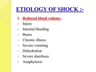
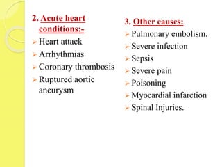
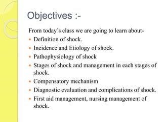
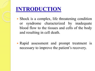
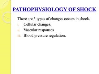
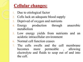
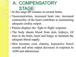
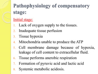
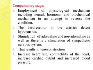
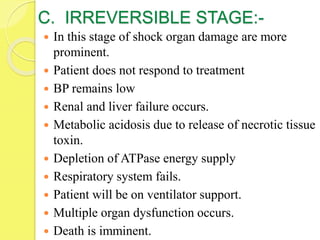
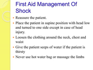
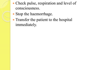
SHOCK
AND ITS
MANAGEMENT
Ms. SAHELI CHAKRABORTY
2ND YEAR MSC NURSING
RINER
Objectives :-
From today’s class we are going to learn about-
 Definition of shock.
 Incidence and Etiology of shock.
 Pathophysiology of shock
 Stages of shock and management in each stages of
shock.
 Compensatory mechanism
 Diagnostic evaluation and complications of shock.
 First aid management, nursing management of
shock.
INTRODUCTION
 Shock is a complex, life threatening condition
or syndrome characterized by inadequate
blood flow to the tissues and cells of the body
and resulting in cell death.
 Rapid assessment and prompt treatment is
necessary to improve the patient’s recovery.
DEFINITION
Shock is a pathological state in which group of
symptoms occurs characterized by-
 decrease tissue perfusion,
 impaired cellular metabolism ,
 increase oxygen and nutrient demand to the
tissues,
 results in cell death.
INCIDENCE OF SHOCK:
 Shock is common in both gender.
 Children and old age group more prone to
shock.
 80% of mortality rate is caused by shock.
 The incidence of septic shock in ICU has a
upward trend across recent years in INDIA
with an estimated mortality of >50%
ETIOLOGY OF SHOCK :-
1. Reduced blood volume-
 Injury
 Internal bleeding
 Burns
 Chronic illness
 Severe vomiting
 Dehydration
 Severe diarrhoea
 Anaphylaxis
2. Acute heart
conditions:-
 Heart attack
 Arrhythmias
 Coronary thrombosis
 Ruptured aortic
aneurysm
3. Other causes:
 Pulmonary embolism.
 Severe infection
 Sepsis
 Severe pain
 Poisoning
 Myocardial infarction
 Spinal Injuries.
PATHOPHYSIOLOGY OF SHOCK
There are 3 types of changes occurs in shock.
i. Cellular changes.
ii. Vascular responses
iii. Blood pressure regulation.
Cellular changes:
1. Due to etiological factor
2. Cells lack an adequate blood supply
3. Deprived of oxygen and nutrients
4. Energy production through anaerobic
metabolism
5. Low energy yields from nutrients and an
acidotic intracellular environment
6. Normal cell function ceases
7. The cells swells and the cell membrane
becomes more permeable , allowing
electrolyte and fluids to seep out of and into
the cell.
Contd..
8. Impairment in Na-K pump, damage of
mitochondria and cell structures, and death of
the cell occurs.
9. This state is called as shock state.
10. catecholamine, cortisol, glucagon and
inflammatory cytokines and mediators are
released in response to stress.
11. Hyperglycemia occurs and insulin resistance to
mobilize glucose for cellular metabolism.
12. Gluconeogenesis, glycogenolysis
13. Depletion of glycogen stores.
14. Increased proteolysis and eventual organ failure.
Vascular responses:
1. Local regulatory mechanism, autoregulation.
2. Stimulate vasodilation or vasoconstriction in
response to biochemical mediators released by
the cell.
3. The substance triggers an action at a cell site or
travels in the blood stream.
4. Need for oxygen and nutrients.
5. Deprived oxygen and nutrient in the cell.
6. Decreased tissue perfusion.
7. Shock.
Blood pressure regulation:
1. When blood pressure drops, catecholamine
releases from the adrenal gland.
2. Increased heart rate
3. Vasoconstriction
4. Restore blood pressure and respiratory rate.
5. Meanwhile catecholamine activates the renin
angiotensin and aldosterone mechanism.
6. Conversion of angiotensin I to angiotensin II and
thus results in vasoconstriction.
7. Angiotensin II activates the aldosterone.
8. Release of ADH by the pituitary gland.
9. Retention of sodium and water
10. Increase blood volume and blood pressure.
11. Continues to drop blood pressure and blood
volume
12. Decrease oxygenation to the tissues.
13. Hypo perfusion to the tissues and vital organs.
14. Multisystem organ failure.
STAGES OF SHOCK:
A convenient way to understand the physiological
responses and subsequent clinical signs and
symptoms of shock is to divide the continuum
into separate stages. It include the following:
A. Compensatory stage
B. Progressive stage
C. Irreversible stage
A. COMPENSATORY
STAGE:
In this stage BP remains in normal limits.
Vasoconstriction, increased heart rate, increased
contractility of the heart contribute to maintaining
adequate cardiac output.
Patient displays the ‘fight or flight’ response.
The body shunts blood from skin, kidneys, GI
tract to the brain, heart and lungs; to maintain the
adequate blood supply.
Skin becomes cool, clammy, hypoactive bowel
sounds and urine output decreases in response to
ADH and aldosterone.
Pathophysiology of compensatory
stage:
Initial stage:
1. Lack of oxygen supply to the tissues.
2. Inadequate tissue perfusion
3. Tissue hypoxia
4. Mitochondria unable to produce the ATP
5. Cell membrane damage because of hypoxia,
leakage of cell content to extracellular fluid.
6. Tissue performs anerobic respiration
7. Formation of pyruvic acid and lactic acid
8. Systemic metabolic acidosis.
Compensatory stage:
1. Employment of physiological mechanism
including neural, hormonal and biochemical
mechanism in an attempt to reverse the
condition.
2. The baroreceptor in the arteries detect
hypotension.
3. Stimulation of adrenaline and nor-adrenaline as
well as there is a stimulation of sympathetic
nervous system.
4. That results in vasoconstriction
5. Increase heart rate, contractility of the heart,
increase cardiac output and increased blood
pressure.
6. Shunting of blood from other organ to ensure
adequate blood supply.
7. Cold, clammy skin, hypoactive bowel sound,
decrease urine output in response to ADH
Clinical manifestation of
compensatory stage of shock:
 Normal BP.
 Inadequate organ perfusion.
 Build up of lactic acid produces metabolic
acidosis.
 Rapid respiratory state.
 Increase blood carbondioxide level and blood pH
level
 Respiratory alkalosis
 Changes in blood status.
 Confusion.
Medical and nursing management:
a) Monitoring tissue perfusion:
 Check for level of consciousness, vital signs,
pulse pressure.
 Check urine output.
 Laboratory values ( eg. Base deficit and lactic
acid level) should be checked.
 The nurse should monitor the patient’s
hemodynamic status.
 Administer prescribed fluids and medications
 Promote patient safety
 The nurse should report a systolic BP less than
90mmHg or a drop in 40mmHg from baseline.
 Pulse pressure should be monitored.
 Continuous central venous oxymetry should be
checked.
b. Reducing anxiety: patient and their families
often become anxious and apprehensive when
they face major threat to health and well being.
 Brief explanation of the patient’s health should
be given.
 Use gentle therapeutic touch.
 Speak in a calm reassuring voice and also help
ease the patient’s concern.
c. Promoting safety:
 The nurse must be vigilant for potential
threats to the patient’s safety because a high
anxiety level and altered mental status impair
judgement.
 Close monitoring and frequent reorientation
intervention are essential.
B. Progressive stage of shock
 In the second stage of shock the mechanism
that regulate blood pressure can no longer
compensate, and the mean arterial pressure
falls below normal limits.
 Patients are clinically hypotensive, systolic
blood pressure less than 90mmHg or a
decrease in systolic blood pressure of 40mmhg
from baseline.
Pathophysiology of 2nd stage of
shock:
1. Decrease cardiac output
2. Tissue hypoperfusion
3. Cells switch from aerobic to anerobic
metabolism
4. Lactic acid production
5. Cell function ceases and swells
6. Membranes becomes more permeable
7. Electrolyte and fluid seep in and out of the cell
8. Sodium build up and potassium leak out.
9. Damage of mitochondria
10. Cell death.
Clinical manifestation Of 2nd stage of
Shock:
1. Respiratory effects:
 Subsequent decompensation of the lungs
increases.
 Rapid and shallow respiration
 Crackles present
 Decrease pulmonary blood flow causes arterial
oxygen levels to decrease and carbon dioxide
level to increase.
 Hypoxemia and released chemical mediator
causes intense inflammatory response and
pulmonary vasoconstriction.
 Pulmonary capillary hypoperfusion.
 Pulmonary capillary begins to leak, causing
pulmonary edema and additional alveolar collapse.
 Acute lung injury.
 Acute respiratory distress syndrome.
2. Cardiovascular effects:
 Dysarrhythmias and ischemia.
 Rapid heart rate.
 The patient may complaints of chest pain and even
suffer a MI
 Levels of cardiac enzyme increases
 Impairs the heart’s ability to pump the blood.
 B-type Natriuretic peptide (BNP) increases due to
over distension of the ventricle
3. Neurologic effects:
 Changes in mental status occur with decreased
cerebral perfusion, hypoxia.
 Agitation
 Confusion
 Lethargy increases
 Loss of consciousness
4. Renal effects:
 Acute renal failure
 Increase BUN and serum creatinine level
 Fluid and electrolyte shift.
 Acid base imbalances
 Decrease urinary output less than 30 ml/kg/hour.
5. Hepatic effects:
 Decrease blood flow to the liver
 Impairment in phagocytic and metabolic function
 Impairment in gluconeogenesis and
glycogenolysis.
 The patient become more susceptible to infection
as the liver fails to filter the blood
 Increase level of bilirubin
 Jaundice
 Elevated liver enzyme level
6. GI Effects:
 Stress ulcer due to GI ischemia
 Risk for GI bleeding
 Bloody diarrhoea as necrosis of the mucosa.
 Ischemia results in increase bacterial toxins
which results in cardiac depression, vasodilation
and an intense inflammatory response with
activation of additional biochemical mediators.
7. Hematologic effects:
 Hypotension
 Sluggish blood flow
 Coagulation system imbalance
 DIC occur as a complication of shock
 Bruises and bleeding
 Prolonged coagulation time
Medical and Nursing
Management :
a) Preventing complication:
 The nurse should monitor the early signs of
complication
 Blood level of medication should be checked.
 Check the neurovascular status if arterial lines
are inserted.
 Maintain correct aseptic technique
 VAP should be prevented.
 Frequent oral care.
 Follow aseptic suction technique
 Turning and elevating the head at least 30 degree
to prevent aspiration
 Positioning of the patient
b) Promoting rest and comfort
 Efforts are made to minimize the cardiac
workload by reducing the patient’s physical
activity and treating pain and anxiety
 Promote rest to the patient
 Maintain temperature because elevated
temperature may increase the metabolic rate and
thus increases the workload of the heart.
 Avoid blankets to prevent vasodilation
c. Supporting family member
 The family member should be informed about
the patient’s status.
 Families need advice from the health care
team to get some rest.
C. IRREVERSIBLE STAGE:-
 In this stage of shock organ damage are more
prominent.
 Patient does not respond to treatment
 BP remains low
 Renal and liver failure occurs.
 Metabolic acidosis due to release of necrotic tissue
toxin.
 Depletion of ATPase energy supply
 Respiratory system fails.
 Patient will be on ventilator support.
 Multiple organ dysfunction occurs.
 Death is imminent.
Medical and Nursing
Management:
 Antibiotic therapy
 Immunomodulation therapy
 Monitor patient and prevent complication,
provide comfort to the patient.
 Reassurance should be given
 The family must be informed about the prognosis
of the patient.
 Counselling with all members of the health care
team and family member promotes better
understanding about the patient’s condition.
COMPENSATORY MECHANISM
1. Posture:
 A patient in acute circulatory failure falls down.
 Patient should lie flat on the floor or in head
down position to improve circulation towards
heart.
2. Contraction of skin vessels:
 Contraction of arterioles and venules of the skin
is usual to improve the blood supply.
 Application of heat to the skin should be
avoided.
3. Insensitivity:
 A much collapse patient has little pain.
 Large quantities of pain relieving drugs are not
effective in this stage unless given by
intravenous route.
4. Urinary secretion:
 These are diminished to conserve fluid in the
body but it is also a sign that tissue perfusion is
inadequate.
5. Heart rate accelerates:
 It is an attempt to ensure that remaining fluid is
circulated as early as possible to provide
sufficient oxygen to the tissues.
6. Subnormal temperature:
It helps in reduction in the requirement of the
tissues for the diminishing amount of oxygen
available.
Diagnostic evaluation
 History collection
 Physical examination
 12 lead ECG
 X ray chart.
 Continuous pulse
oxymetry
 Continuous cardiac
monitoring
 Hemodynamic
monitoring
 Laboratory
investigation.
 CBC
 Decrease
hemoglobin
 Decrease hematocrit
 Decrease/increase
WBC
 Decrease/increase
glucose
 Decrease/increase
potassium
 Decrease calcium
 Increase lactate.
COMPLICATION
 Multiple organ dysfunction syndrome
 Acute respiratory distress syndrome
 Acute renal failure
 Gastrointestinal ulceration
 DIC
 Death
First Aid Management Of
Shock
 Reassure the patient.
 Place the patient in supine position with head low
and turned to one side except in case of head
injury.
 Loosen the clothing around the neck, chest and
waist
 Give the patient seeps of water if the patient is
thirsty
 Never use hot water bag or massage the limbs
 Check pulse, respiration and level of
consciousness.
 Stop the haemorrhage.
 Transfer the patient to the hospital
immediately.
NURSING MANAGEMENT:
 Maintain airway, breathing, circulation of the
patient.
 Continuous monitoring of the vital sign.
 Provide supplemental oxygen therapy to the
patient. In severe cases put the patient in
mechanical ventilator.
 Monitor for ABG values,
 Check for urine output
 Maintain nutritional status of the patient
 Administer prescribed medication to the patient
 Give psychological support to the patient and
family.

Shock and its management

  • 1.
    SHOCK AND ITS MANAGEMENT Ms. SAHELICHAKRABORTY 2ND YEAR MSC NURSING RINER
  • 2.
    Objectives :- From today’sclass we are going to learn about-  Definition of shock.  Incidence and Etiology of shock.  Pathophysiology of shock  Stages of shock and management in each stages of shock.  Compensatory mechanism  Diagnostic evaluation and complications of shock.  First aid management, nursing management of shock.
  • 3.
    INTRODUCTION  Shock isa complex, life threatening condition or syndrome characterized by inadequate blood flow to the tissues and cells of the body and resulting in cell death.  Rapid assessment and prompt treatment is necessary to improve the patient’s recovery.
  • 4.
    DEFINITION Shock is apathological state in which group of symptoms occurs characterized by-  decrease tissue perfusion,  impaired cellular metabolism ,  increase oxygen and nutrient demand to the tissues,  results in cell death.
  • 5.
    INCIDENCE OF SHOCK: Shock is common in both gender.  Children and old age group more prone to shock.  80% of mortality rate is caused by shock.  The incidence of septic shock in ICU has a upward trend across recent years in INDIA with an estimated mortality of >50%
  • 6.
    ETIOLOGY OF SHOCK:- 1. Reduced blood volume-  Injury  Internal bleeding  Burns  Chronic illness  Severe vomiting  Dehydration  Severe diarrhoea  Anaphylaxis
  • 7.
    2. Acute heart conditions:- Heart attack  Arrhythmias  Coronary thrombosis  Ruptured aortic aneurysm 3. Other causes:  Pulmonary embolism.  Severe infection  Sepsis  Severe pain  Poisoning  Myocardial infarction  Spinal Injuries.
  • 8.
    PATHOPHYSIOLOGY OF SHOCK Thereare 3 types of changes occurs in shock. i. Cellular changes. ii. Vascular responses iii. Blood pressure regulation.
  • 9.
    Cellular changes: 1. Dueto etiological factor 2. Cells lack an adequate blood supply 3. Deprived of oxygen and nutrients 4. Energy production through anaerobic metabolism 5. Low energy yields from nutrients and an acidotic intracellular environment 6. Normal cell function ceases 7. The cells swells and the cell membrane becomes more permeable , allowing electrolyte and fluids to seep out of and into the cell.
  • 10.
    Contd.. 8. Impairment inNa-K pump, damage of mitochondria and cell structures, and death of the cell occurs. 9. This state is called as shock state. 10. catecholamine, cortisol, glucagon and inflammatory cytokines and mediators are released in response to stress. 11. Hyperglycemia occurs and insulin resistance to mobilize glucose for cellular metabolism. 12. Gluconeogenesis, glycogenolysis 13. Depletion of glycogen stores. 14. Increased proteolysis and eventual organ failure.
  • 11.
    Vascular responses: 1. Localregulatory mechanism, autoregulation. 2. Stimulate vasodilation or vasoconstriction in response to biochemical mediators released by the cell. 3. The substance triggers an action at a cell site or travels in the blood stream. 4. Need for oxygen and nutrients. 5. Deprived oxygen and nutrient in the cell. 6. Decreased tissue perfusion. 7. Shock.
  • 12.
    Blood pressure regulation: 1.When blood pressure drops, catecholamine releases from the adrenal gland. 2. Increased heart rate 3. Vasoconstriction 4. Restore blood pressure and respiratory rate. 5. Meanwhile catecholamine activates the renin angiotensin and aldosterone mechanism. 6. Conversion of angiotensin I to angiotensin II and thus results in vasoconstriction. 7. Angiotensin II activates the aldosterone. 8. Release of ADH by the pituitary gland.
  • 13.
    9. Retention ofsodium and water 10. Increase blood volume and blood pressure. 11. Continues to drop blood pressure and blood volume 12. Decrease oxygenation to the tissues. 13. Hypo perfusion to the tissues and vital organs. 14. Multisystem organ failure.
  • 14.
    STAGES OF SHOCK: Aconvenient way to understand the physiological responses and subsequent clinical signs and symptoms of shock is to divide the continuum into separate stages. It include the following: A. Compensatory stage B. Progressive stage C. Irreversible stage
  • 15.
    A. COMPENSATORY STAGE: In thisstage BP remains in normal limits. Vasoconstriction, increased heart rate, increased contractility of the heart contribute to maintaining adequate cardiac output. Patient displays the ‘fight or flight’ response. The body shunts blood from skin, kidneys, GI tract to the brain, heart and lungs; to maintain the adequate blood supply. Skin becomes cool, clammy, hypoactive bowel sounds and urine output decreases in response to ADH and aldosterone.
  • 16.
    Pathophysiology of compensatory stage: Initialstage: 1. Lack of oxygen supply to the tissues. 2. Inadequate tissue perfusion 3. Tissue hypoxia 4. Mitochondria unable to produce the ATP 5. Cell membrane damage because of hypoxia, leakage of cell content to extracellular fluid. 6. Tissue performs anerobic respiration 7. Formation of pyruvic acid and lactic acid 8. Systemic metabolic acidosis.
  • 17.
    Compensatory stage: 1. Employmentof physiological mechanism including neural, hormonal and biochemical mechanism in an attempt to reverse the condition. 2. The baroreceptor in the arteries detect hypotension. 3. Stimulation of adrenaline and nor-adrenaline as well as there is a stimulation of sympathetic nervous system. 4. That results in vasoconstriction 5. Increase heart rate, contractility of the heart, increase cardiac output and increased blood pressure.
  • 18.
    6. Shunting ofblood from other organ to ensure adequate blood supply. 7. Cold, clammy skin, hypoactive bowel sound, decrease urine output in response to ADH
  • 19.
    Clinical manifestation of compensatorystage of shock:  Normal BP.  Inadequate organ perfusion.  Build up of lactic acid produces metabolic acidosis.  Rapid respiratory state.  Increase blood carbondioxide level and blood pH level  Respiratory alkalosis  Changes in blood status.  Confusion.
  • 20.
    Medical and nursingmanagement: a) Monitoring tissue perfusion:  Check for level of consciousness, vital signs, pulse pressure.  Check urine output.  Laboratory values ( eg. Base deficit and lactic acid level) should be checked.  The nurse should monitor the patient’s hemodynamic status.  Administer prescribed fluids and medications  Promote patient safety
  • 21.
     The nurseshould report a systolic BP less than 90mmHg or a drop in 40mmHg from baseline.  Pulse pressure should be monitored.  Continuous central venous oxymetry should be checked. b. Reducing anxiety: patient and their families often become anxious and apprehensive when they face major threat to health and well being.  Brief explanation of the patient’s health should be given.  Use gentle therapeutic touch.  Speak in a calm reassuring voice and also help ease the patient’s concern.
  • 22.
    c. Promoting safety: The nurse must be vigilant for potential threats to the patient’s safety because a high anxiety level and altered mental status impair judgement.  Close monitoring and frequent reorientation intervention are essential.
  • 23.
    B. Progressive stageof shock  In the second stage of shock the mechanism that regulate blood pressure can no longer compensate, and the mean arterial pressure falls below normal limits.  Patients are clinically hypotensive, systolic blood pressure less than 90mmHg or a decrease in systolic blood pressure of 40mmhg from baseline.
  • 24.
    Pathophysiology of 2ndstage of shock: 1. Decrease cardiac output 2. Tissue hypoperfusion 3. Cells switch from aerobic to anerobic metabolism 4. Lactic acid production 5. Cell function ceases and swells 6. Membranes becomes more permeable 7. Electrolyte and fluid seep in and out of the cell 8. Sodium build up and potassium leak out. 9. Damage of mitochondria 10. Cell death.
  • 25.
    Clinical manifestation Of2nd stage of Shock: 1. Respiratory effects:  Subsequent decompensation of the lungs increases.  Rapid and shallow respiration  Crackles present  Decrease pulmonary blood flow causes arterial oxygen levels to decrease and carbon dioxide level to increase.  Hypoxemia and released chemical mediator causes intense inflammatory response and pulmonary vasoconstriction.
  • 26.
     Pulmonary capillaryhypoperfusion.  Pulmonary capillary begins to leak, causing pulmonary edema and additional alveolar collapse.  Acute lung injury.  Acute respiratory distress syndrome. 2. Cardiovascular effects:  Dysarrhythmias and ischemia.  Rapid heart rate.  The patient may complaints of chest pain and even suffer a MI  Levels of cardiac enzyme increases  Impairs the heart’s ability to pump the blood.  B-type Natriuretic peptide (BNP) increases due to over distension of the ventricle
  • 27.
    3. Neurologic effects: Changes in mental status occur with decreased cerebral perfusion, hypoxia.  Agitation  Confusion  Lethargy increases  Loss of consciousness 4. Renal effects:  Acute renal failure  Increase BUN and serum creatinine level  Fluid and electrolyte shift.  Acid base imbalances  Decrease urinary output less than 30 ml/kg/hour.
  • 28.
    5. Hepatic effects: Decrease blood flow to the liver  Impairment in phagocytic and metabolic function  Impairment in gluconeogenesis and glycogenolysis.  The patient become more susceptible to infection as the liver fails to filter the blood  Increase level of bilirubin  Jaundice  Elevated liver enzyme level
  • 29.
    6. GI Effects: Stress ulcer due to GI ischemia  Risk for GI bleeding  Bloody diarrhoea as necrosis of the mucosa.  Ischemia results in increase bacterial toxins which results in cardiac depression, vasodilation and an intense inflammatory response with activation of additional biochemical mediators.
  • 30.
    7. Hematologic effects: Hypotension  Sluggish blood flow  Coagulation system imbalance  DIC occur as a complication of shock  Bruises and bleeding  Prolonged coagulation time
  • 31.
    Medical and Nursing Management: a) Preventing complication:  The nurse should monitor the early signs of complication  Blood level of medication should be checked.  Check the neurovascular status if arterial lines are inserted.  Maintain correct aseptic technique  VAP should be prevented.  Frequent oral care.  Follow aseptic suction technique
  • 32.
     Turning andelevating the head at least 30 degree to prevent aspiration  Positioning of the patient b) Promoting rest and comfort  Efforts are made to minimize the cardiac workload by reducing the patient’s physical activity and treating pain and anxiety  Promote rest to the patient  Maintain temperature because elevated temperature may increase the metabolic rate and thus increases the workload of the heart.  Avoid blankets to prevent vasodilation
  • 33.
    c. Supporting familymember  The family member should be informed about the patient’s status.  Families need advice from the health care team to get some rest.
  • 34.
    C. IRREVERSIBLE STAGE:- In this stage of shock organ damage are more prominent.  Patient does not respond to treatment  BP remains low  Renal and liver failure occurs.  Metabolic acidosis due to release of necrotic tissue toxin.  Depletion of ATPase energy supply  Respiratory system fails.  Patient will be on ventilator support.  Multiple organ dysfunction occurs.  Death is imminent.
  • 35.
    Medical and Nursing Management: Antibiotic therapy  Immunomodulation therapy  Monitor patient and prevent complication, provide comfort to the patient.  Reassurance should be given  The family must be informed about the prognosis of the patient.  Counselling with all members of the health care team and family member promotes better understanding about the patient’s condition.
  • 36.
    COMPENSATORY MECHANISM 1. Posture: A patient in acute circulatory failure falls down.  Patient should lie flat on the floor or in head down position to improve circulation towards heart. 2. Contraction of skin vessels:  Contraction of arterioles and venules of the skin is usual to improve the blood supply.  Application of heat to the skin should be avoided.
  • 37.
    3. Insensitivity:  Amuch collapse patient has little pain.  Large quantities of pain relieving drugs are not effective in this stage unless given by intravenous route. 4. Urinary secretion:  These are diminished to conserve fluid in the body but it is also a sign that tissue perfusion is inadequate. 5. Heart rate accelerates:  It is an attempt to ensure that remaining fluid is circulated as early as possible to provide sufficient oxygen to the tissues.
  • 38.
    6. Subnormal temperature: Ithelps in reduction in the requirement of the tissues for the diminishing amount of oxygen available.
  • 39.
    Diagnostic evaluation  Historycollection  Physical examination  12 lead ECG  X ray chart.  Continuous pulse oxymetry  Continuous cardiac monitoring  Hemodynamic monitoring  Laboratory investigation.  CBC  Decrease hemoglobin  Decrease hematocrit  Decrease/increase WBC  Decrease/increase glucose  Decrease/increase potassium  Decrease calcium  Increase lactate.
  • 40.
    COMPLICATION  Multiple organdysfunction syndrome  Acute respiratory distress syndrome  Acute renal failure  Gastrointestinal ulceration  DIC  Death
  • 41.
    First Aid ManagementOf Shock  Reassure the patient.  Place the patient in supine position with head low and turned to one side except in case of head injury.  Loosen the clothing around the neck, chest and waist  Give the patient seeps of water if the patient is thirsty  Never use hot water bag or massage the limbs
  • 42.
     Check pulse,respiration and level of consciousness.  Stop the haemorrhage.  Transfer the patient to the hospital immediately.
  • 43.
    NURSING MANAGEMENT:  Maintainairway, breathing, circulation of the patient.  Continuous monitoring of the vital sign.  Provide supplemental oxygen therapy to the patient. In severe cases put the patient in mechanical ventilator.  Monitor for ABG values,  Check for urine output  Maintain nutritional status of the patient  Administer prescribed medication to the patient  Give psychological support to the patient and family.