Fluid and electrolyte
Disturbance
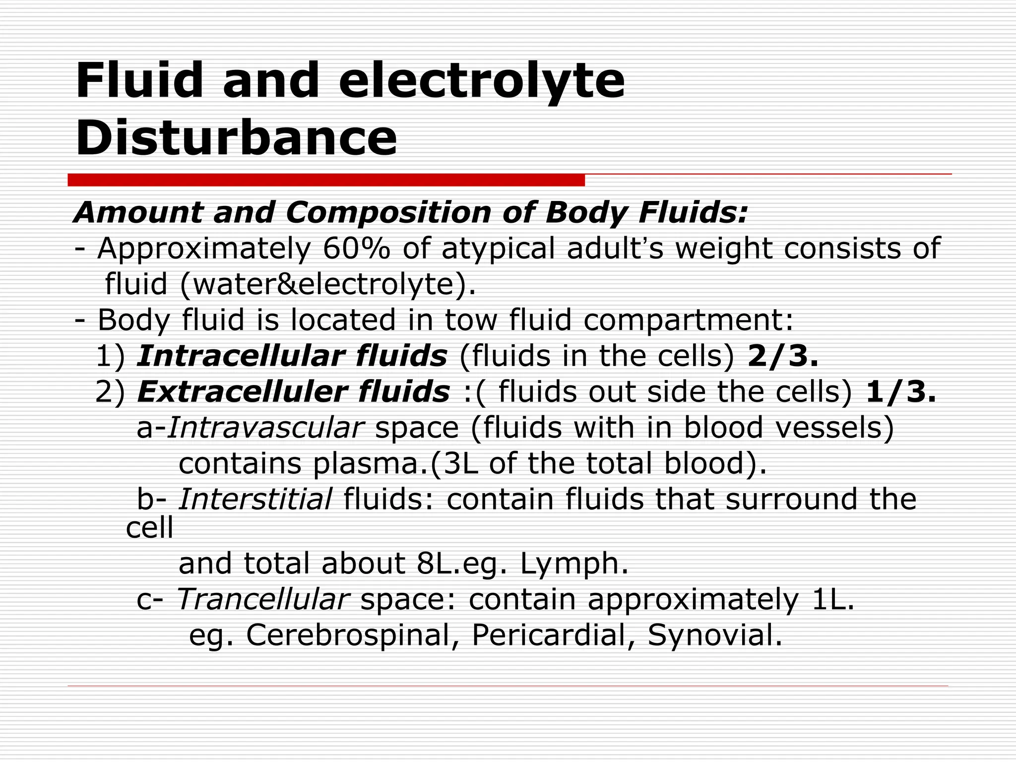
Amountand Composition of Body Fluids:
- Approximately 60% of atypical adult’s weight consists of
fluid (water&electrolyte).
- Body fluid is located in tow fluid compartment:
1) Intracellular fluids (fluids in the cells) 2/3.
2) Extracelluler fluids :( fluids out side the cells) 1/3.
a-Intravascular space (fluids with in blood vessels)
contains plasma.(3L of the total blood).
b- Interstitial fluids: contain fluids that surround the
cell
and total about 8L.eg. Lymph.
c- Trancellular space: contain approximately 1L.
eg. Cerebrospinal, Pericardial, Synovial.
3.
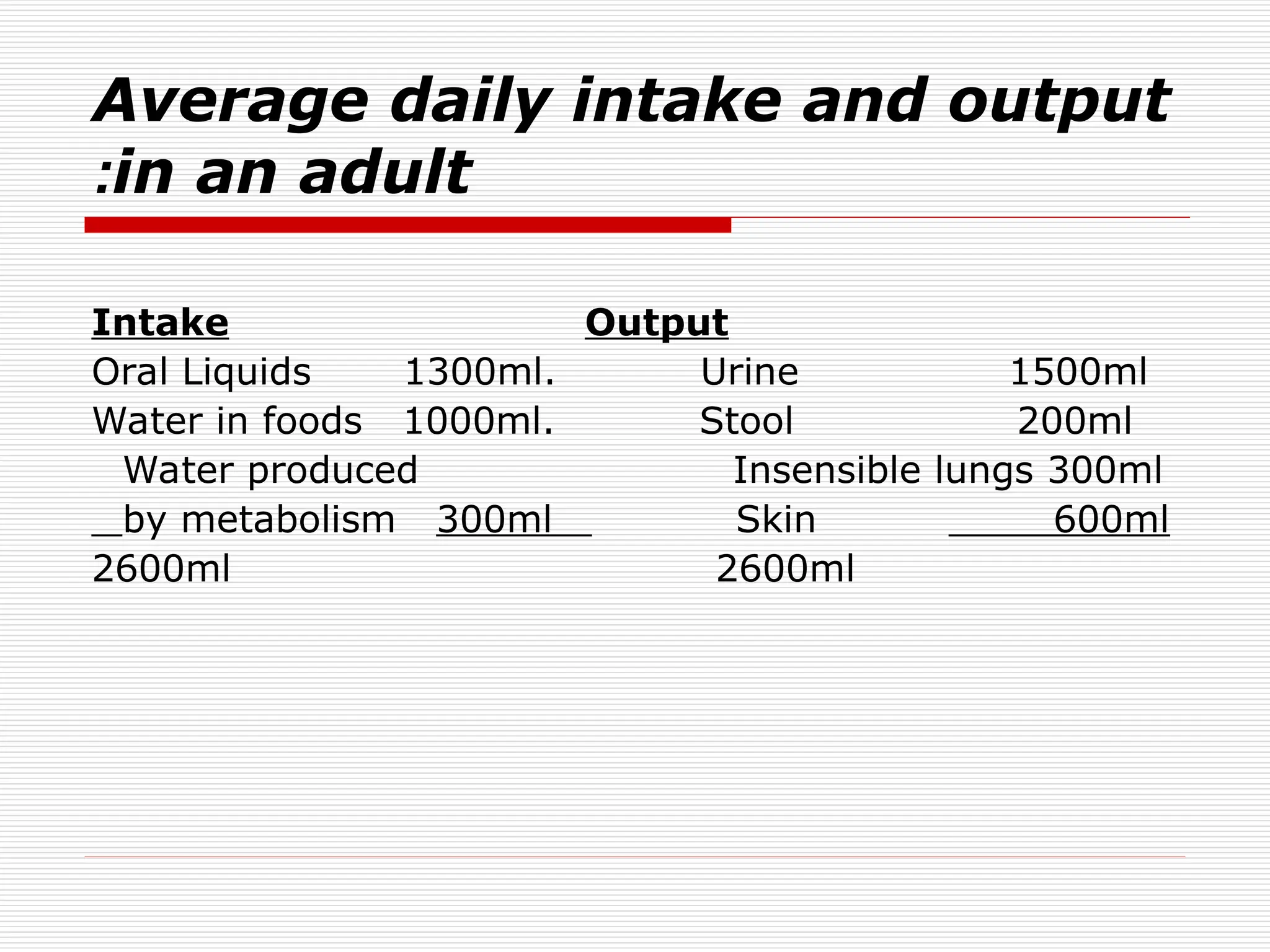
Average daily intakeand output
in an adult
:
Intake Output
Oral Liquids 1300ml. Urine 1500ml
Water in foods 1000ml. Stool 200ml
Water produced Insensible lungs 300ml
by metabolism 300ml Skin 600ml
2600ml 2600ml
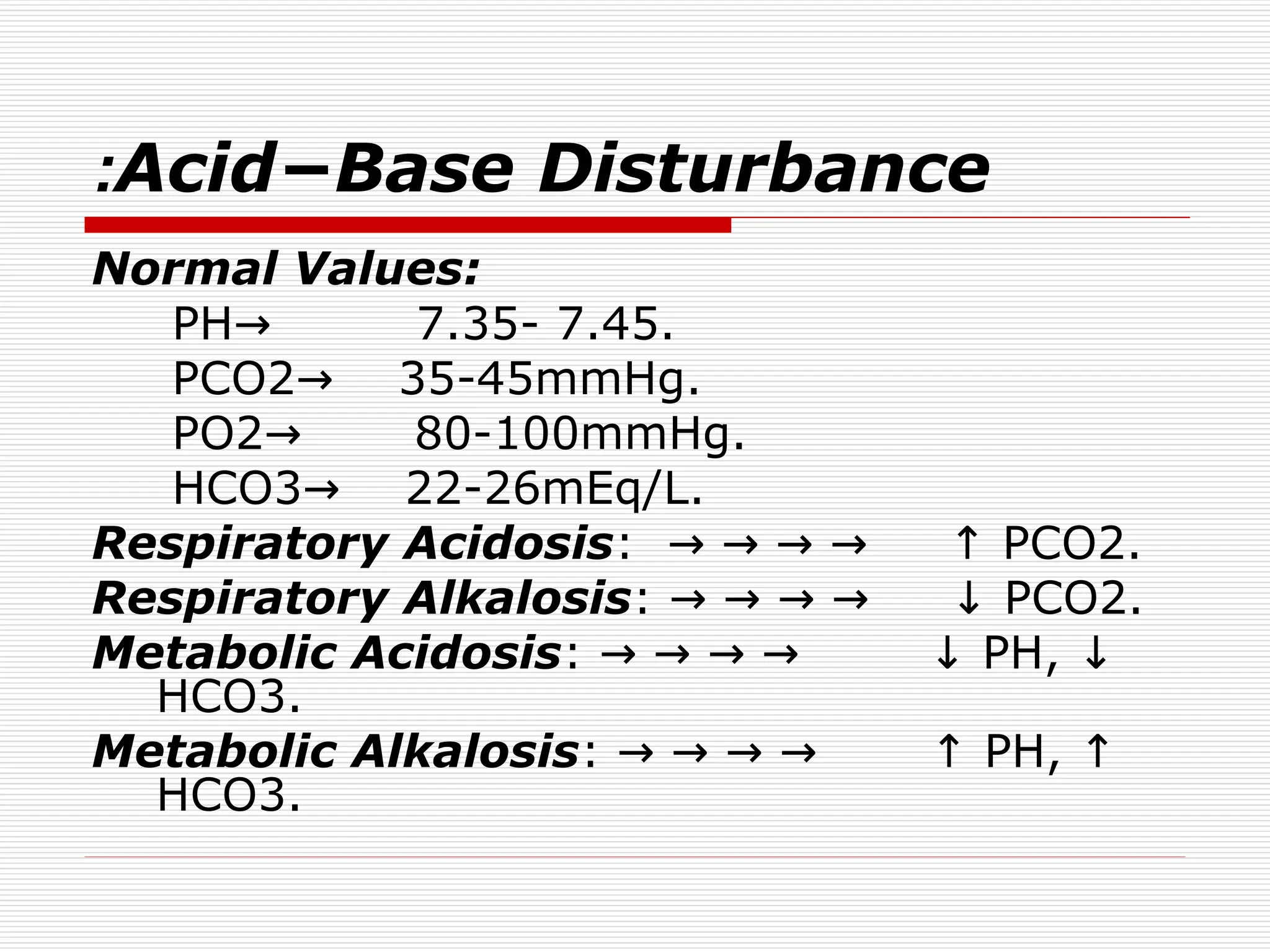
Fluid Volume Disturbance
:
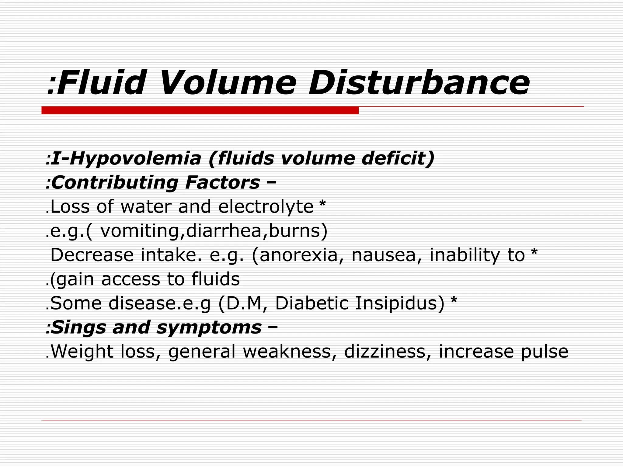
I-Hypovolemia(fluids volume deficit)
:
−
Contributing Factors
:
*
Loss of water and electrolyte
.
e.g.( vomiting,diarrhea,burns)
.
*
Decrease intake. e.g. (anorexia, nausea, inability to
gain access to fluids
.)
*
Some disease.e.g (D.M, Diabetic Insipidus)
.
−
Sings and symptoms
:
Weight loss, general weakness, dizziness, increase pulse
.
6.
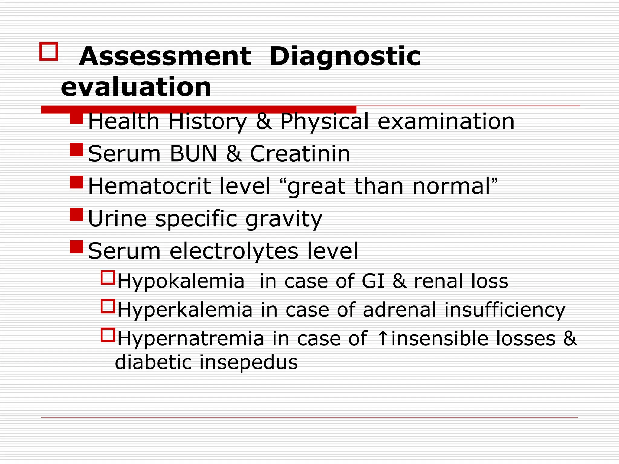
Assessment Diagnostic
evaluation
HealthHistory & Physical examination
Serum BUN & Creatinin
Hematocrit level “great than normal”
Urine specific gravity
Serum electrolytes level
Hypokalemia in case of GI & renal loss
Hyperkalemia in case of adrenal insufficiency
Hypernatremia in case of insensible losses &
↑
diabetic insepedus
8.
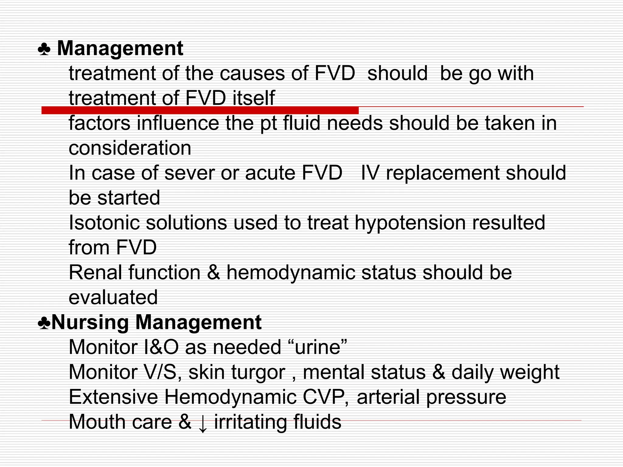
♣ Management
treatment ofthe causes of FVD should be go with
treatment of FVD itself
factors influence the pt fluid needs should be taken in
consideration
In case of sever or acute FVD IV replacement should
be started
Isotonic solutions used to treat hypotension resulted
from FVD
Renal function & hemodynamic status should be
evaluated
♣Nursing Management
Monitor I&O as needed “urine”
Monitor V/S, skin turgor , mental status & daily weight
Extensive Hemodynamic CVP, arterial pressure
Mouth care & ↓ irritating fluids
9.
Fluid Volume Disturbance
:
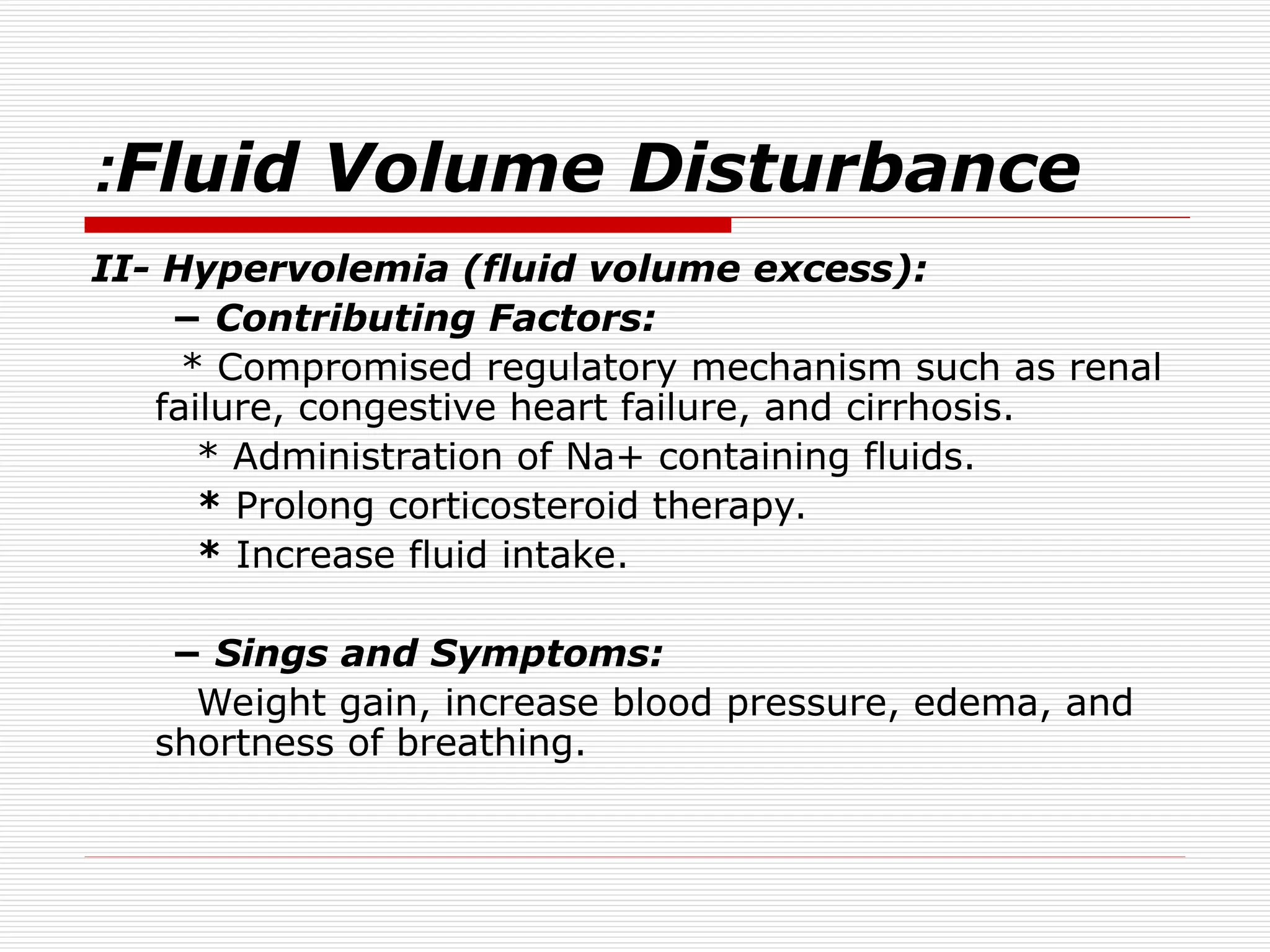
II-Hypervolemia (fluid volume excess):
− Contributing Factors:
* Compromised regulatory mechanism such as renal
failure, congestive heart failure, and cirrhosis.
* Administration of Na+ containing fluids.
* Prolong corticosteroid therapy.
* Increase fluid intake.
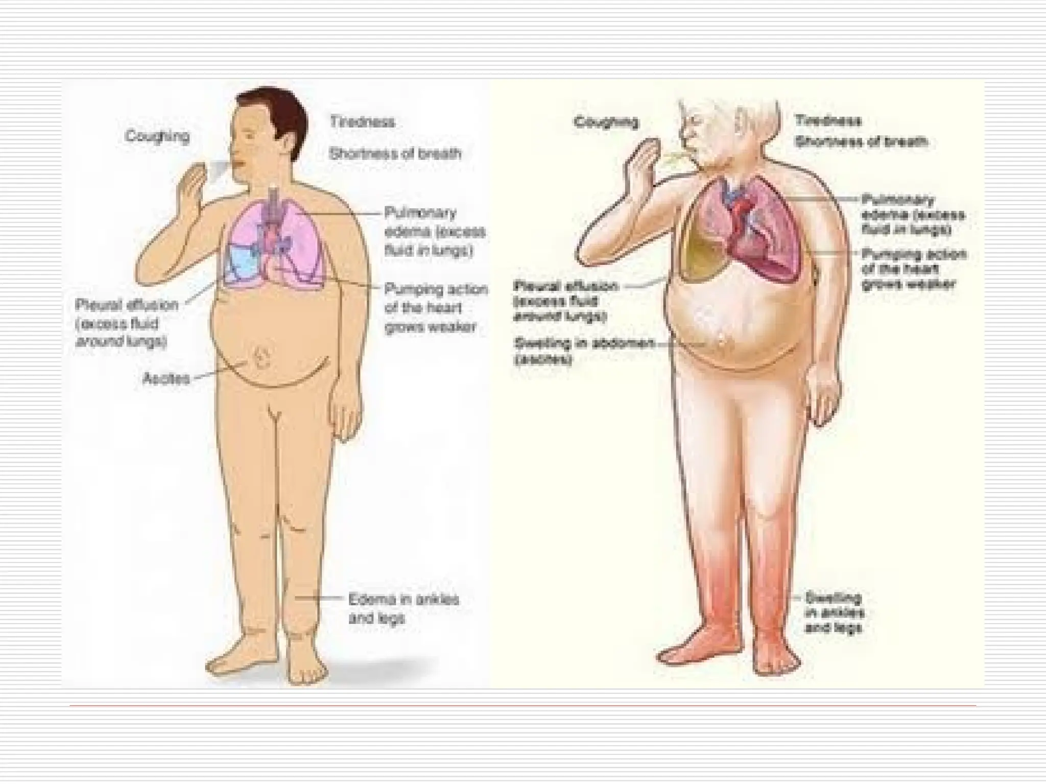
− Sings and Symptoms:
Weight gain, increase blood pressure, edema, and
shortness of breathing.
10.
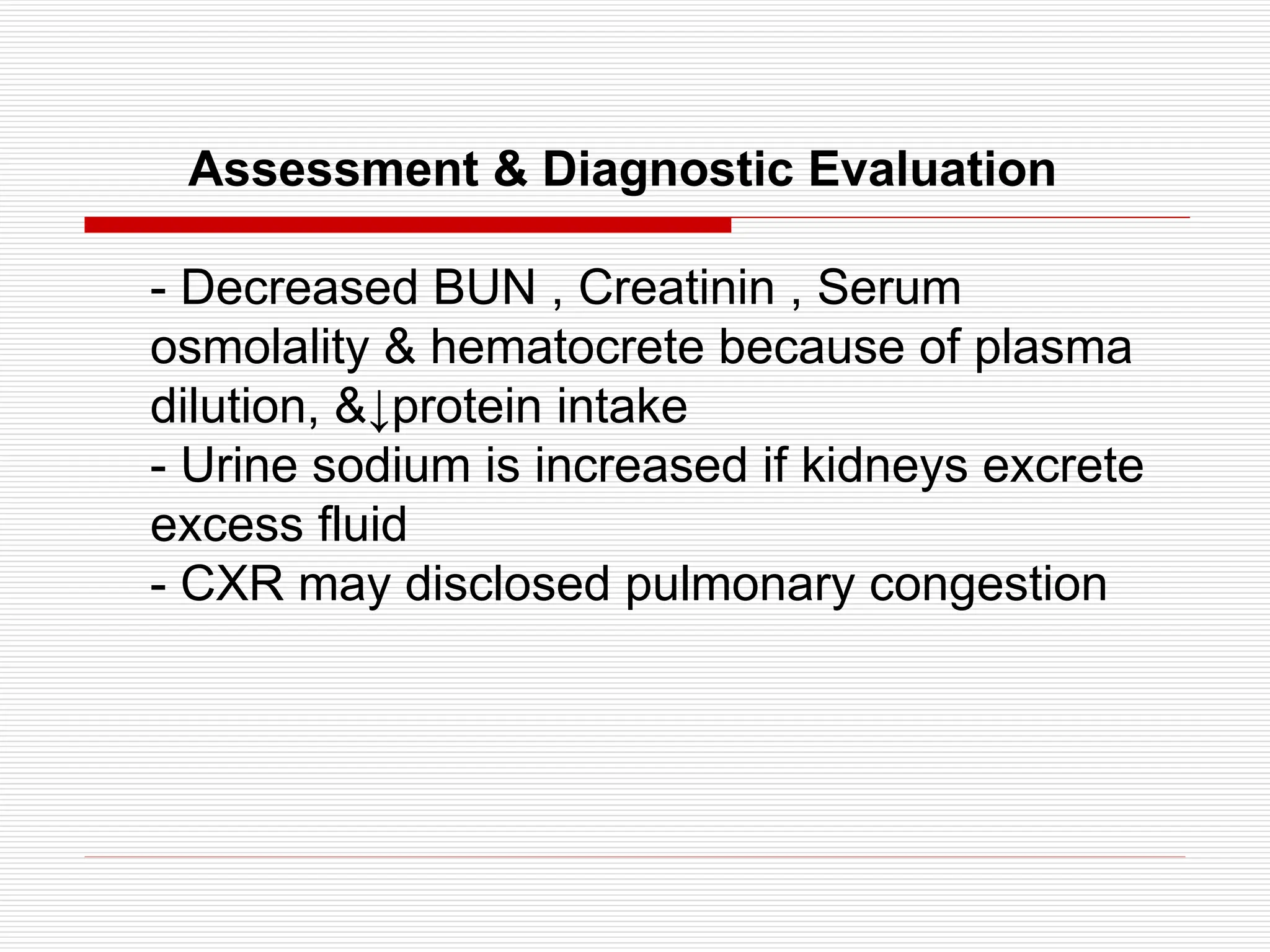
Assessment & DiagnosticEvaluation
- Decreased BUN , Creatinin , Serum
osmolality & hematocrete because of plasma
dilution, &↓protein intake
- Urine sodium is increased if kidneys excrete
excess fluid
- CXR may disclosed pulmonary congestion
12.
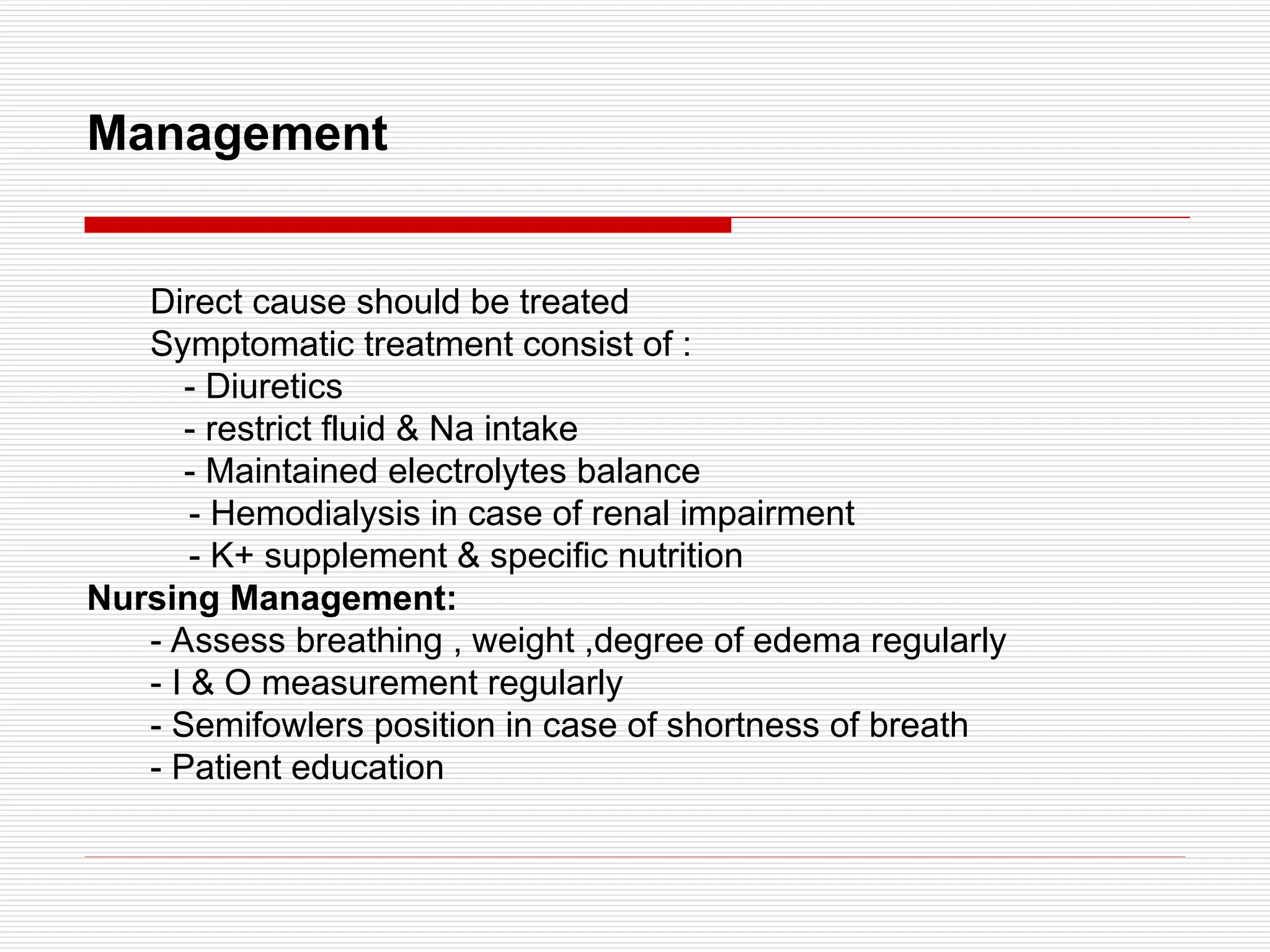
Management
Direct cause shouldbe treated
Symptomatic treatment consist of :
- Diuretics
- restrict fluid & Na intake
- Maintained electrolytes balance
- Hemodialysis in case of renal impairment
- K+ supplement & specific nutrition
Nursing Management:
- Assess breathing , weight ,degree of edema regularly
- I & O measurement regularly
- Semifowlers position in case of shortness of breath
- Patient education
13.
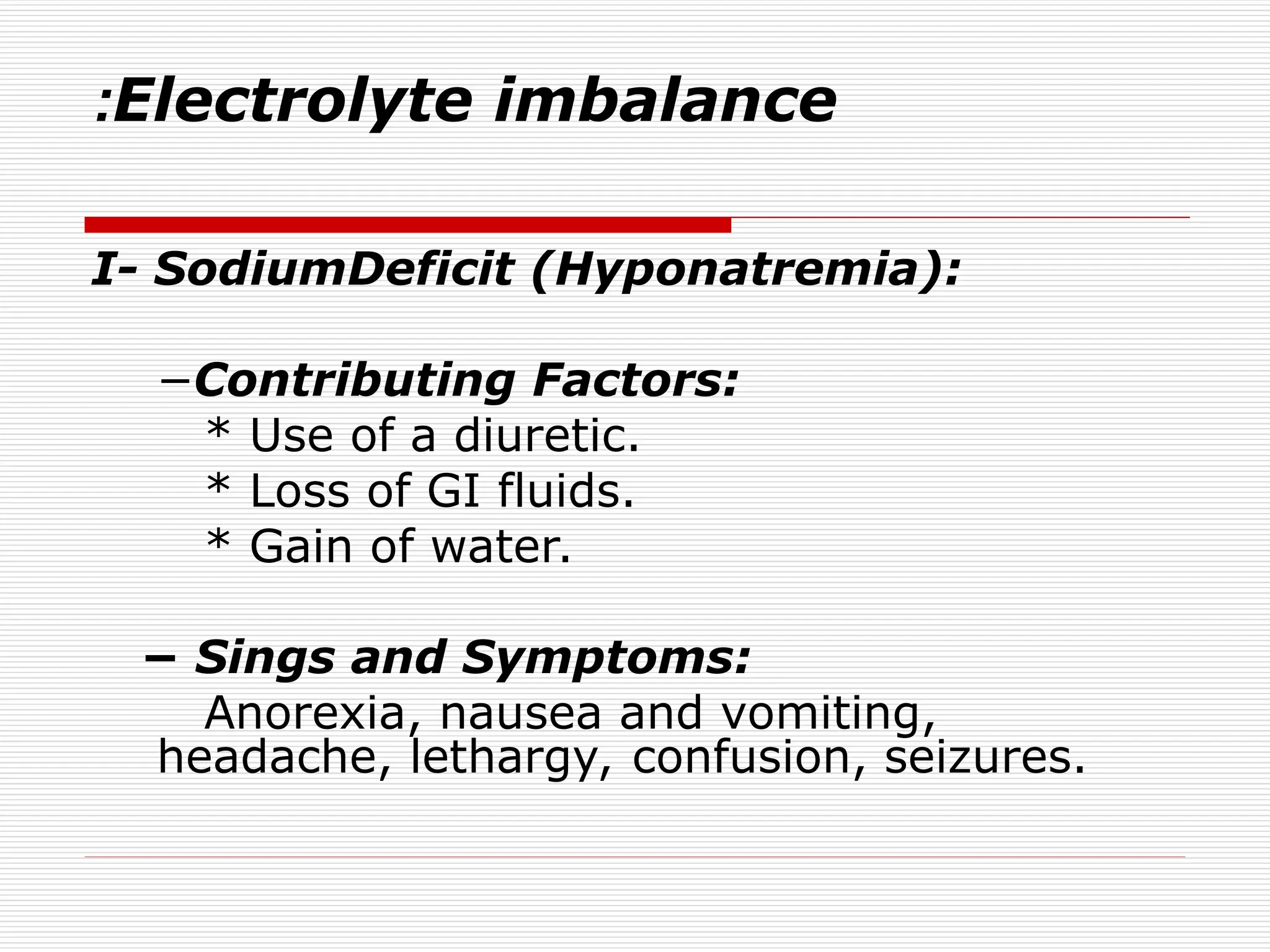
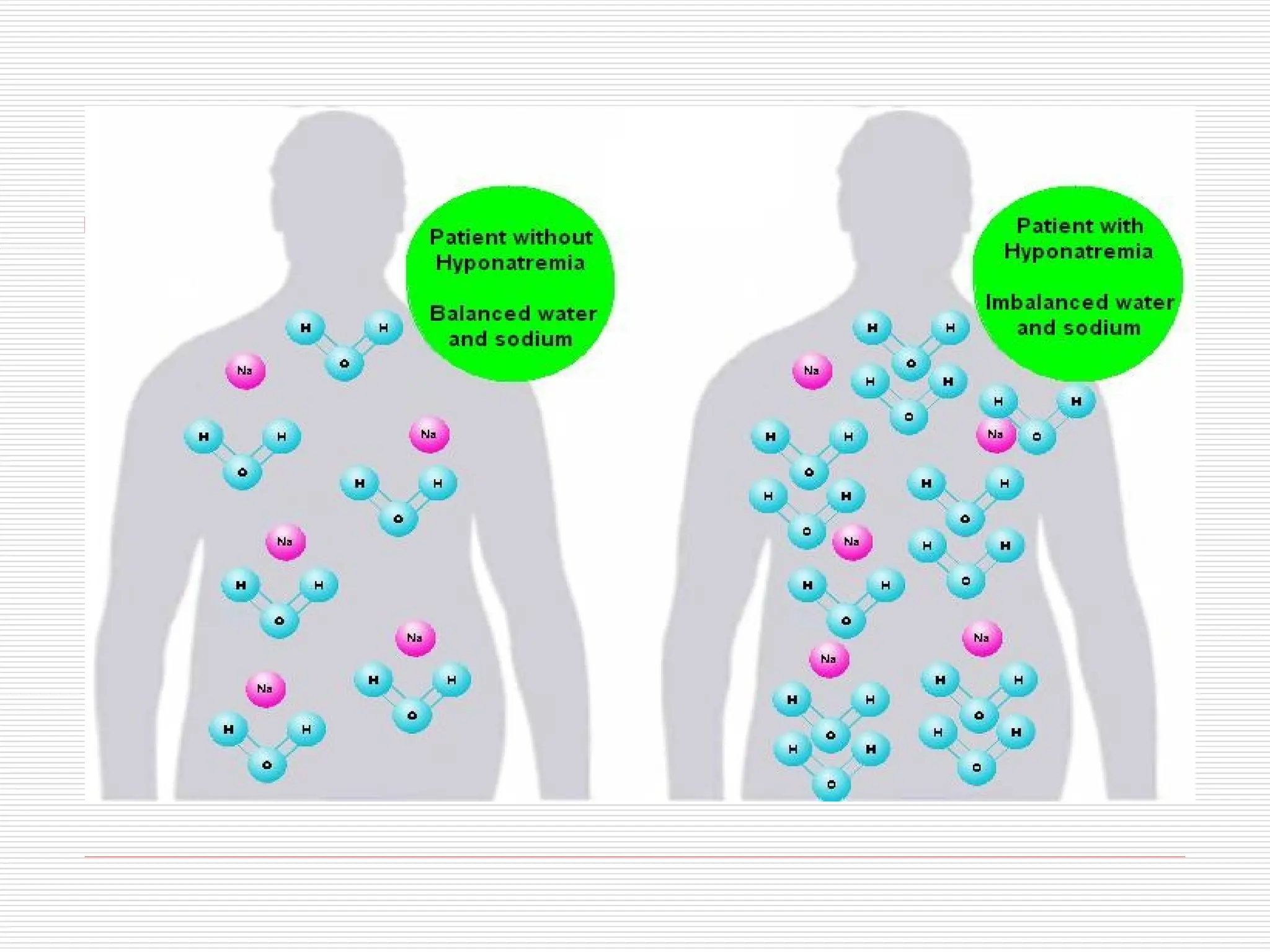
Electrolyte imbalance
:
I- SodiumDeficit(Hyponatremia):
−Contributing Factors:
* Use of a diuretic.
* Loss of GI fluids.
* Gain of water.
− Sings and Symptoms:
Anorexia, nausea and vomiting,
headache, lethargy, confusion, seizures.
15.
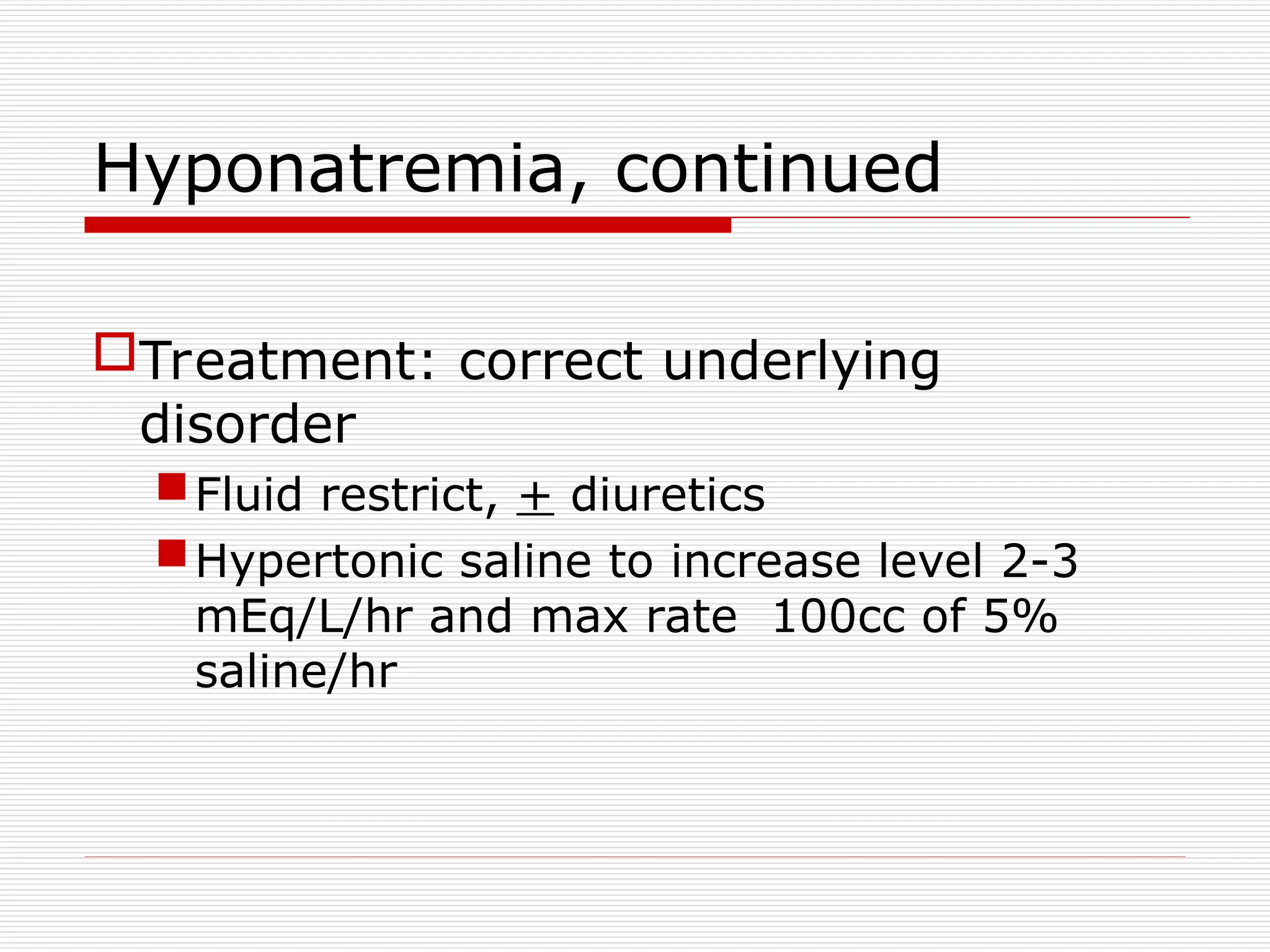
Hyponatremia, continued
Treatment: correctunderlying
disorder
Fluid restrict, + diuretics
Hypertonic saline to increase level 2-3
mEq/L/hr and max rate 100cc of 5%
saline/hr
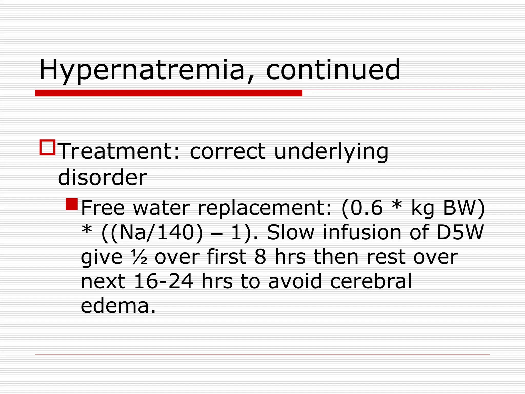
Hypernatremia, continued
Treatment: correctunderlying
disorder
Free water replacement: (0.6 * kg BW)
* ((Na/140) – 1). Slow infusion of D5W
give ½ over first 8 hrs then rest over
next 16-24 hrs to avoid cerebral
edema.
19.
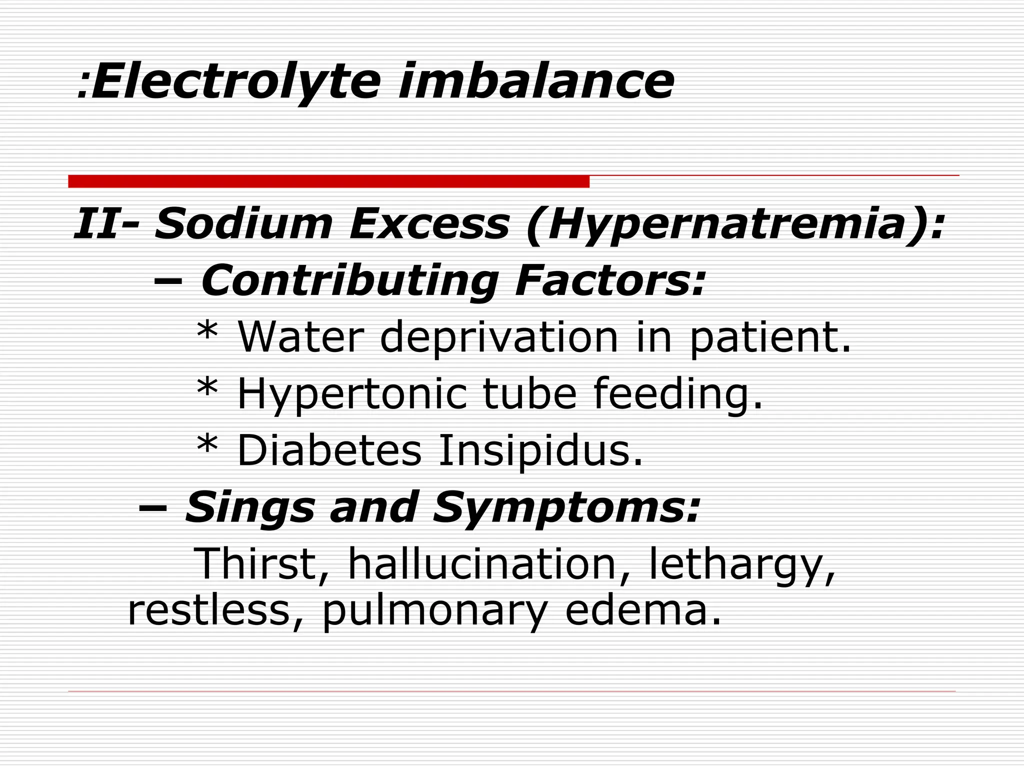
Electrolyte imbalance
:
III- PotassiumDeficit (Hypokalemia):
− Contributing factors:
* Dirrhea, vomiting, gastric suctions.
* Corticosteroid administration.
* Diuretics.
− Sings and symptoms:
Fatigue, anorexia, nausea, vomiting,
muscle weakness, change in ECG.
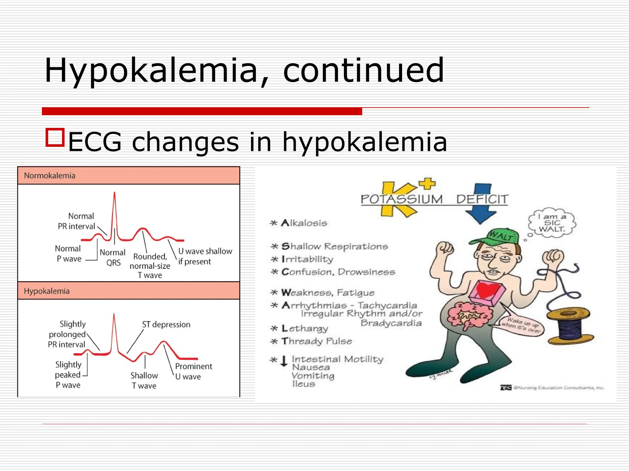
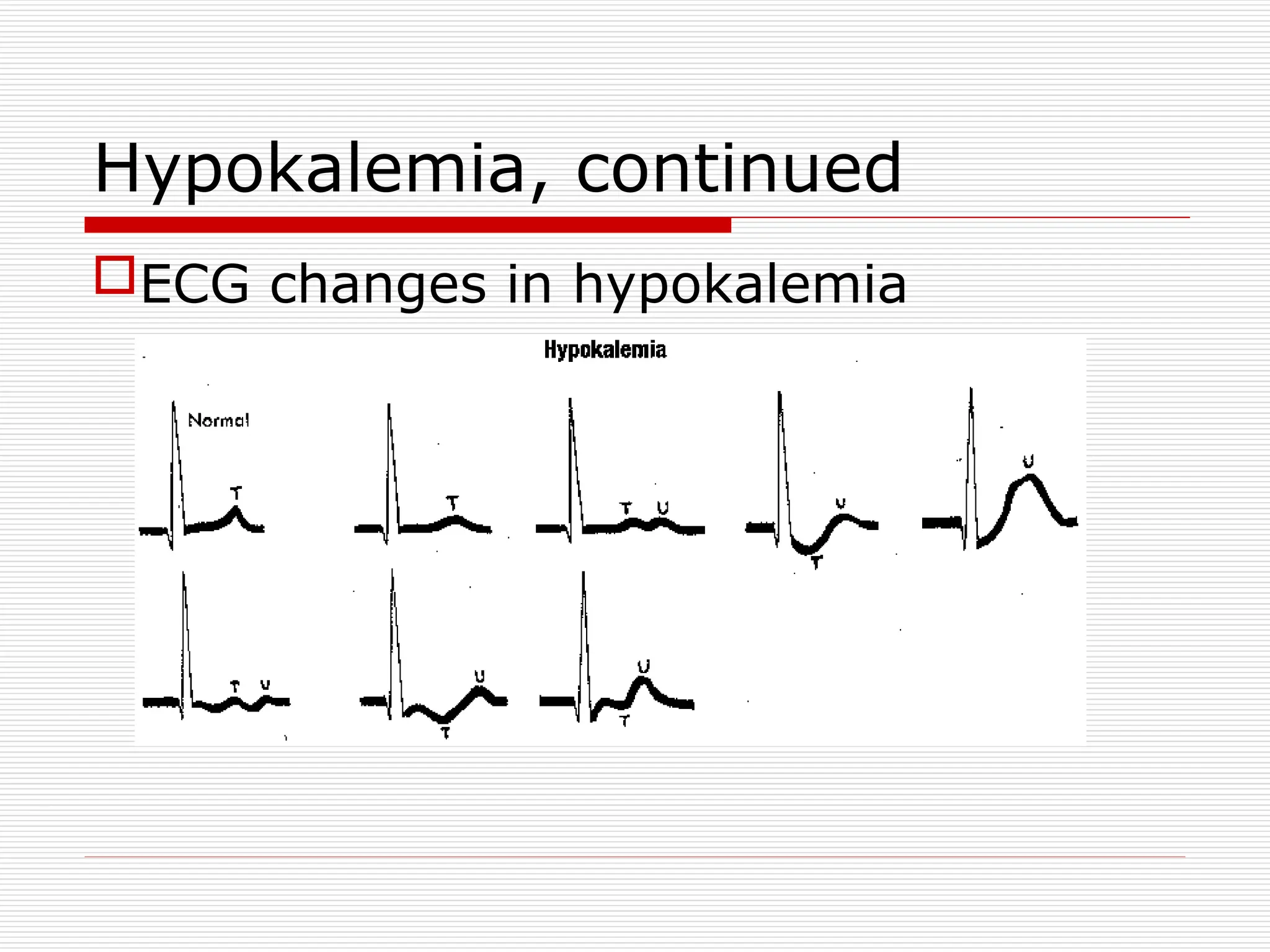
EKG: low, flat T-waves, ST depression, and U
waves
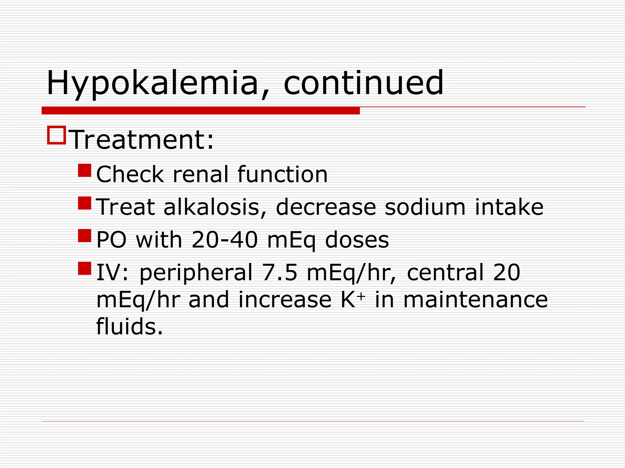
Hypokalemia, continued
Treatment:
Check renalfunction
Treat alkalosis, decrease sodium intake
PO with 20-40 mEq doses
IV: peripheral 7.5 mEq/hr, central 20
mEq/hr and increase K+
in maintenance
fluids.
23.
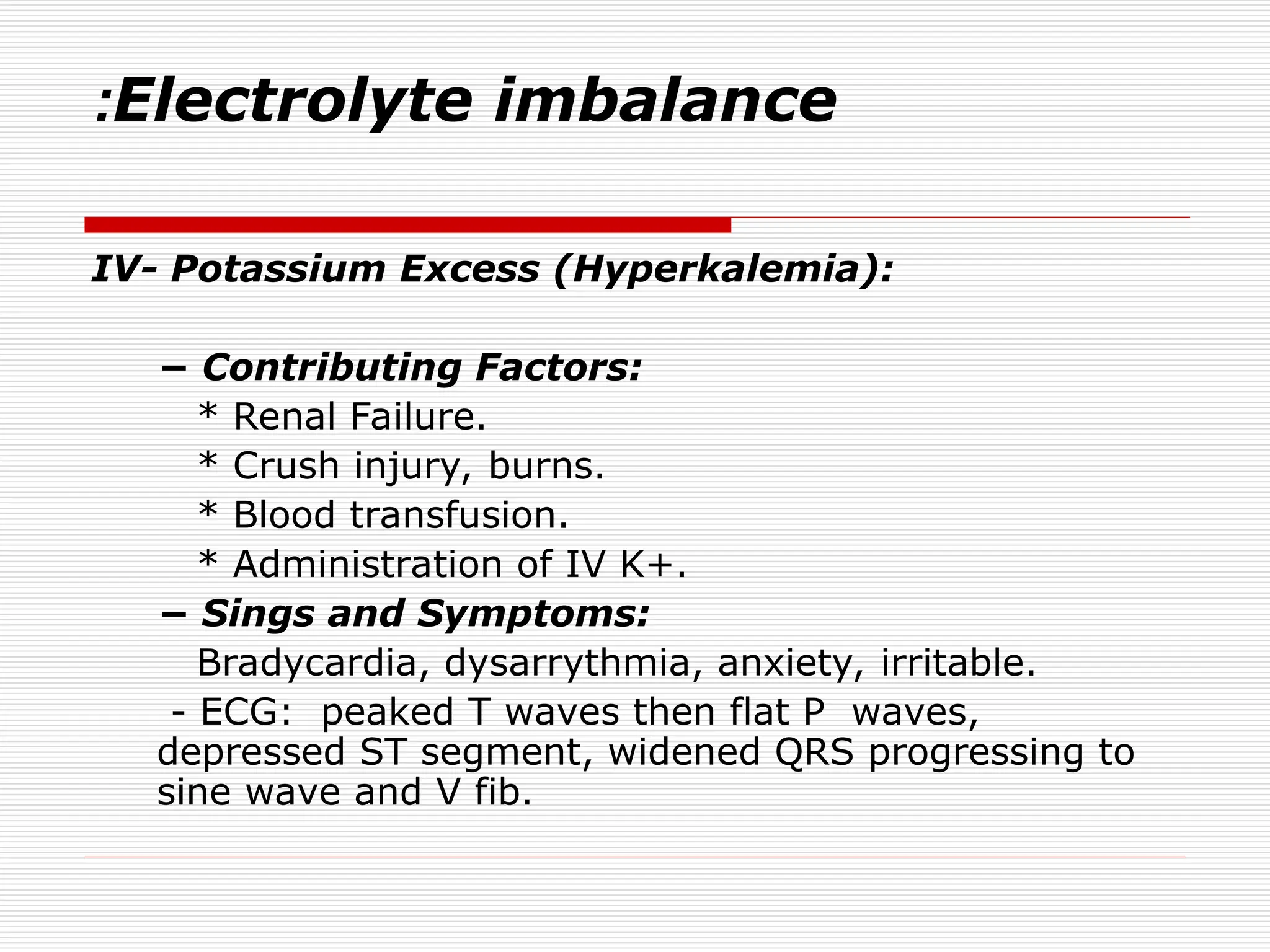
Electrolyte imbalance
:
IV- PotassiumExcess (Hyperkalemia):
− Contributing Factors:
* Renal Failure.
* Crush injury, burns.
* Blood transfusion.
* Administration of IV K+.
− Sings and Symptoms:
Bradycardia, dysarrythmia, anxiety, irritable.
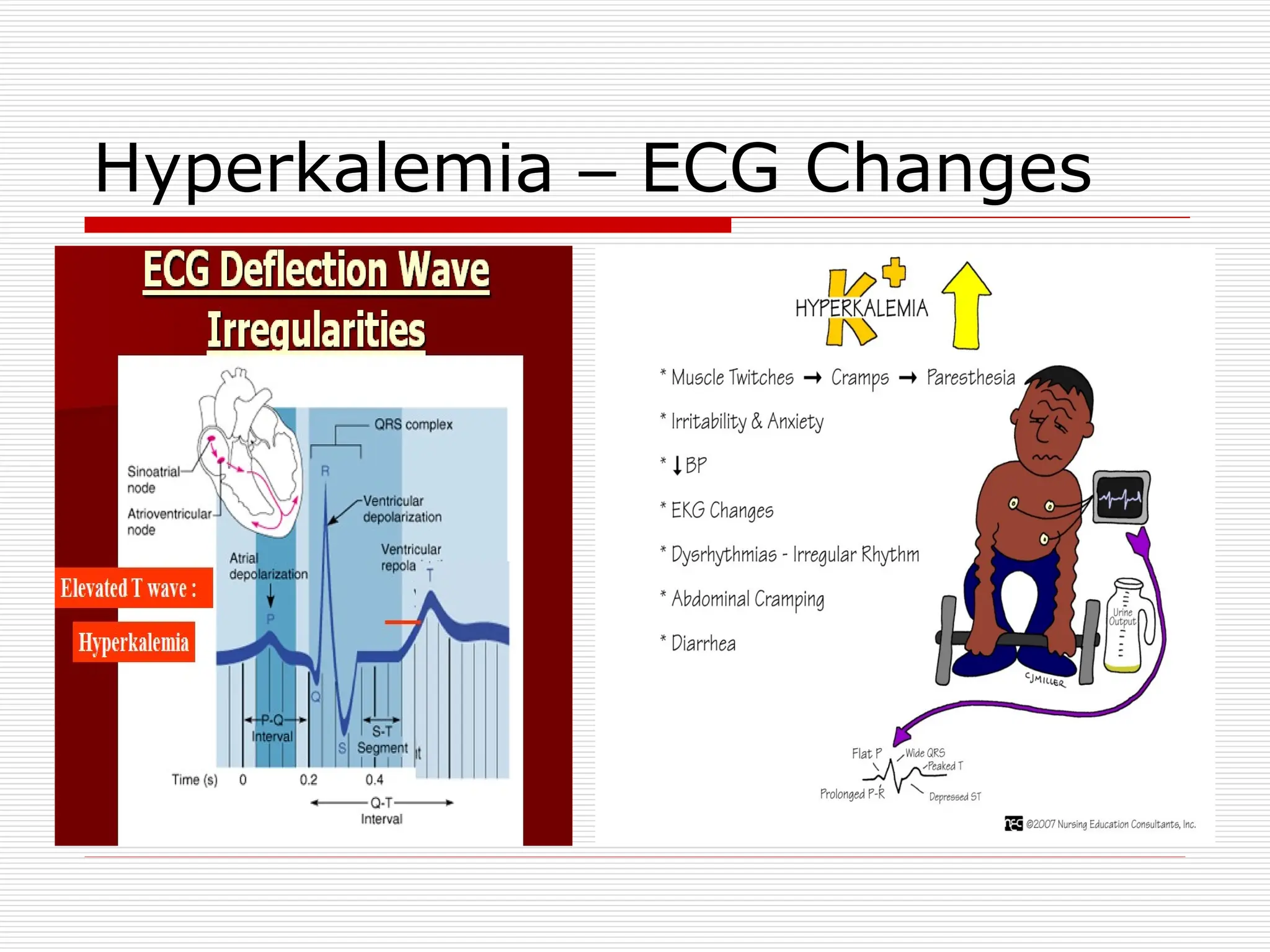
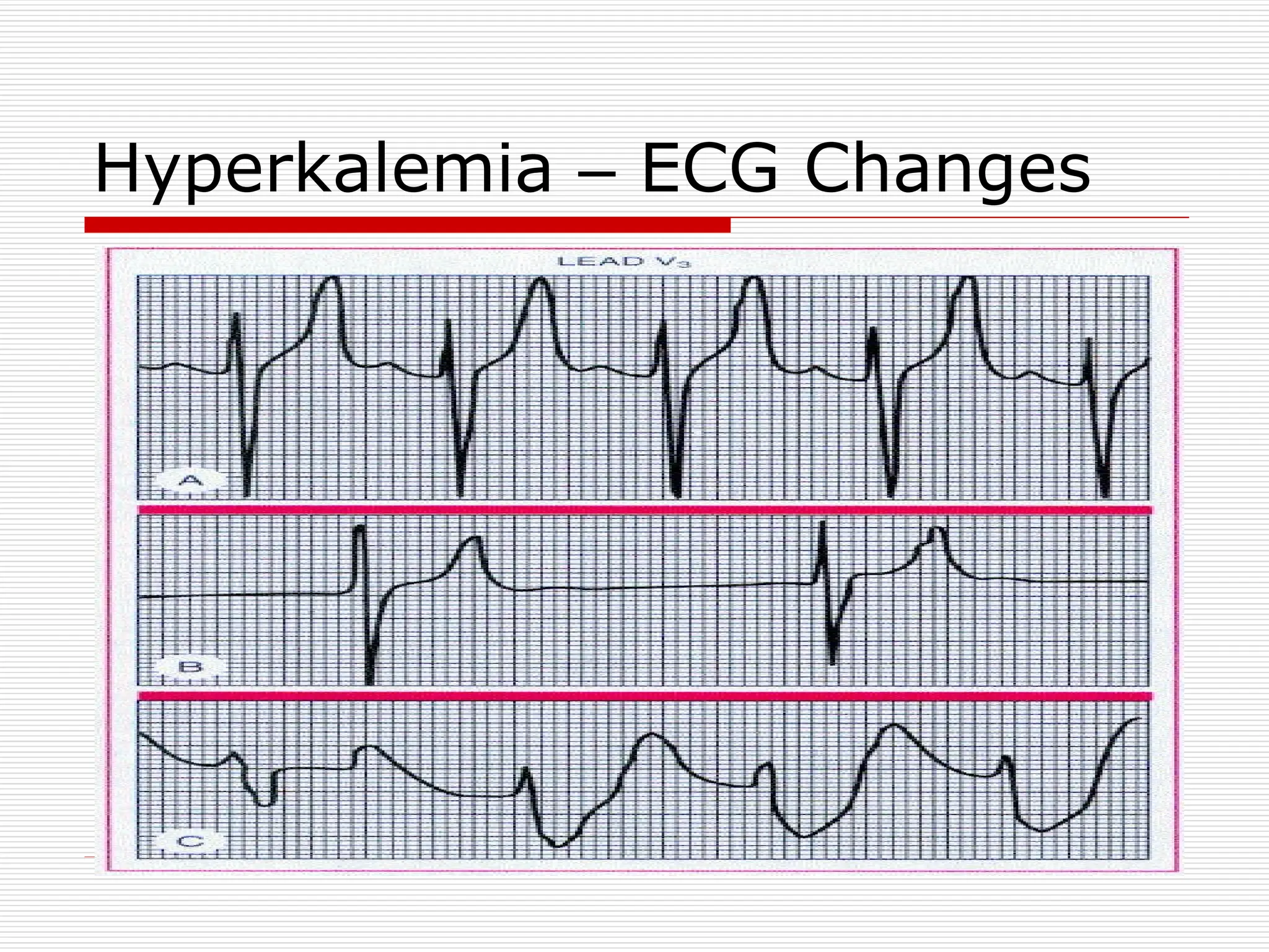
- ECG: peaked T waves then flat P waves,
depressed ST segment, widened QRS progressing to
sine wave and V fib.
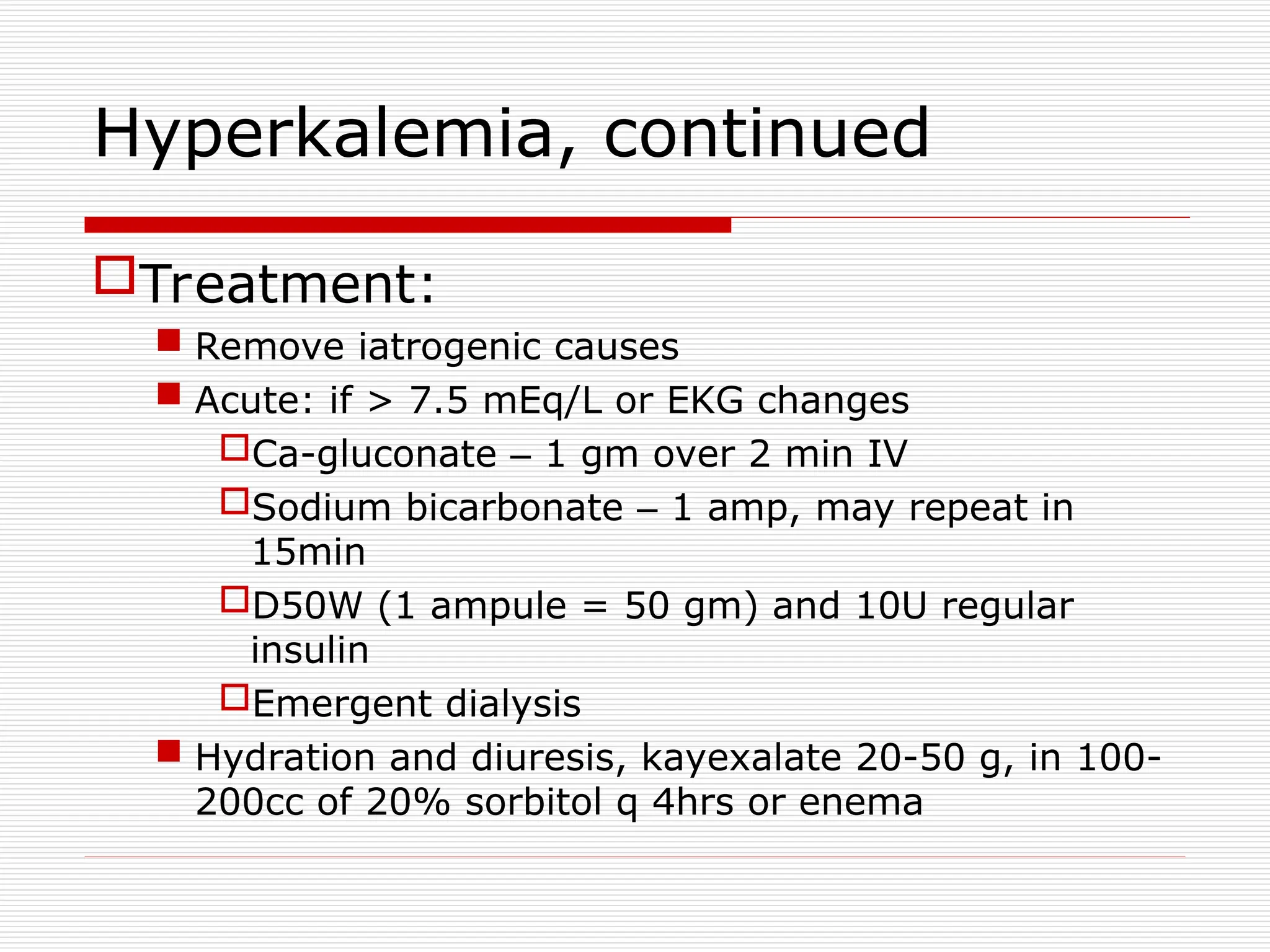
Hyperkalemia, continued
Treatment:
Removeiatrogenic causes
Acute: if > 7.5 mEq/L or EKG changes
Ca-gluconate – 1 gm over 2 min IV
Sodium bicarbonate – 1 amp, may repeat in
15min
D50W (1 ampule = 50 gm) and 10U regular
insulin
Emergent dialysis
Hydration and diuresis, kayexalate 20-50 g, in 100-
200cc of 20% sorbitol q 4hrs or enema
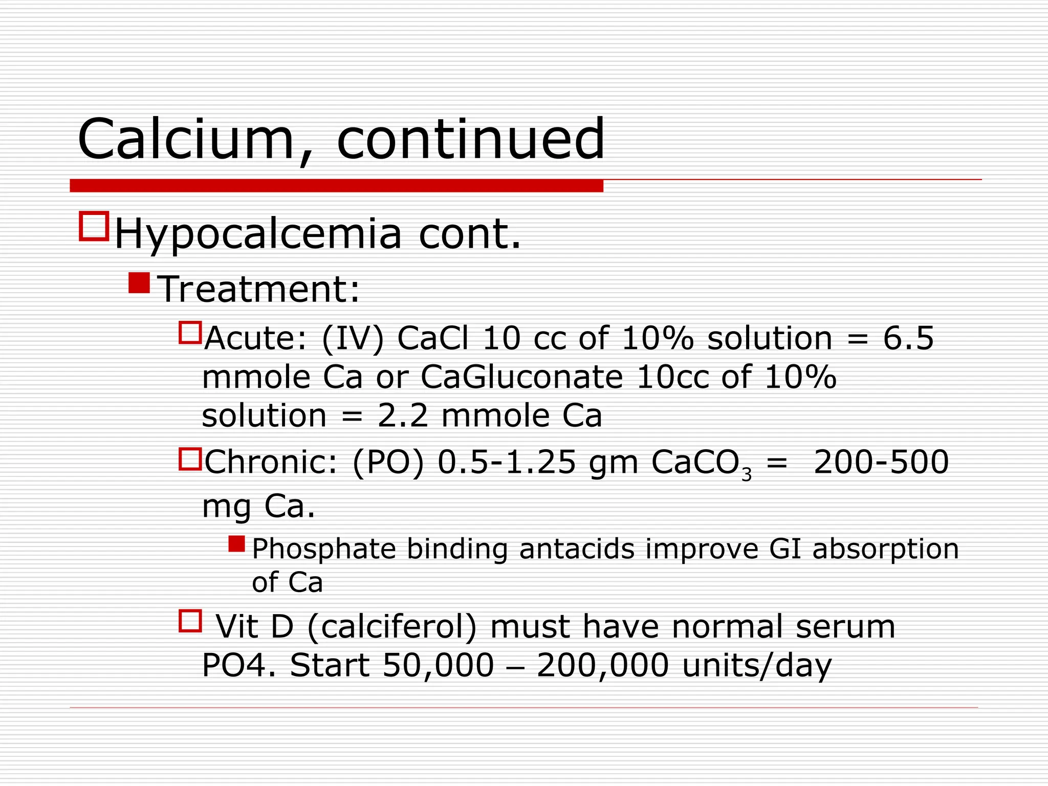
Calcium, continued
Hypocalcemia cont.
Treatment:
Acute:(IV) CaCl 10 cc of 10% solution = 6.5
mmole Ca or CaGluconate 10cc of 10%
solution = 2.2 mmole Ca
Chronic: (PO) 0.5-1.25 gm CaCO3 = 200-500
mg Ca.
Phosphate binding antacids improve GI absorption
of Ca
Vit D (calciferol) must have normal serum
PO4. Start 50,000 – 200,000 units/day
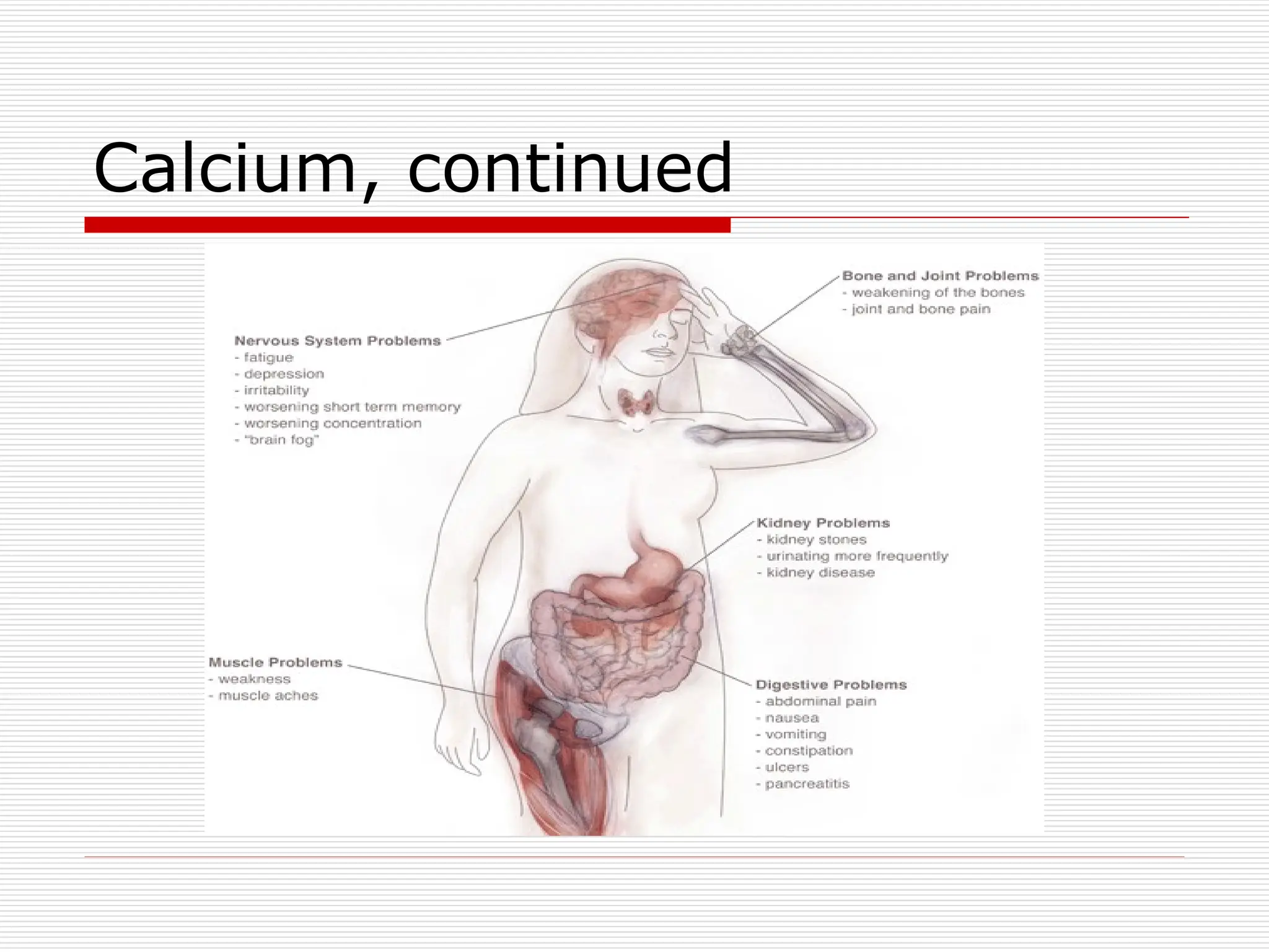
Calcium, continued
Hypercalcemia
Usuallysecondary to hyperparathyroidism or
malignancy. Other causes are thiazides, milk-
alkali syndrome, granulomatous disease, acute
adrenal insufficiency
Acute crisis is serum Ca> 12mg/dL. Critical at 16-
20mg/dL
S/Sx: N/V, anorexia, abdominal pain, confusion,
lethargy MS changes= “Bones, stone, abdominal
groans and psychic overtones.”
33.
Calcium, continued
Treatment: Hydrationwith NS then loop
diuretic. Steroids for lymphoma, multiple
myeloma, adrenal insufficiency, bone
mets, Vit D intoxication. May need
Hemodialysis.
Mithramycin for malignancy induced
hyperCa refractory to other treatment. Give
15-25 mcg/kg IVP
Calcitonin in malignant PTH syndromes
34.
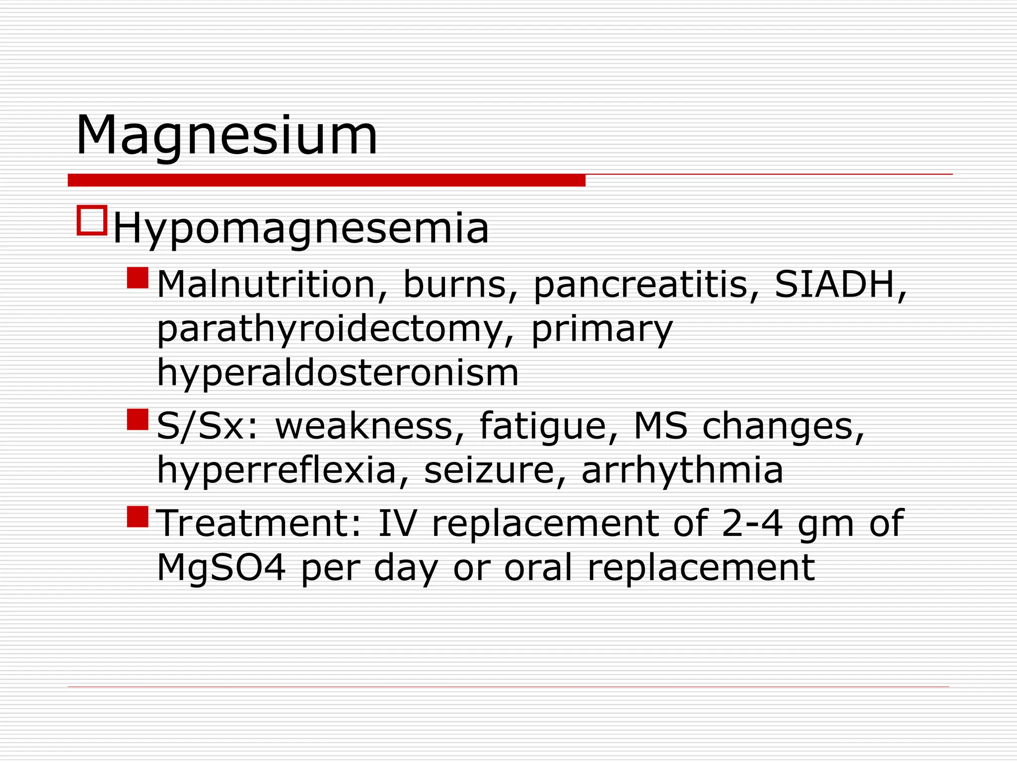
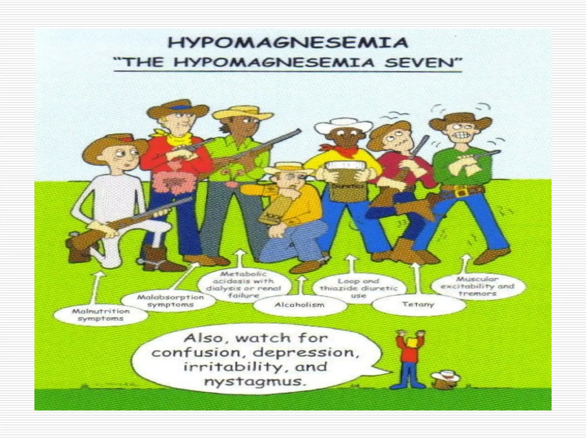
Magnesium
Hypomagnesemia
Malnutrition, burns, pancreatitis,SIADH,
parathyroidectomy, primary
hyperaldosteronism
S/Sx: weakness, fatigue, MS changes,
hyperreflexia, seizure, arrhythmia
Treatment: IV replacement of 2-4 gm of
MgSO4 per day or oral replacement
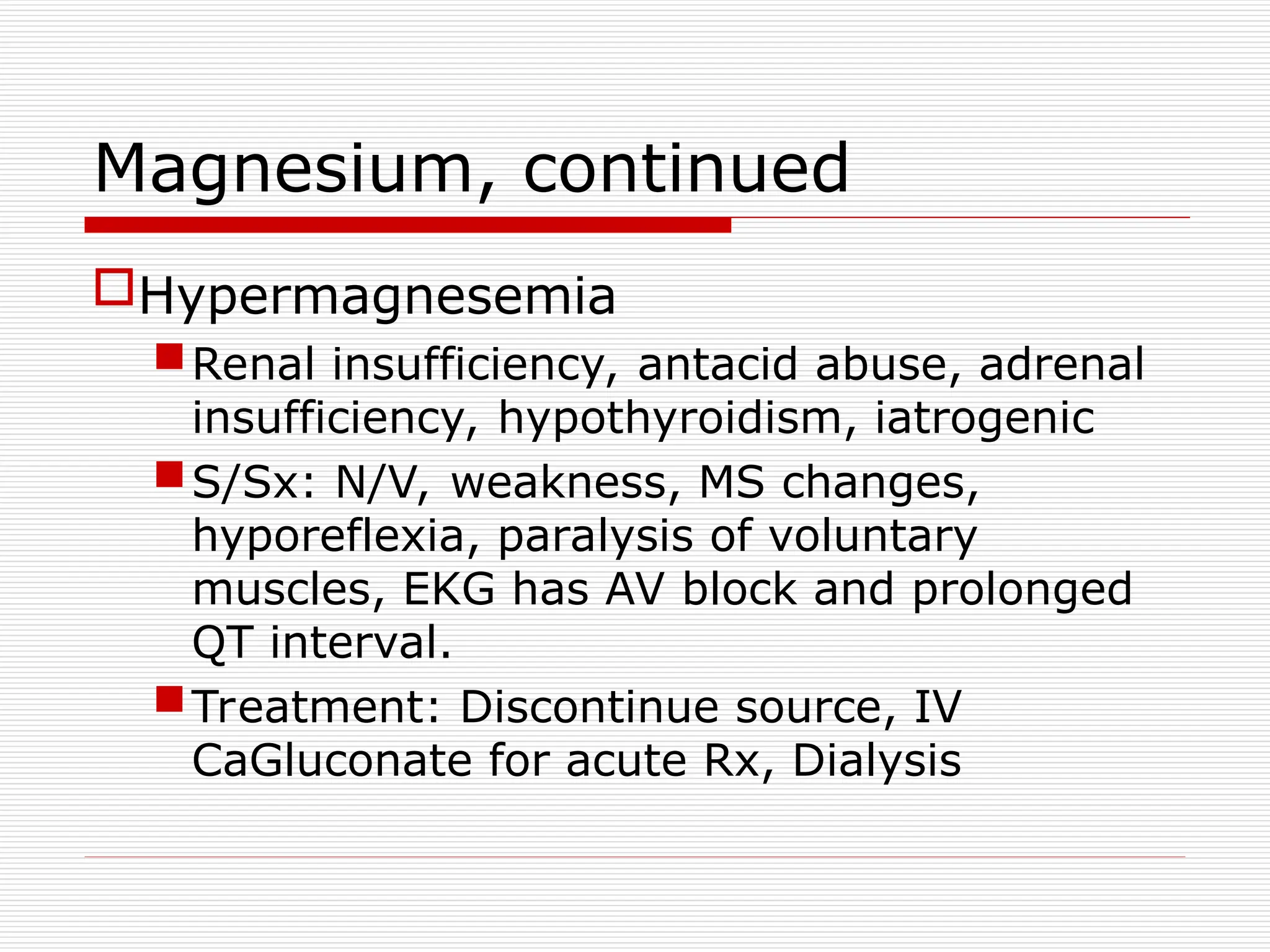
Magnesium, continued
Hypermagnesemia
Renal insufficiency,antacid abuse, adrenal
insufficiency, hypothyroidism, iatrogenic
S/Sx: N/V, weakness, MS changes,
hyporeflexia, paralysis of voluntary
muscles, EKG has AV block and prolonged
QT interval.
Treatment: Discontinue source, IV
CaGluconate for acute Rx, Dialysis
37.
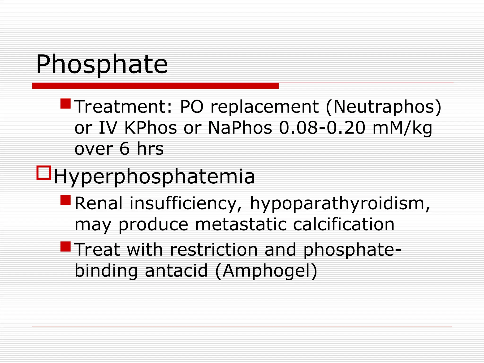
Phosphate
Treatment: PO replacement(Neutraphos)
or IV KPhos or NaPhos 0.08-0.20 mM/kg
over 6 hrs
Hyperphosphatemia
Renal insufficiency, hypoparathyroidism,
may produce metastatic calcification
Treat with restriction and phosphate-
binding antacid (Amphogel)
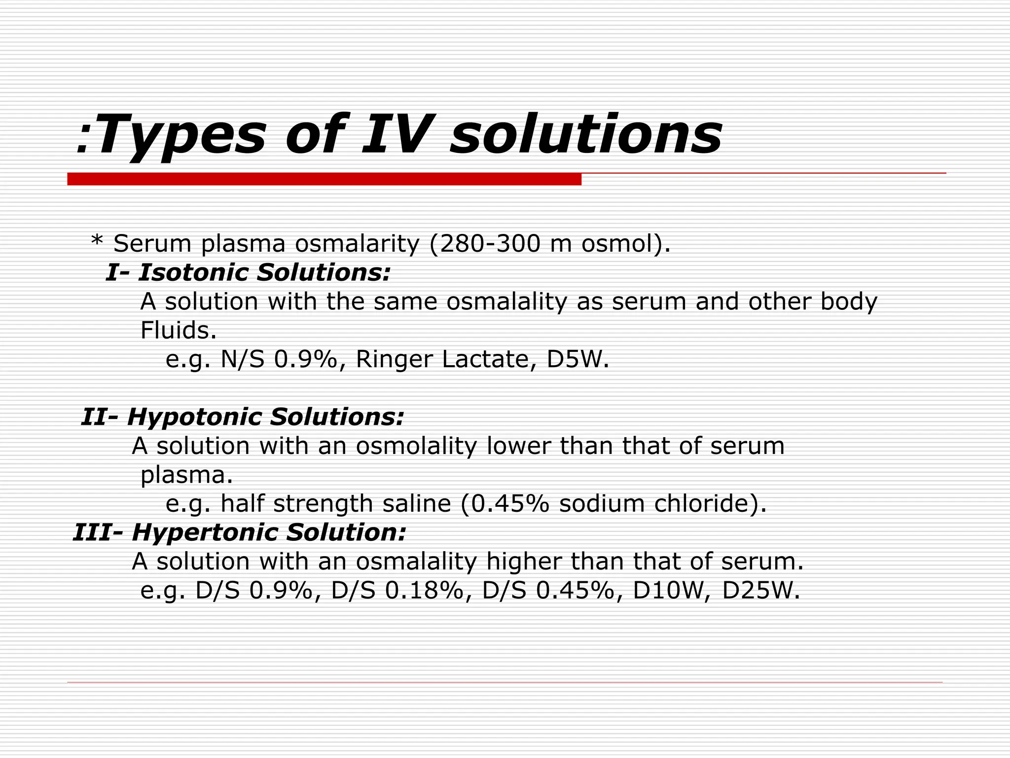
Types of IVsolutions
:
* Serum plasma osmalarity (280-300 m osmol).
I- Isotonic Solutions:
A solution with the same osmalality as serum and other body
Fluids.
e.g. N/S 0.9%, Ringer Lactate, D5W.
II- Hypotonic Solutions:
A solution with an osmolality lower than that of serum
plasma.
e.g. half strength saline (0.45% sodium chloride).
III- Hypertonic Solution:
A solution with an osmalality higher than that of serum.
e.g. D/S 0.9%, D/S 0.18%, D/S 0.45%, D10W, D25W.
40.
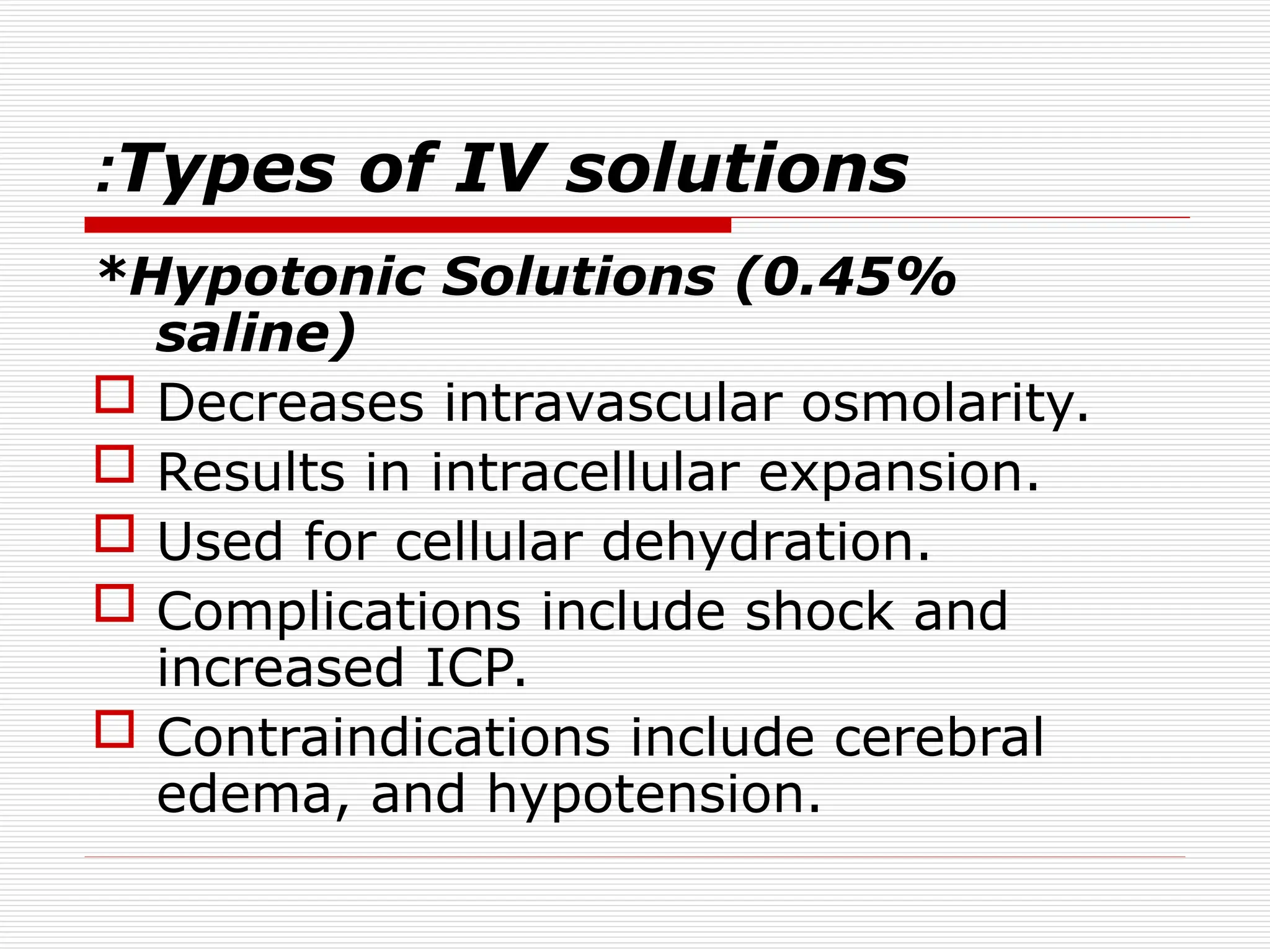
Types of IVsolutions
:
*Hypotonic Solutions (0.45%
saline)
Decreases intravascular osmolarity.
Results in intracellular expansion.
Used for cellular dehydration.
Complications include shock and
increased ICP.
Contraindications include cerebral
edema, and hypotension.
41.
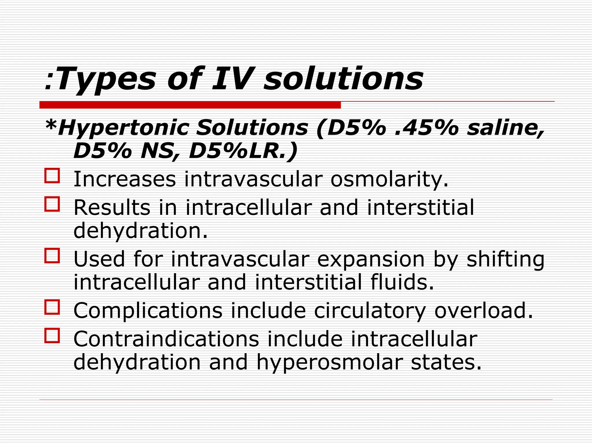
Types of IVsolutions
:
*Hypertonic Solutions (D5% .45% saline,
D5% NS, D5%LR.)
Increases intravascular osmolarity.
Results in intracellular and interstitial
dehydration.
Used for intravascular expansion by shifting
intracellular and interstitial fluids.
Complications include circulatory overload.
Contraindications include intracellular
dehydration and hyperosmolar states.
42.
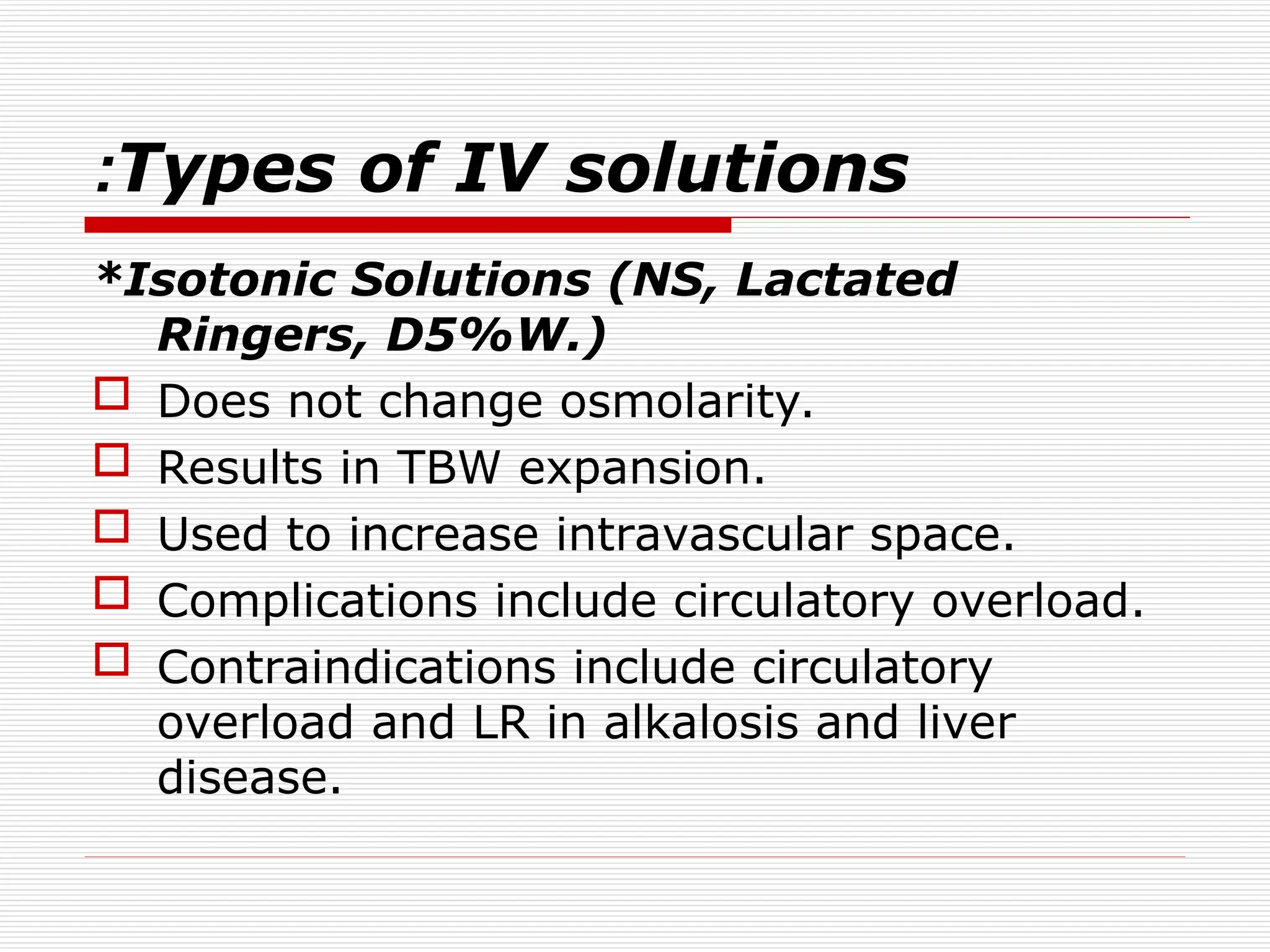
Types of IVsolutions
:
*Isotonic Solutions (NS, Lactated
Ringers, D5%W.)
Does not change osmolarity.
Results in TBW expansion.
Used to increase intravascular space.
Complications include circulatory overload.
Contraindications include circulatory
overload and LR in alkalosis and liver
disease.


![WILLIAM__FLUID_AND_ELECTROLYTE[1].pptx](https://cdn.slidesharecdn.com/ss_thumbnails/williamfluidandelectrolyte1-230310182617-481b32fd-thumbnail.jpg?width=640&height=640&fit=bounds)


