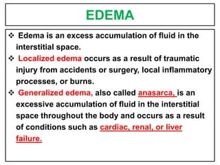
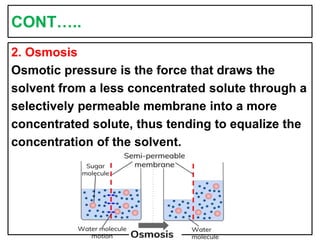
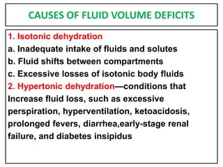
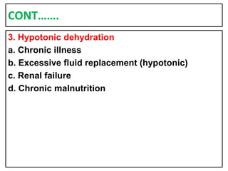
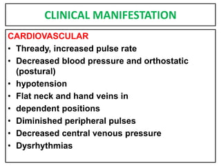
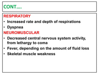
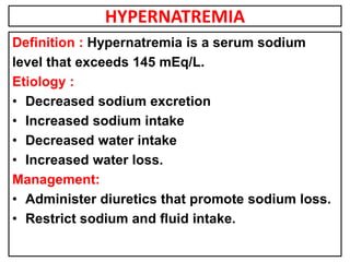
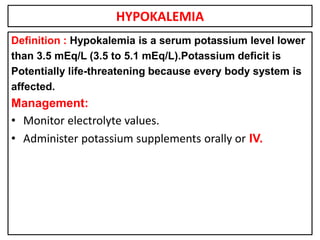
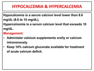
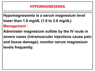
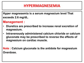
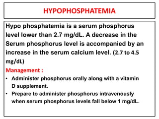
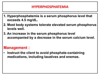
FLUID AND ELECTROLYTE
BALANCE
Prepared By; Mr.Mihir.M.Patel,
M.Sc.Nursing,
Critical Care Nursing.
ELECTROLYTES
A substance that is dissolved in solution and
Some of its molecules split or dissociate into
Electrically charged atoms or ions.
MEASUREMENT
• The metric system is used to measure volumes
of fluids—liters (L) or milliliters (mL).
• The unit of measure that expresses the
combining activity of an electrolyte is the
milliequivalent (mEq).
CONT…….
• The atom is composed of particles known as
the proton(positive charge), neutron (neutral),
and electron (negative charge).
Cation:
• A cation is an ion that carries a positive charge
and has given away or lost electrons.
Anion:
• An anion is an ion that has gained electrons
and therefore carries a negative charge.
BODY FLUID COMPARTMENT
• Intravascular compartment: Refers to fluid
inside a blood vessel
• Intracellular compartment: The intracellular
compartment refers to all fluid inside the cell.
• The extracellular compartment : Refers to fluid
outside the cell.e.g. The extracellular
compartment includes the interstitial fluid,
which is fluid between cells (sometimes called
the third space), blood, lymph, bone,
connective tissue, water, and transcellular fluid.
EDEMA
 Edema is an excess accumulation of fluid in the
interstitial space.
 Localized edema occurs as a result of traumatic
injury from accidents or surgery, local inflammatory
processes, or burns.
 Generalized edema, also called anasarca, is an
excessive accumulation of fluid in the interstitial
space throughout the body and occurs as a result
of conditions such as cardiac, renal, or liver
failure.
BODY FLUID
Total body fluid (intracellular and extracellular)
amounts to about 60% of body weight in the
adult, 55% in the older adult, and 80% in the
infant.
BODY FLUID TRANSPORT
1. Diffusion
Diffusion is the process whereby a solute
(substance that is dissolved) may spread through
a solution or solvent (solution in which the solute
is dissolved).
Diffusion of a solute spreads the molecules from
an area of higher concentration to an area of
lower concentration.
CONT…..
2. Osmosis
Osmotic pressure is the force that draws the
solvent from a less concentrated solute through a
selectively permeable membrane into a more
concentrated solute, thus tending to equalize the
concentration of the solvent.
CONT…..
3. Filtration
Filtration is the movement of solutes and solvents
by hydrostatic pressure.
4. Hydrostatic Pressure
Hydrostatic pressure is the force exerted by
the weight of a solution.
• osmotic pressure is measured in milliosmoles (mOsm).
• The normal osmolality of plasma is 270 to 300
milliosmoles/kilogram (mOsm/kg) water.
MOVEMENT OF BODY FLUID
1. Isotonic solutions
Both sides of a selectively permeable membrane
have established equilibrium or are equal in
concentration, they are isotonic.
CONT…..
2. Hypotonic solutions
solution contains a lower concentration of salt or
Solute than another more concentrated solution,
it is considered hypotonic. Less salt or more
water than an isotonic solution.
CONT….
3. Hypertonic solutions
A solution that has a higher concentration of
solutes than another less concentrated solution
is hypertonic.
BODY FLUID INTAKE AND OUT PUT
CONT….
• Water lost through the skin is called insensible
loss (the individual is unaware of losing that
water).
• The adrenal glands, through the secretion of
aldosterone, also aid in controlling extracellular
fluid volume by regulating the amount of
sodium reabsorbed by the kidneys.
CONT….
Antidiuretic hormone from the pituitary gland
regulates the osmotic pressure of extracellular
fluid by regulating the amount of water
Reabsorbed by the kidney.
FLUID VOLUME DEFICIT /HYPOVOLEMIA
DEFINITION :
Dehydration occurs when the fluid intake of the
body is not sufficient to meet the fluid needs of
the body.
TYPES OF FLUID VOLUME DEFICITS
1. Isotonic dehydration
Water and dissolved electrolytes are lost in equal
proportions.
2. Hypertonic dehydration
Water loss exceeds electrolyte loss.
3. Hypotonic dehydration
Electrolyte loss exceeds water loss.
CAUSES OF FLUID VOLUME DEFICITS
1. Isotonic dehydration
a. Inadequate intake of fluids and solutes
b. Fluid shifts between compartments
c. Excessive losses of isotonic body fluids
2. Hypertonic dehydration—conditions that
Increase fluid loss, such as excessive
perspiration, hyperventilation, ketoacidosis,
prolonged fevers, diarrhea,early-stage renal
failure, and diabetes insipidus
CONT…….
3. Hypotonic dehydration
a. Chronic illness
b. Excessive fluid replacement (hypotonic)
c. Renal failure
d. Chronic malnutrition
CLINICAL MANIFESTATION
CARDIOVASCULAR
• Thready, increased pulse rate
• Decreased blood pressure and orthostatic
(postural)
• hypotension
• Flat neck and hand veins in
• dependent positions
• Diminished peripheral pulses
• Decreased central venous pressure
• Dysrhythmias
CONT….
RESPIRATORY
• Increased rate and depth of respirations
• Dyspnea
NEUROMUSCULAR
• Decreased central nervous system activity,
from lethargy to coma
• Fever, depending on the amount of fluid loss
• Skeletal muscle weakness
CONT…..
RENAL
Decreased urine output
INTEGUMENTARY
Dry skin
Poor turgor, tenting
Dry mouth
GASTROINTESTINAL
Decreased motility and diminished bowel sounds
Constipation
Thirst
Decreased body weight
LABORATORY FINDINGS
 Increased serum osmolality
 Increased hematocrit.
 Increased blood urea nitrogen (BUN) level
 Increased serum sodium level
 Increased urinary specific gravity
FLUID VOLUME EXCESS/HYPERVOLEMIA
DEFINITION:
Fluid intake or fluid retention exceeds the fluid
needs of the body. Fluid volume excess is also
called over hydration or fluid overload.
ETIOLOGY
1. Isotonic over hydration
a. Inadequately controlled IV therapy
b. Renal failure
c. Long-term corticosteroid therapy
2. Hypertonic over hydration
a. Excessive sodium ingestion
b. Rapid infusion of hypertonic saline
c. Excessive sodium bicarbonate therapy
Cont…
3. Hypotonic over hydration
a. Early renal failure
b. Congestive heart failure
c. Syndrome of inappropriate anti-diuretic
hormone secretion
d. Inadequately controlled IV therapy
e. Replacement of isotonic fluid loss with
hypotonic fluids
f. Irrigation of wounds and body cavities with
hypotonic fluids
LABORATORY FINDING
 Decreased serum osmolality
 Decreased hematocrit
 Decreased BUN level
 Decreased serum sodium level
 Decreased urine specific gravity
Management
• Administer diuretics; osmotic diuretics typically
are prescribed first to prevent severe electrolyte
imbalances.
• Restrict fluid and sodium intake as prescribed.
• Monitor intake and output; monitor weight.
HYPONATREMIA
Definition : Hyponatremia is a serum sodium level lower
than 135 mEq/L (135 to 145 mEq/L)
Etiology :
1. Increased sodium excretion
2. Inadequate sodium intake
3. Dilution of serum sodium
Management:
1. Sodium chloride infusions are administered to restore
sodium content and fluid volume.
2. Increase oral sodium intake.
HYPERNATREMIA
Definition : Hypernatremia is a serum sodium
level that exceeds 145 mEq/L.
Etiology :
• Decreased sodium excretion
• Increased sodium intake
• Decreased water intake
• Increased water loss.
Management:
• Administer diuretics that promote sodium loss.
• Restrict sodium and fluid intake.
HYPOKALEMIA
Definition : Hypokalemia is a serum potassium level lower
than 3.5 mEq/L (3.5 to 5.1 mEq/L).Potassium deficit is
Potentially life-threatening because every body system is
affected.
Management:
• Monitor electrolyte values.
• Administer potassium supplements orally or IV.
HYPERKALEMIA
• Hyperkalemia is a serum potassium level that
exceeds 5.1 mEq/L.
Management:
• Discontinue Oral & IV potassium.
• Initiate a potassium-restricted diet.
• Prepare the client for dialysis if potassium
levels are critically high.
• Prepare for the IV administration of hypertonic
glucose with regular insulin to move excess
potassium into the cells.
HYPOCALCEMIA & HYPERCALCEMIA
Hypocalcemia is a serum calcium level lower than 8.6
mg/dL (8.6 to 10 mg/dL).
Hypercalcemia is a serum calcium level that exceeds 10
mg/dL.
Management:
• Administer calcium supplements orally or calcium
intravenously.
• Keep 10% calcium gluconate available for treatment
of acute calcium deficit.
HYPOMAGNESEMIA
Hypomagnesemia is a serum magnesium level
lower than 1.6 mg/dL (1.6 to 2.6 mg/dL)
Management :
Administer magnesium sulfate by the IV route in
severe cases (intramuscular injections cause pain
and tissue damage); monitor serum magnesium
levels frequently.
HYPERMAGNESEMIA
Hyper magnesemia is a serum magnesium level That
exceeds 2.6 mg/dL.
Management :
• Diuretics are prescribed to increase renal excretion of
magnesium.
• Intravenously administered calcium chloride or calcium
gluconate may be prescribed to reverse the effects of
magnesium on cardiac muscle.
Note : Calcium gluconate is the antidote for magnesium
Overdose.
HYPOPHOSPHATEMIA
Hypo phosphatemia is a serum phosphorus
level lower than 2.7 mg/dL. A decrease in the
Serum phosphorus level is accompanied by an
increase in the serum calcium level. (2.7 to 4.5
mg/dL)
Management :
• Administer phosphorus orally along with a vitamin
D supplement.
• Prepare to administer phosphorus intravenously
when serum phosphorus levels fall below 1 mg/dL.
HYPERPHOSPHATEMIA
1. Hyperphosphatemia is a serum phosphorus level that
exceeds 4.5 mg/dL.
2. Most body systems tolerate elevated serum phosphorus
levels well.
3. An increase in the serum phosphorus level
accompanied by a decrease in the serum calcium level.
Management :
• Instruct the client to avoid phosphate-containing
medications, including laxatives and enemas.
Thank You

Fluid and electrolyte balance PPT

  • 1.
    FLUID AND ELECTROLYTE BALANCE PreparedBy; Mr.Mihir.M.Patel, M.Sc.Nursing, Critical Care Nursing.
  • 2.
    ELECTROLYTES A substance thatis dissolved in solution and Some of its molecules split or dissociate into Electrically charged atoms or ions. MEASUREMENT • The metric system is used to measure volumes of fluids—liters (L) or milliliters (mL). • The unit of measure that expresses the combining activity of an electrolyte is the milliequivalent (mEq).
  • 3.
    CONT……. • The atomis composed of particles known as the proton(positive charge), neutron (neutral), and electron (negative charge). Cation: • A cation is an ion that carries a positive charge and has given away or lost electrons. Anion: • An anion is an ion that has gained electrons and therefore carries a negative charge.
  • 4.
    BODY FLUID COMPARTMENT •Intravascular compartment: Refers to fluid inside a blood vessel • Intracellular compartment: The intracellular compartment refers to all fluid inside the cell. • The extracellular compartment : Refers to fluid outside the cell.e.g. The extracellular compartment includes the interstitial fluid, which is fluid between cells (sometimes called the third space), blood, lymph, bone, connective tissue, water, and transcellular fluid.
  • 5.
    EDEMA  Edema isan excess accumulation of fluid in the interstitial space.  Localized edema occurs as a result of traumatic injury from accidents or surgery, local inflammatory processes, or burns.  Generalized edema, also called anasarca, is an excessive accumulation of fluid in the interstitial space throughout the body and occurs as a result of conditions such as cardiac, renal, or liver failure.
  • 6.
    BODY FLUID Total bodyfluid (intracellular and extracellular) amounts to about 60% of body weight in the adult, 55% in the older adult, and 80% in the infant.
  • 7.
    BODY FLUID TRANSPORT 1.Diffusion Diffusion is the process whereby a solute (substance that is dissolved) may spread through a solution or solvent (solution in which the solute is dissolved). Diffusion of a solute spreads the molecules from an area of higher concentration to an area of lower concentration.
  • 8.
    CONT….. 2. Osmosis Osmotic pressureis the force that draws the solvent from a less concentrated solute through a selectively permeable membrane into a more concentrated solute, thus tending to equalize the concentration of the solvent.
  • 9.
    CONT….. 3. Filtration Filtration isthe movement of solutes and solvents by hydrostatic pressure. 4. Hydrostatic Pressure Hydrostatic pressure is the force exerted by the weight of a solution. • osmotic pressure is measured in milliosmoles (mOsm). • The normal osmolality of plasma is 270 to 300 milliosmoles/kilogram (mOsm/kg) water.
  • 10.
    MOVEMENT OF BODYFLUID 1. Isotonic solutions Both sides of a selectively permeable membrane have established equilibrium or are equal in concentration, they are isotonic.
  • 11.
    CONT….. 2. Hypotonic solutions solutioncontains a lower concentration of salt or Solute than another more concentrated solution, it is considered hypotonic. Less salt or more water than an isotonic solution.
  • 12.
    CONT…. 3. Hypertonic solutions Asolution that has a higher concentration of solutes than another less concentrated solution is hypertonic.
  • 13.
    BODY FLUID INTAKEAND OUT PUT
  • 14.
    CONT…. • Water lostthrough the skin is called insensible loss (the individual is unaware of losing that water). • The adrenal glands, through the secretion of aldosterone, also aid in controlling extracellular fluid volume by regulating the amount of sodium reabsorbed by the kidneys.
  • 15.
    CONT…. Antidiuretic hormone fromthe pituitary gland regulates the osmotic pressure of extracellular fluid by regulating the amount of water Reabsorbed by the kidney.
  • 16.
    FLUID VOLUME DEFICIT/HYPOVOLEMIA DEFINITION : Dehydration occurs when the fluid intake of the body is not sufficient to meet the fluid needs of the body.
  • 17.
    TYPES OF FLUIDVOLUME DEFICITS 1. Isotonic dehydration Water and dissolved electrolytes are lost in equal proportions. 2. Hypertonic dehydration Water loss exceeds electrolyte loss. 3. Hypotonic dehydration Electrolyte loss exceeds water loss.
  • 18.
    CAUSES OF FLUIDVOLUME DEFICITS 1. Isotonic dehydration a. Inadequate intake of fluids and solutes b. Fluid shifts between compartments c. Excessive losses of isotonic body fluids 2. Hypertonic dehydration—conditions that Increase fluid loss, such as excessive perspiration, hyperventilation, ketoacidosis, prolonged fevers, diarrhea,early-stage renal failure, and diabetes insipidus
  • 19.
    CONT……. 3. Hypotonic dehydration a.Chronic illness b. Excessive fluid replacement (hypotonic) c. Renal failure d. Chronic malnutrition
  • 20.
    CLINICAL MANIFESTATION CARDIOVASCULAR • Thready,increased pulse rate • Decreased blood pressure and orthostatic (postural) • hypotension • Flat neck and hand veins in • dependent positions • Diminished peripheral pulses • Decreased central venous pressure • Dysrhythmias
  • 21.
    CONT…. RESPIRATORY • Increased rateand depth of respirations • Dyspnea NEUROMUSCULAR • Decreased central nervous system activity, from lethargy to coma • Fever, depending on the amount of fluid loss • Skeletal muscle weakness
  • 22.
    CONT….. RENAL Decreased urine output INTEGUMENTARY Dryskin Poor turgor, tenting Dry mouth GASTROINTESTINAL Decreased motility and diminished bowel sounds Constipation Thirst Decreased body weight
  • 23.
    LABORATORY FINDINGS  Increasedserum osmolality  Increased hematocrit.  Increased blood urea nitrogen (BUN) level  Increased serum sodium level  Increased urinary specific gravity
  • 24.
    FLUID VOLUME EXCESS/HYPERVOLEMIA DEFINITION: Fluidintake or fluid retention exceeds the fluid needs of the body. Fluid volume excess is also called over hydration or fluid overload.
  • 25.
    ETIOLOGY 1. Isotonic overhydration a. Inadequately controlled IV therapy b. Renal failure c. Long-term corticosteroid therapy 2. Hypertonic over hydration a. Excessive sodium ingestion b. Rapid infusion of hypertonic saline c. Excessive sodium bicarbonate therapy
  • 26.
    Cont… 3. Hypotonic overhydration a. Early renal failure b. Congestive heart failure c. Syndrome of inappropriate anti-diuretic hormone secretion d. Inadequately controlled IV therapy e. Replacement of isotonic fluid loss with hypotonic fluids f. Irrigation of wounds and body cavities with hypotonic fluids
  • 27.
    LABORATORY FINDING  Decreasedserum osmolality  Decreased hematocrit  Decreased BUN level  Decreased serum sodium level  Decreased urine specific gravity
  • 28.
    Management • Administer diuretics;osmotic diuretics typically are prescribed first to prevent severe electrolyte imbalances. • Restrict fluid and sodium intake as prescribed. • Monitor intake and output; monitor weight.
  • 29.
    HYPONATREMIA Definition : Hyponatremiais a serum sodium level lower than 135 mEq/L (135 to 145 mEq/L) Etiology : 1. Increased sodium excretion 2. Inadequate sodium intake 3. Dilution of serum sodium Management: 1. Sodium chloride infusions are administered to restore sodium content and fluid volume. 2. Increase oral sodium intake.
  • 30.
    HYPERNATREMIA Definition : Hypernatremiais a serum sodium level that exceeds 145 mEq/L. Etiology : • Decreased sodium excretion • Increased sodium intake • Decreased water intake • Increased water loss. Management: • Administer diuretics that promote sodium loss. • Restrict sodium and fluid intake.
  • 31.
    HYPOKALEMIA Definition : Hypokalemiais a serum potassium level lower than 3.5 mEq/L (3.5 to 5.1 mEq/L).Potassium deficit is Potentially life-threatening because every body system is affected. Management: • Monitor electrolyte values. • Administer potassium supplements orally or IV.
  • 32.
    HYPERKALEMIA • Hyperkalemia isa serum potassium level that exceeds 5.1 mEq/L. Management: • Discontinue Oral & IV potassium. • Initiate a potassium-restricted diet. • Prepare the client for dialysis if potassium levels are critically high. • Prepare for the IV administration of hypertonic glucose with regular insulin to move excess potassium into the cells.
  • 33.
    HYPOCALCEMIA & HYPERCALCEMIA Hypocalcemiais a serum calcium level lower than 8.6 mg/dL (8.6 to 10 mg/dL). Hypercalcemia is a serum calcium level that exceeds 10 mg/dL. Management: • Administer calcium supplements orally or calcium intravenously. • Keep 10% calcium gluconate available for treatment of acute calcium deficit.
  • 34.
    HYPOMAGNESEMIA Hypomagnesemia is aserum magnesium level lower than 1.6 mg/dL (1.6 to 2.6 mg/dL) Management : Administer magnesium sulfate by the IV route in severe cases (intramuscular injections cause pain and tissue damage); monitor serum magnesium levels frequently.
  • 35.
    HYPERMAGNESEMIA Hyper magnesemia isa serum magnesium level That exceeds 2.6 mg/dL. Management : • Diuretics are prescribed to increase renal excretion of magnesium. • Intravenously administered calcium chloride or calcium gluconate may be prescribed to reverse the effects of magnesium on cardiac muscle. Note : Calcium gluconate is the antidote for magnesium Overdose.
  • 36.
    HYPOPHOSPHATEMIA Hypo phosphatemia isa serum phosphorus level lower than 2.7 mg/dL. A decrease in the Serum phosphorus level is accompanied by an increase in the serum calcium level. (2.7 to 4.5 mg/dL) Management : • Administer phosphorus orally along with a vitamin D supplement. • Prepare to administer phosphorus intravenously when serum phosphorus levels fall below 1 mg/dL.
  • 37.
    HYPERPHOSPHATEMIA 1. Hyperphosphatemia isa serum phosphorus level that exceeds 4.5 mg/dL. 2. Most body systems tolerate elevated serum phosphorus levels well. 3. An increase in the serum phosphorus level accompanied by a decrease in the serum calcium level. Management : • Instruct the client to avoid phosphate-containing medications, including laxatives and enemas.
  • 38.