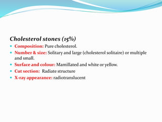
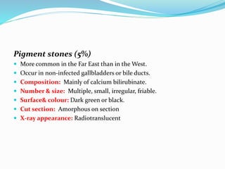
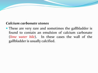
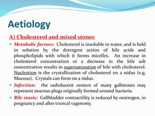
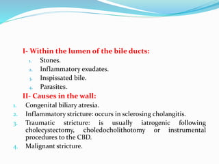
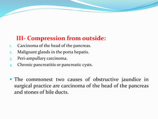
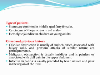
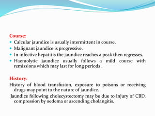
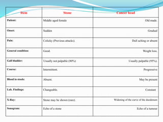
2. The term chronic cholecystitis is applied clinically to all chronic
affections of the gall bladder which include calculi, chronic
inflammation, metabolic disorders (cholesterosis) and neurogenic
dysfunction ( dyskinesia).
Chronic calcular cholecystitis (gall stones) is the most common (95%).
The other forms of chronic cholecystitis includes:
1. Chronic Non-Calcular Cholecystitis:
2. Cholesterolosis of the gall bladder: Strawberry Bladder
3. Biliary dyskinesia: surgery aggrevates symptoms
3. CHRONIC CALCULAR CHOLECYSTITIS
GALL STONES
Gallstones are the commonest biliary pathology.
In 95% of cases of chronic cholecystitis, stones are found in
the gall bladder .
Gall stones are more common in western countries. (may be
related to diet) .
It is more common in female 3 : 1 .
The main varieties of gall stones recognized are:
1. Mixed stones (80%)
2. Cholesterol stones (15%)
3. Pigment stones (5%)
4. Calcium carbonate stones
4. Mixed stones (80%)
Occur in infected gallbladders
Composition: Concentric laminae of cholesterol (the major component),
calcium carbonate, calcium phosphate, calcium bilirubinate, calcium
palmitate and proteins around an organic nucleus.
Number & size: Always multiple and often occurs in groups of similarly
sized stones, each group representing an acute attack of cholecystitis.
Surface and colour: Faceted and brown.
Cut surface: Laminated with alternate dark and light zones of pigment
and cholesterol.
X-ray appearance: Typical “signet-ring” shadow on X-ray.
5. Cholesterol stones (15%)
Composition: Pure cholesterol.
Number & size: Solitary and large (cholesterol solitaire) or multiple
and small.
Surface and colour: Mamillated and white or yellow.
Cut section: Radiate structure
X-ray appearance: radiotranslucent
6. Pigment stones (5%)
More common in the Far East than in the West.
Occur in non-infected gallbladders or bile ducts.
Composition: Mainly of calcium bilirubinate.
Number & size: Multiple, small, irregular, friable.
Surface& colour: Dark green or black.
Cut section: Amorphous on section
X-ray appearance: Radiotranslucent
7. Calcium carbonate stones
These are very rare and sometimes the gallbladder is
found to contain an emulsion of calcium carbonate
(lime water bile). In these cases the wall of the
gallbladder is usually calcified.
8. Aetiology
A) Cholesterol and mixed stones
Metabolic factors: Cholesterol is insoluble in water, and is held
in solution by the detergent action of bile acids and
phospholipids with which it forms micelles. An increase in
cholesterol concentration or a decrease in the bile salt
concentration results in supersaturation of bile with cholesterol.
Nucleation is the crystallization of cholesterol on a nidus (e.g.
Mucous). Crystals can form on a nidus.
Infection: the radiolucent centers of many gallstones may
represent mucous plugs originally formed around bacteria.
Bile stasis: Gallbladder contractility is reduced by oestrogen, in
pregnancy and after truncal vagotomy.
9. B) Pigment stones
Haemolysis due to increased bilirubin production.
Strictures (found in the ducts) due to stasis.
Infestations of the biliary tree by Ascaris Lumbricoides (most
common in oriental countries). E. Coli is often found in the bile of
these patients and produces the enzyme glucuronidase, which
converts the bilirubin into its unconjugated insoluble form, which
precipitates as the calcium salt.
10. Complications of gall stones:
1. Migration: Obstruction of the cystic duct or common
bile duct.
2. Inflammation: (cholecystitis, cholangitis or
pancreatitis).
3. Ulceration and fistula formation with duodenum →
gall stone ileus.
4. Malignant change in long-standing cases.
11. Clinical Features of Chronic Cholecystitis:
It is common in middle-aged females who are usually
fatty, fertile, filthy, forty female. (5 F)
Symptoms:
Sometime, Asymptomatic, and gall stones discovered accidentally
by US, However, if there is symptoms, they are aggravated by
fatty foods, these symptoms includes:
Flatulent dyspepsia: This is commonly associated with heart
burn and water brushing.
Pain: This may be fixed to the right hypochondrium or may be
referred to the right shoulder or to the back.
In calcular cases, biliary colic may occur if a stone migrates to the
bile duct or obstructs the cystic duct.
12. Jaundice: Uncomplicated stones of the gall bladder are
rarely the cause of jaundice. Jaundice is liable to occur in:
1. If infection develops and causes cholangitis.
2. If a stone migrates and obstructs the common bile duct.
3. Rarely a big stone in Hartmann's pouch 'presses on the
common bile duct from the outside and causes
obstruction, This may be helped by associated oedema.
Toxic features: A patient with long standing gall bladder
infection may complain of myositis, fibrositis or
myocarditis.
13. Signs:
Murphy's sign: There is tenderness below the tip of the ninth right
costal cartilage, and if pressure is applied while the patient is in
inspiration, she catches her breath.
Boas' sign: may be positive.
A palpable gall bladder: This may be detected in the right
hypochondrium. It represents a mucocele of the gall bladder. It is
caused by obstruction of the cystic duct usually by a stone, and
distention of the gall bladder with mucus.
14. Differential Diagnosis:
Sometimes chronic cholecystitis gives rise to features
similar to the features of duodenal ulcer or chronic
appendicitis and it is not infrequent to find the three
conditions associated together as an abdominal triad
in one patient (Wilkies triad).
Saint's triad refers to cholecystitis, diverticulitis and
hiatus hernia.
The history is of great importance. The following
diagnostic measure is helpful: ultrasonography
15. Treatment of chronic gall bladder
disease:
A. Medical Treatment: For mild non-calcular cases, the
items are:
1. Regulation of diet with restriction of fats.
2. Antibiotics.
3. Cholagogues as magnesium sulphate to help
drainage of bladder.
4. Choleretics to increase the flow of the bile (bile salts
and olive oil).
5. Antispasmodics to help drainage and relieve pain,
16. B. Surgical treatment: cholecystectomy either open or
laparoscopic
Open cholecystectomy: Steps of the operation:
Incision;
Abdominal exploration:
Identification of the anatomy: (Callot's triangle),
Removal of the gall bladder.
Drainage:
17.
18. Fundus first cholecystectomy: Indications
1. In the presence of marked adhesions; the anatomy of
the structures in the porta hepatis cannot be easily
identified.
2. When the cystic duct is congenitally absent.
3. If cholecystostomy was previously done.
19. Exploration of the common bile duct:
After cholecystectomy, the common bile duct should be opened
and probed in the following conditions:
I. If there is history of recurrent jaundice.
II. If the duct is unduely dilated.
III. If a stone or stricture is felt from the outside.
20. Laparoscopic cholecystectomy (LC):
LC has become the "gold standard" in treatment of
symptomatic gall stone disease. It has the following
advantages over open cholecystectomy:
Less postoperative pain.
Shorter hospital stay.
Earlier return to work.
Better cosmetic result.
21. Complications of cholecystectomy:
A- General:
1. Haemorrhage and shock.
2. Pulmonary complications (bronchopneumonia and lung
collapse).
3. Acute dilatation of the stomach and paralytic ileus.
B- Local:
1) Injury to important structures in the porta hepatis such as the
right branch of the hepatic artery, the common bile duct
(causing biliary fistula or stricture of CBD) and the portal vein.
2) Subphrenic abscess.
3) Post cholecystectomy dyspepsia.
22. The symptoms of chronic cholecystitis may persist
after operation ( post-choecystectomy syndrome ) due
to one of the following factors:
Wrong diagnosis; the dyspepsia was due to another cause.
Wrong time; late after onset of cholangitis or another
complication.
Wrong way; the surgeon has missed a stone in the common
bile duct, caused a stricture or left a big stump of the cystic
duct, which may maintain the infection.
Wrong patient; this is the neurotic patient who always
complains.
23. JAUNDICE
Definition:
It is a state of yellow discoloration of the skin, mucous
membranes and sclera due to retention of bilirubin in
the blood.
The central nervous system, cerebrospinal fluid, milk
and saliva are not discoloured.
Normal bile metabolism:
24.
25. Classification of Jaundice:
Haemolytic jaundice:
There is excessive production of bilirubin due to destruction of large
numbers of RBC's.
It is seen in cases of incompatible blood transfusion, erythroblastosis
foetalis, severe haemolytic anaemia and snake poisoning, but the jaundice
of surgical importance is congenital haemolytic jaundice.
The amount of conjugated bilirubin reaching the intestine is in excess, i.e.
there is an exaggeration of the normal cycle. It follows that urobilinogen is
secreted in excess in stools and in urine, but still there is no billirubin in
urine. Bile salts do not accumulate in the blood.
26. Hepatocellular jaundice:
There is a disturbance of liver cells due to viral hepatitis, liver
cirrhosis or chemical poisons as alcohol, chloroform and carbon
tetrachloride.
There are two elements working together:
Disturbance of the liver cell, which causes inability to
conjugate the normal amount of bilirubin reaching it, i.e.
some will be retained in the blood and cannot be secreted in
urine (attached to protein).
Oedema, which causes partial obstruction of the bile
canaliculi, therefore some of the conjugated bilirubin will be
regurgitated into the blood and can pass through the kidney.
27. In hepatocellular jaundice
bilirubin will appear in urine (but less than in obstructive
jaundice),
stercobilinogen in stools will diminish, but
urobilinogen may increase in urine because the part entering
the enterohepatic cycle will not be completely excreted by the
diseased liver cells and instead it will pass through kidneys.
Bile salts may increase in blood.
28. Obstructive jaundice:
As there is obstruction to the flow of the conjugated (free)
bilirubin formed in the liver, this conjugated bilirubin
regurgitates back to the blood. As it is free of protein it can
pass through the kidney to the urine. As the amount
reaching the intestine is negligible, faecal stercobilinogen
and urinary urobilinogen will be decreased.
29. Obstruction of the CBD due to these causes:
1. Within the lumen of the bile ducts:
1. Causes in the wall:
2. Compression from outside:
30. I- Within the lumen of the bile ducts:
1. Stones.
2. Inflammatory exudates.
3. Inspissated bile.
4. Parasites.
II- Causes in the wall:
1. Congenital biliary atresia.
2. Inflammatory stricture: occurs in sclerosing cholangitis.
3. Traumatic stricture: is usually iatrogenic following
cholecystectomy, choledocholithotomy or instrumental
procedures to the CBD.
4. Malignant stricture.
31. III- Compression from outside:
1. Carcinoma of the head of the pancreas.
2. Malignant glands in the porta hepatis.
3. Peri-ampullary carcinoma.
4. Chronic pancreatitis or pancreatic cysts.
The commonest two causes of obstructive jaundice in
surgical practice are carcinoma of the head of the pancreas
and stones of bile ducts.
32. Type of patient:
Stones are common in middle aged fatty females.
Carcinoma of the pancreas in old males.
Hemolytic jaundice in children or young adults.
Onset and previous history.
Calcular obstruction is usually of sudden onset, associated with
biliary colic, and previous attacks of similar nature are
pathognomonic.
Malignant obstruction is usually insidious and is painless or
associated with dull pain in the upper abdomen.
Infective hepatitis is usually preceded by fever, nausea and pain
in the region of the liver.
33. Course:
Calcular jaundice is usually intermittent in course.
Malignant jaundice is progressive.
In infective hepatitis the jaundice reaches a peak then regresses.
Haemolytic jaundice usually follows a mild course with
remissions which may last for long periods .
History:
History of blood transfusion, exposure to poisons or receiving
drugs may point to the nature of jaundice.
Jaundice following cholecystectomy may be due to injury of CBD,
compression by oedema or ascending cholangitis.
34. Examination:
1. Discolouration:
1. In haemolytic jaundice the skin is mildly discoloured (Lemon Coloured).
2. In hepatocellular jaundice it is moderately discoloured (Orange
Coloured)
3. In obstruction it is deeply coloured (Olive Green).
2. Anaemia is marked in haemolytic jaundice.
3. Itching, haemorrhagic manifestations and bradycardia are features of
obstructive jaundice.
4. The stools are clay coloured in obstructive jaundice and at the peak of
hepatocellular jaundice. They are normal in haemolytic jaundice.
5. The urine is deeply coloured in obstructive jaundice and in infective
hepatitis but in the latter condition it gradually returns to normal. In
haemolytic jaundice it is not coloured due to absence of bilirubin (Acholuric
jaundice).
35. Abdominal examination:
1. The liver is enlarged and tender in infective hepatitis,
enlarged and firm in long standing obstructive jaundice
and slightly enlarged in haemolytic jaundice.
2. The spleen is moderately enlarged in haemolytic
jaundice.
3. Ascites points to malignancy or advanced stage of liver
cirrhosis.
4. The gall bladder is distended in cases of malignant
obstruction (Courvoisier's law). However if the
obstruction is above the cystic duct, it will not be
palpable.
36. In obstruction due to stone from the gall bladder it is
shrunken due to previous fibrosis.
However it may be palpable:
If the obstruction is caused by a cholesterol stone and the
gallbladder is normal.
If a stone obstructs the bile duct causing jaundice and
another stone obstructs the cystic duct causing a mucocele
of the gall bladder.
If a big stone in Hartmann's pouch obstructs both the
cystic duct and the common bile duct (by pressure from the
outside).
If the stone is primarily formed in the bile duct.
37. Investigations:
I. Fragility test for Spherocytosis.
II. Blood in the stools favours obstruction by a malignant
tumour.
III. Assessment of amount of bile pigments in urine and
stools.
IV. Liver function tests:
1. Alk. Phosphatase elevates in obstructive jaundice .
2. SGOT&SGPT are elevated in cholangitis .
38. V- Radiography:
1. Plain X-ray may sow the shadow of stones.
2. Cholangiography. Contraindicated in the presence of
jaundice especially if, level serum bilirubin is above 2 mg %,
because the dye is excreted in the same pathway of excretion
of bilirubin. The diseased liver cell will fail to excrete it;
moreover it may be toxic to the liver.
3. Barium-meal examination may help to diagnose carcinoma
of the head of the pancreas.
4. Ultrasonography (investigation of choice), and
computerized tomography are very helpful in difficult
cases and especially for differentiation between extrahepatic
and intrahepatic obstruction.
39. VI- Endoscopic retrograde cholangiopancreaticography
(ERCP) for lesions in the lower part of the common bile
duct (periampullary carcinoma).
VII- MRCP
VIII- Percutaneous transhepatic cholangiography (PTC)
is useful in diagnosis of lesions in the upper part of the
ducts e.g. postoperative stricture or carcinoma of the
hepatic ducts.
IX- Exploration is indicated in undiagnosed cases.
40. I. Calcular Obstructive Jaundice:
Pathology:
Stone (choledocholithiasis): stones may arise
in the gall bladder (in most of cases), or in common
bile duct (rare).
Bile passages:
Dilated above the stone.
Distended with biliary mud or pus in cases of severe
inflammation.
Sometimes in advanced cases the liver fails to secrete
bile and the bile ducts will contain mucus only (white
bile). This indicates a grave prognosis.
41. Gall Bladder:
Contracted in 80% of cases (Courvoisier's law).
Liver:
If the liver is not infected marked dilatation occurs above the
stone (Hydrohepatosis). The liver becomes brown, firm and
granular . In late cases biliary cirrhosis develops.
If there is infection the bile ducts above the stone will be
distended with pus, multiple abscesses will form in the liver
and due to the associated fibrosis dilatation is not marked.
42. Symptoms:
Charcot's triad:
1. Charcot's intermittent fever: Intermittent fever
associated with chills and rigors. Fever may occur
even in the absence of infection
2. Intermittent pain: it may be dull in the
epigastrium or lumbar region or colicky and
referred.
3. Intermittent jaundice: because the stone may be
dislodged from time to time and therefore the
jaundice is not so severe as in cases of cancer head
of pancreas.
43. Signs:
1) Tenderness in the epigastrium or hypochondrium.
2) Gall bladder is usually impalpable (Courvoisier's law).
44. Treatment of stone in the common bile
duct:
Pre-operative treatment:
1. High carbohydrate diet.
2. Vitamin K.
3. Infusions of glucose and saline.
4. Antibiotics.
This treatment is continued so long there is improvement
because it is better to operate during a remission. However,
early intervention is indicated in:
1. Increasing jaundice.
2. Persistent fever
45. A) If the gall bladder was not removed
previously:
ERCP
Operation:
If the common bile duct is abnormally wide: it
should be drained by one of two methods:
a-Sphincterotomy (transduodenal)
b-Choledocho-duodenostomy: Side to side or end to
side.
47. B)Treatment of Calcular obstructive jaundice that
occurs after cholecystectomy:
Flushing through T-tube, with saline containing heparin
or lignocaine. Pressure should not exceed 30 cm H20 to
avoid bilio-venous reflux and bacteraemia.
Antispasmodic drugs may be given to relax the sphincter.
This method may be successful if the stone is not bigger
than7 mm.
Stone dissolution: the most successful fluid injected
through the T-tube is methyl tertiary butyl ether
(MTBE). Results are satisfactory and the stone may
dissolve within few hours.
48. Extraction by choledochoscope. This needs a delay for 4-6
weeks, until the tract of the tube becomes well formed and
can be dilated to allow introduction of choledochoscope to
extract the stone under direct vision. The above three
methods are possible when the T -tube is still present.
Endoscopic extraction (ERCP): after sphincterotomy, the
stone is extracted by a Dormia basket. This can be done
whether a T-tube is found or not.
Surgical exploration: when all the above methods fail.
49. Missed stones:
Stones discovered within 2 years of operation. They cause
recurrent attacks of cholangitis and pain. A stone less than
7 mm may pass spontaneously and therefore, should have
chance of few weeks of conservative treatment before
resorting to other procedures.
Recurrent stones:
Stones discovered after 2 years. Common predisposing
causes are stricture of the duct or presence of non-
absorbable sutures (better avoided in surgery on the bile
ducts). The best treatment is choledochoduodenostomy
50. Item Stone Cancer head
Patient: Middle aged female Old made.
Onset: Sudden Gradual
Pain: Colicky (Previous attacks). Dull aching or absent
General condition: Good. Weight loss.
Gall bladder: Usually not palpable (80%) Usually palpable (95%).
Course: Intermittent. Progressive
Blood in stools: Absent. May be present
Lab. Findings: Changeable. Constant
X-Ray: Stone may be shown (rare). Widening of the curve of the duodenum
Sonogram: Echo of a stone Echo of a tumour