
Downloaded 1,132 times


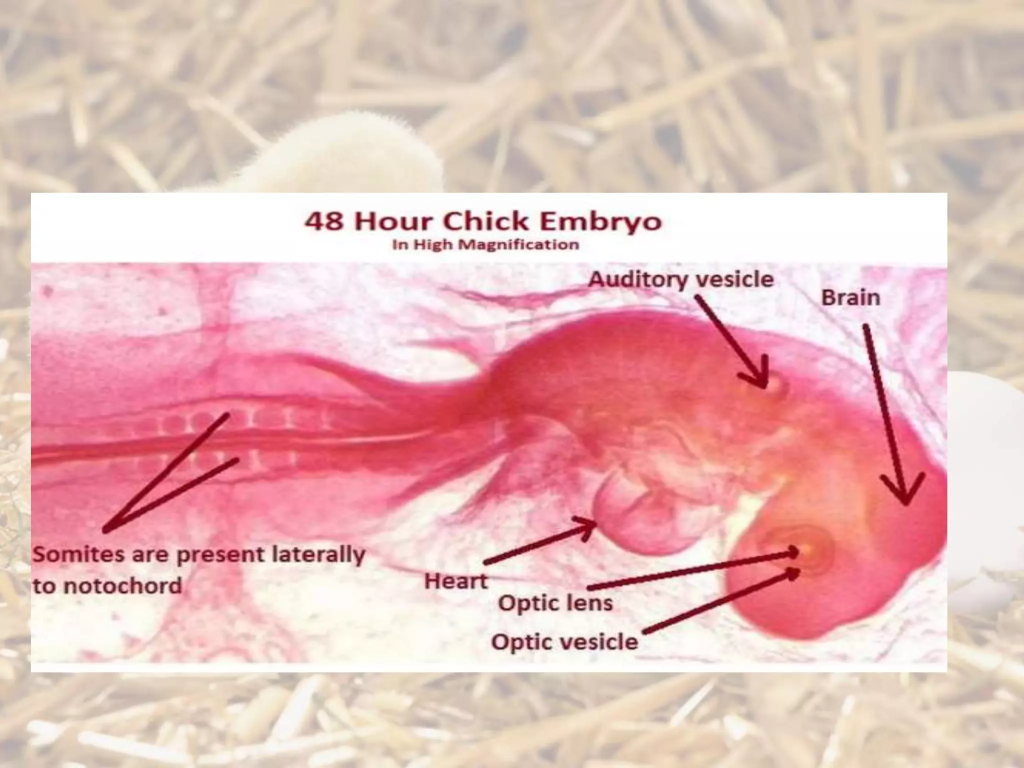


The document summarizes chicken embryonic development from fertilization through organogenesis. It discusses key stages including cleavage, blastulation, gastrulation forming the three germ layers, primitive streak formation, somite formation, neural tube formation, and organogenesis. Diagrams of chicken embryos at various developmental stages from 18 hours to 96 hours are included to illustrate structures as they develop.
Presentation by Arooba Baig and team introducing the topic of chick embryonic development.
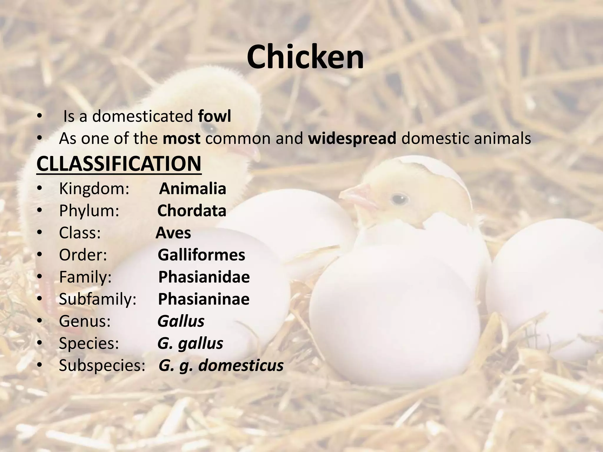
Details on the classification of chickens as domesticated animals in the Animalia kingdom.
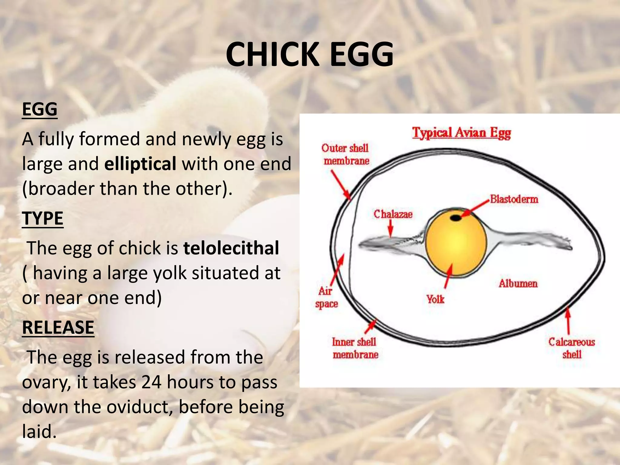
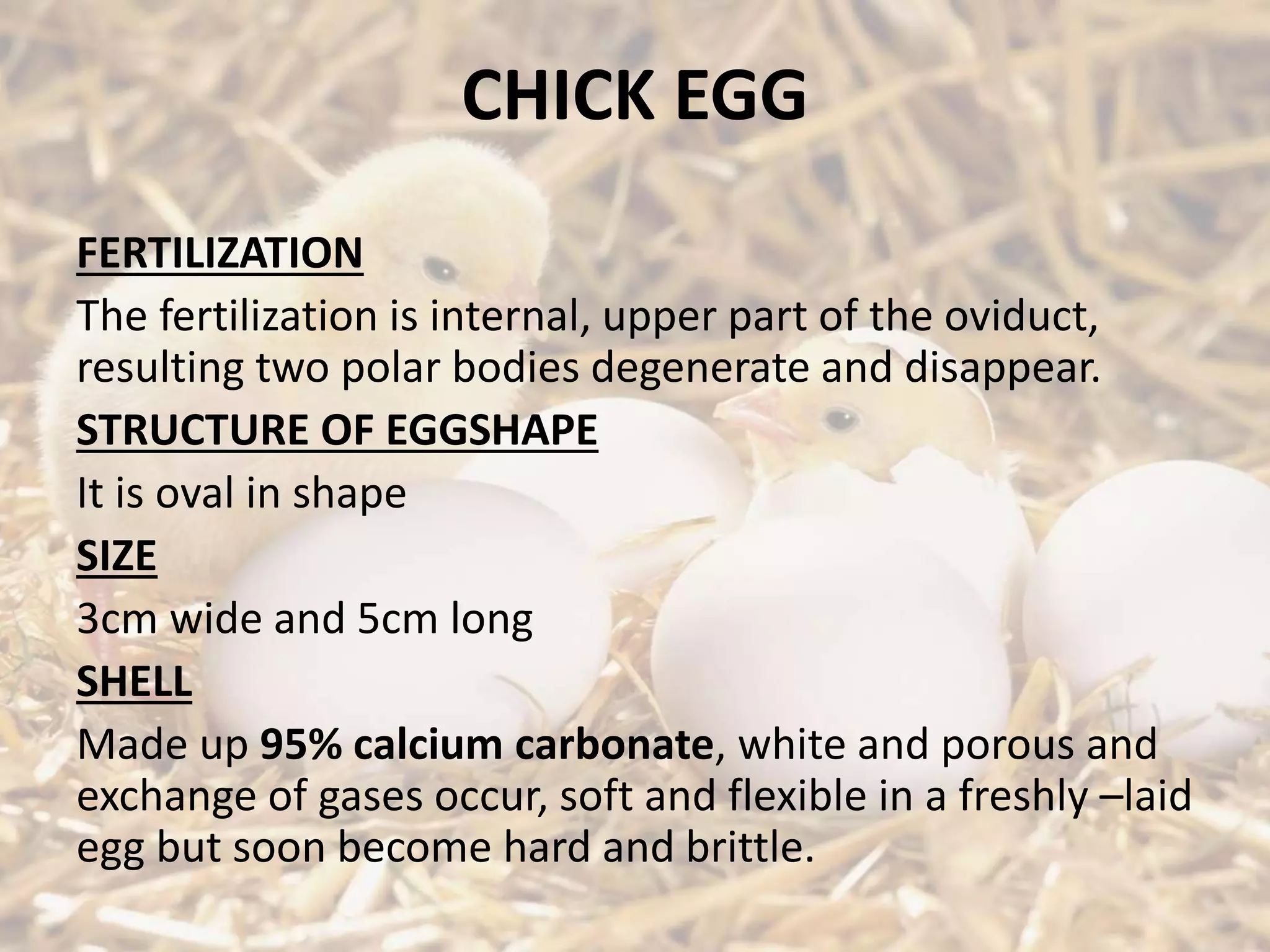
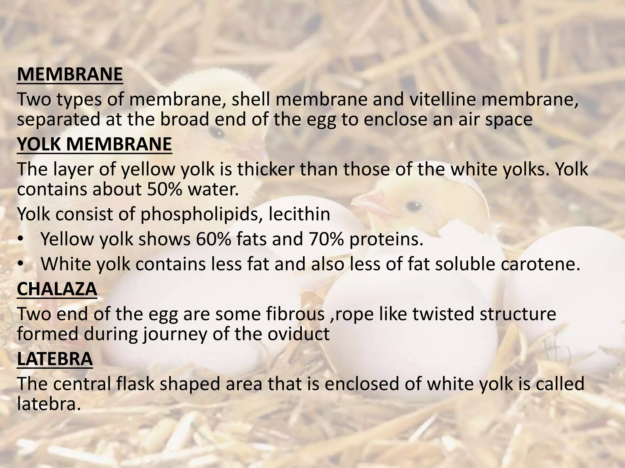
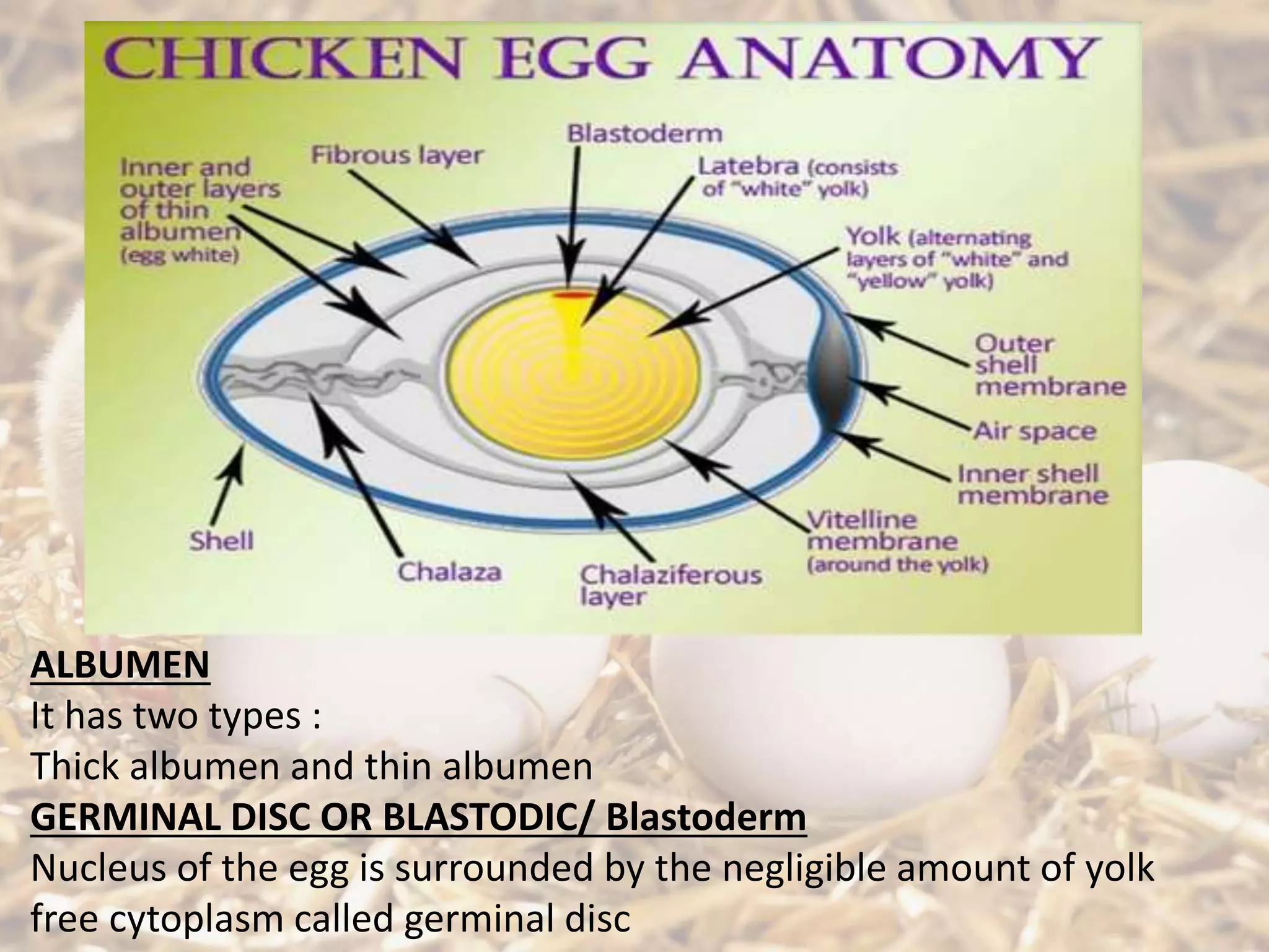
Description of chick egg structure, types of membranes, fertilization process, and yolk composition.
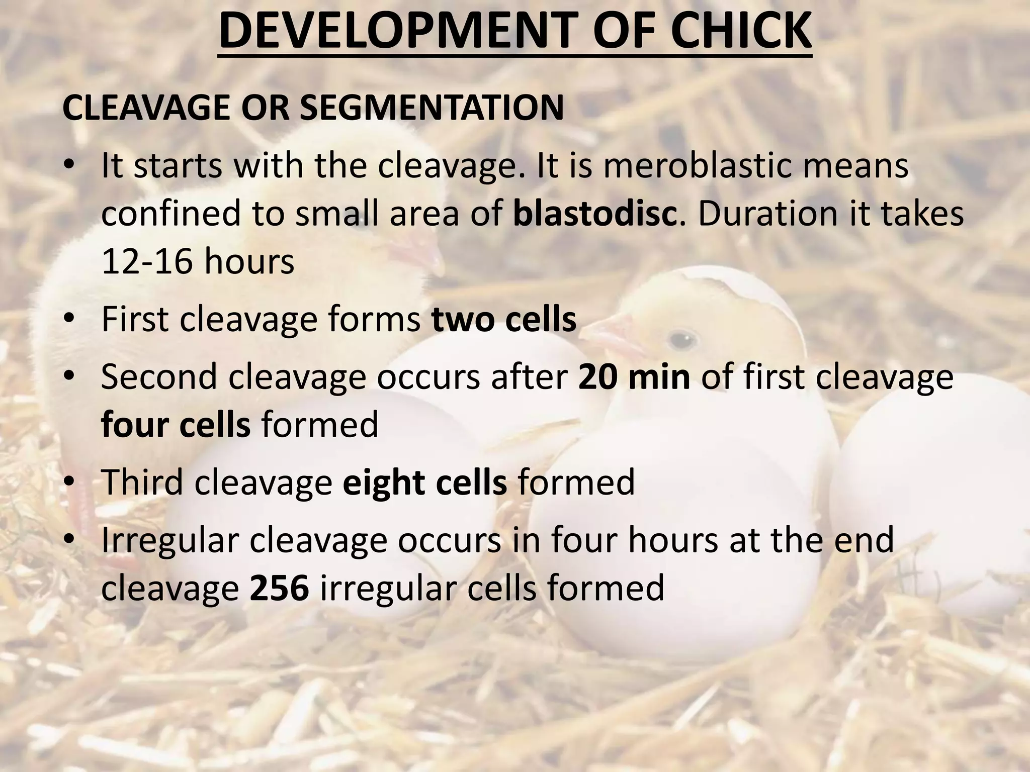
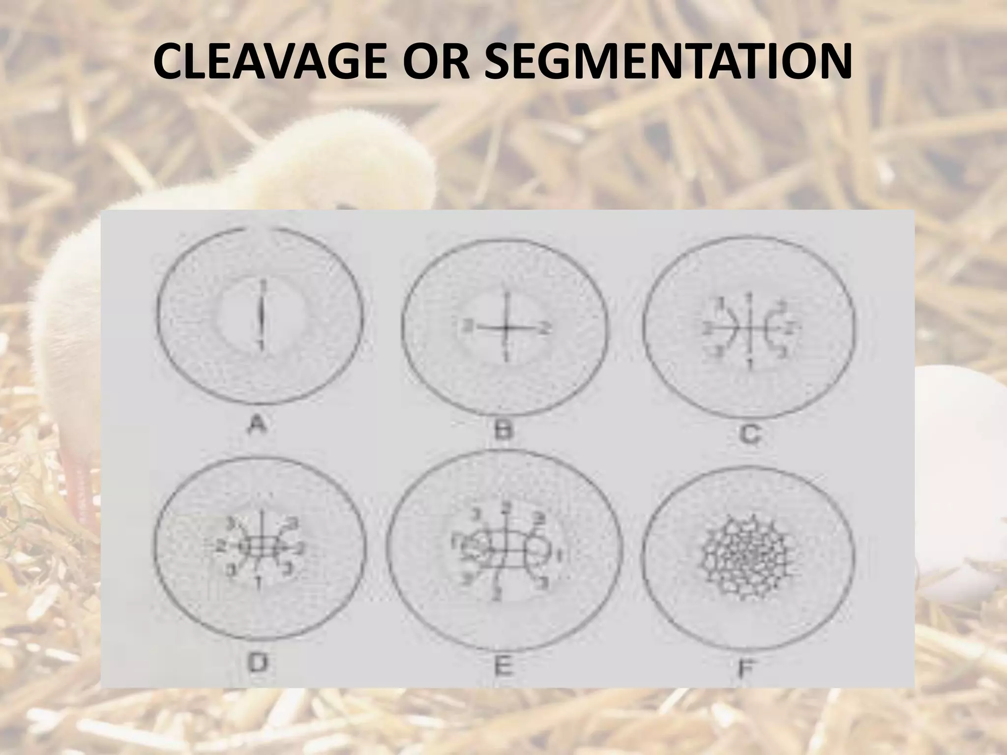
Discussion on the cleavage stages of the chick embryo including the timing and resulting cell formations.
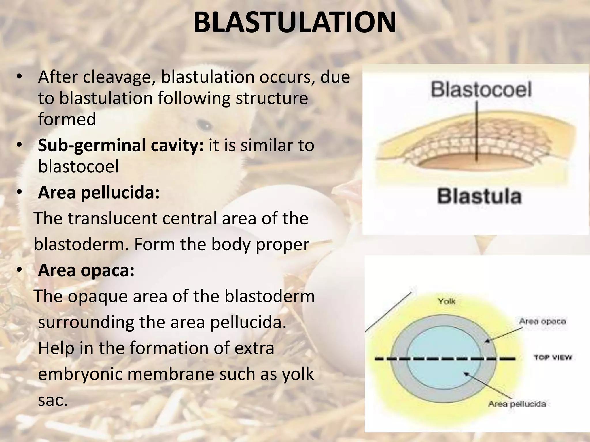
Explains the blastulation stage and formation of structures like sub-germinal cavity and areas of the blastoderm.
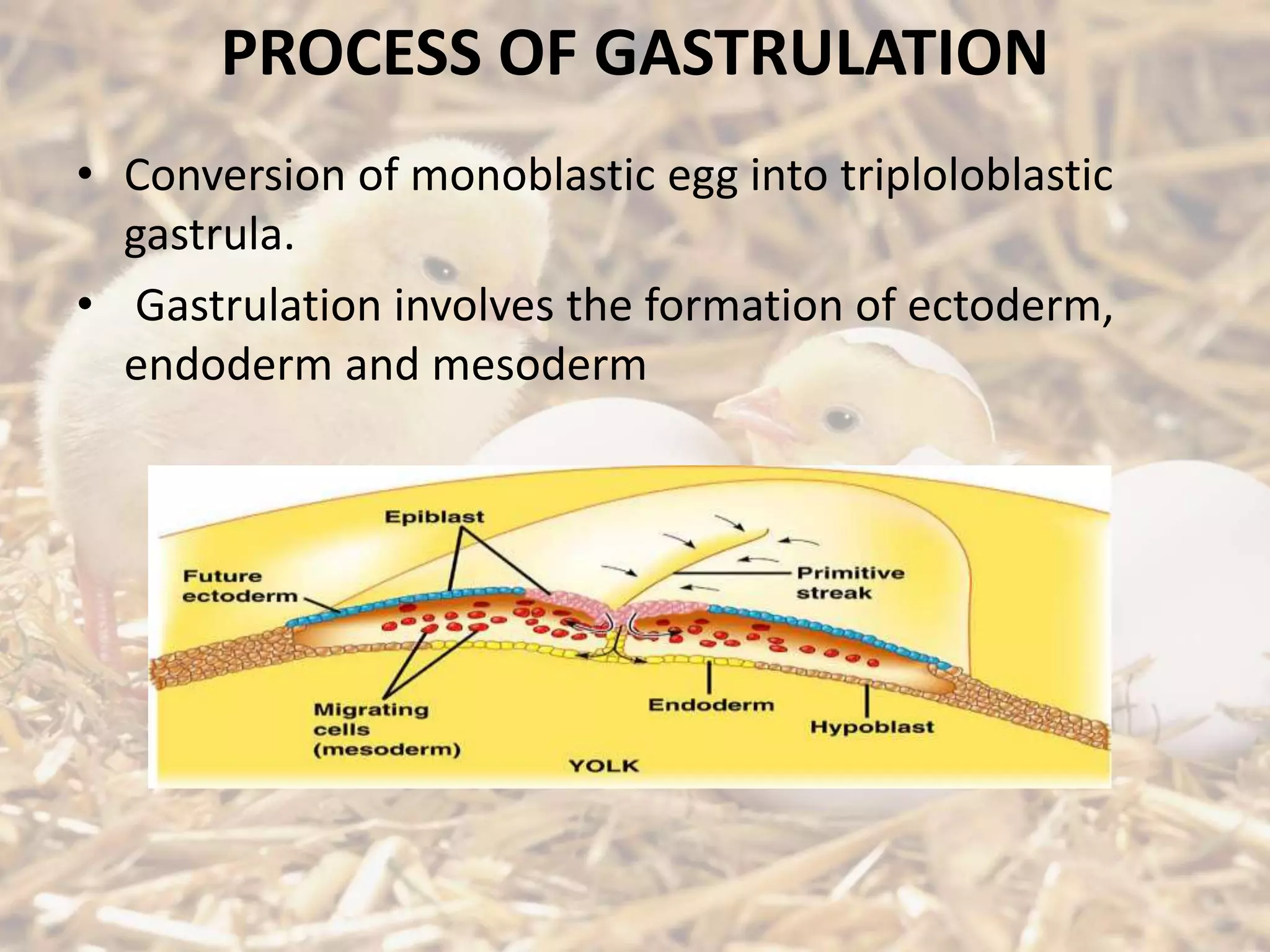
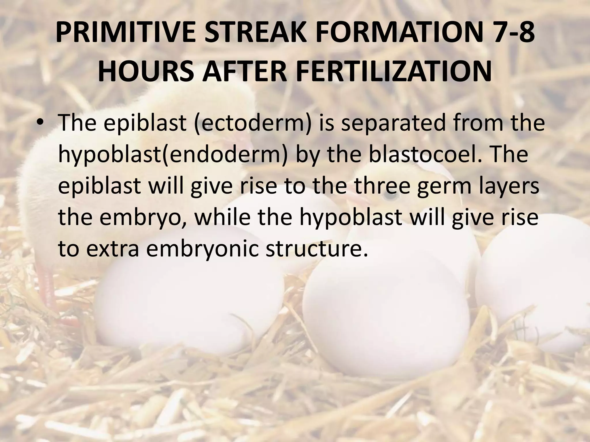
The conversion to triploblastic gastrula and formation of germ layers including ectoderm, mesoderm, and endoderm.
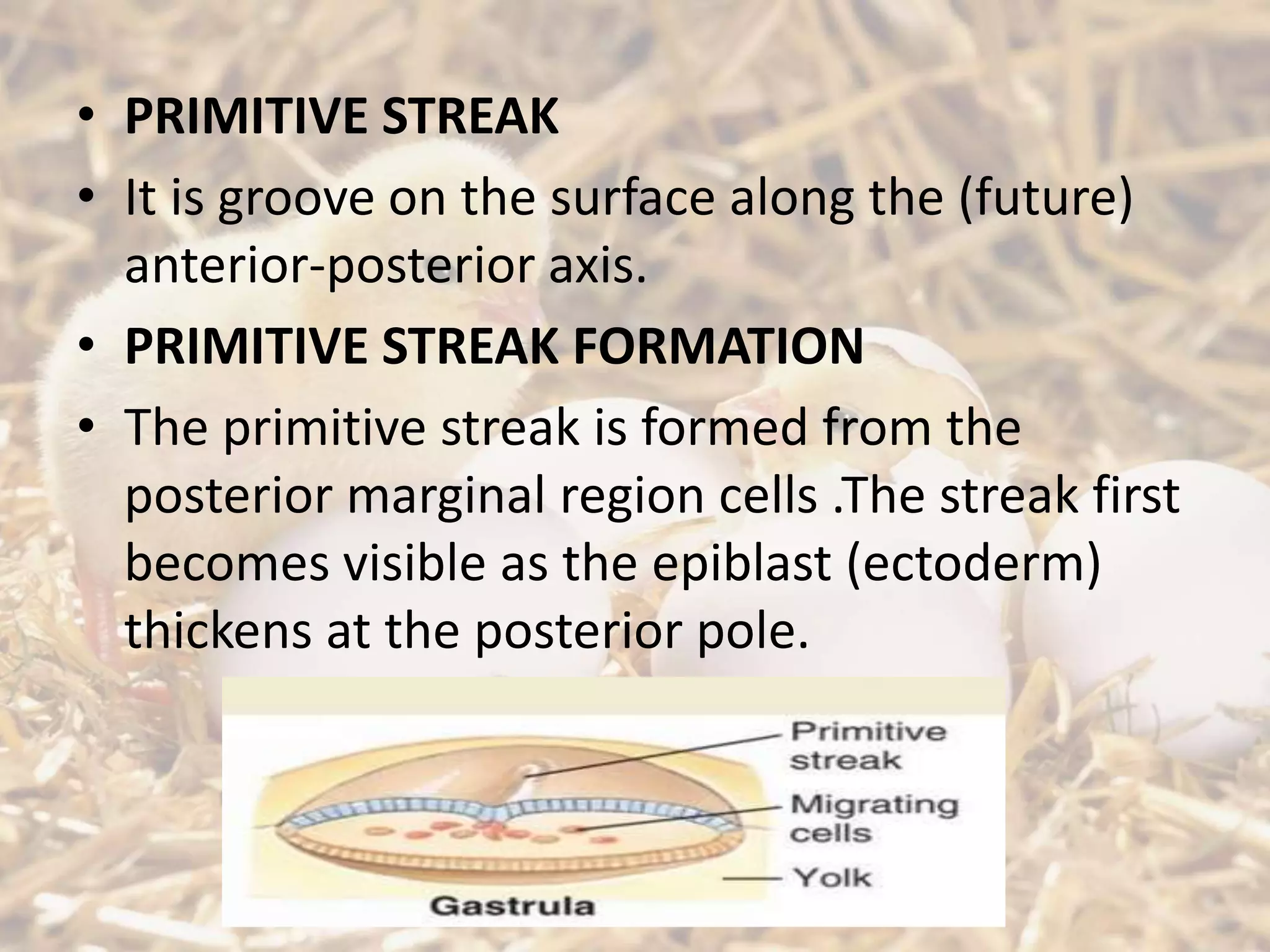
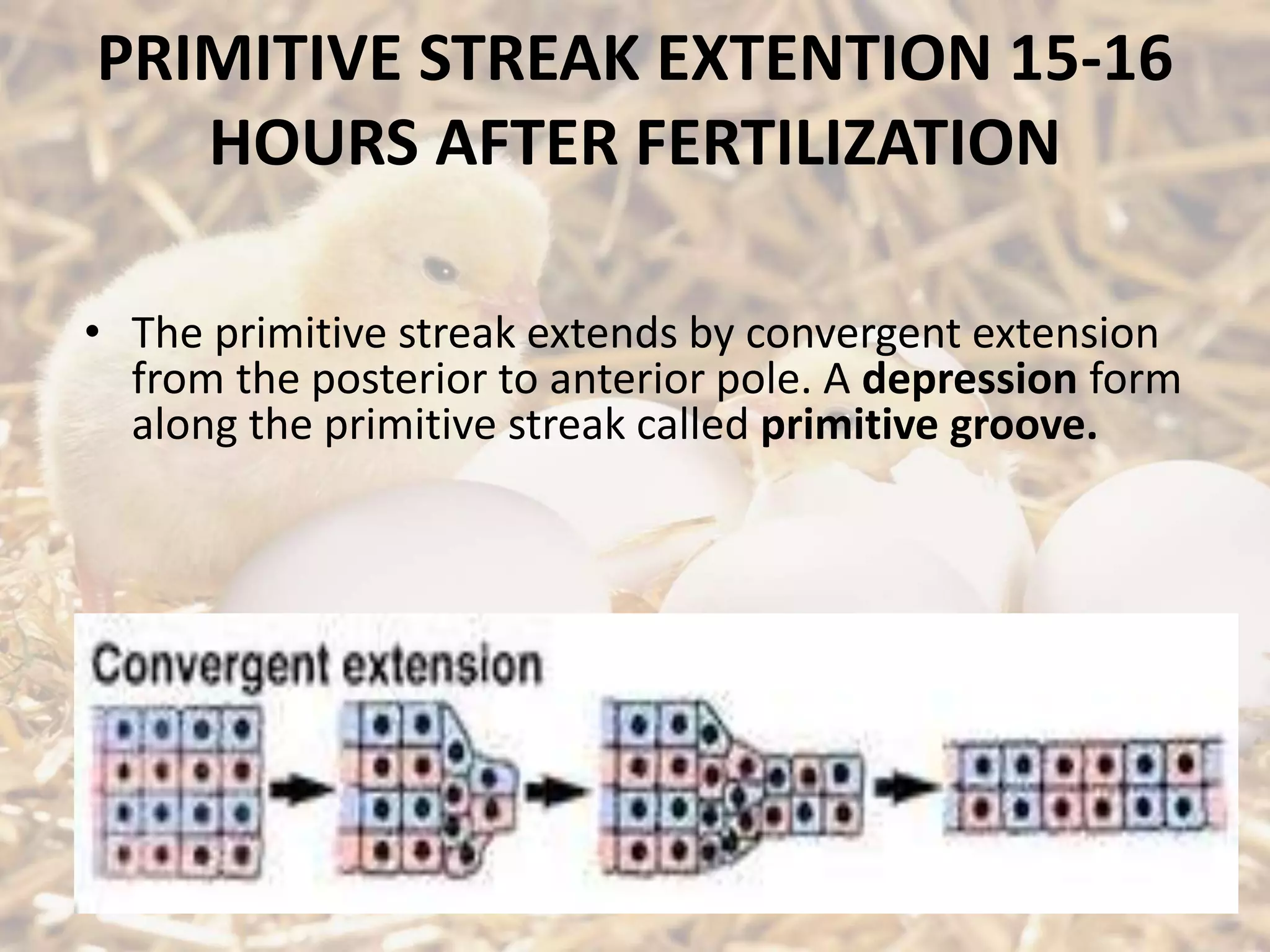
Describes the formation and extension of the primitive streak in the developing embryo, indicating a key developmental phase.
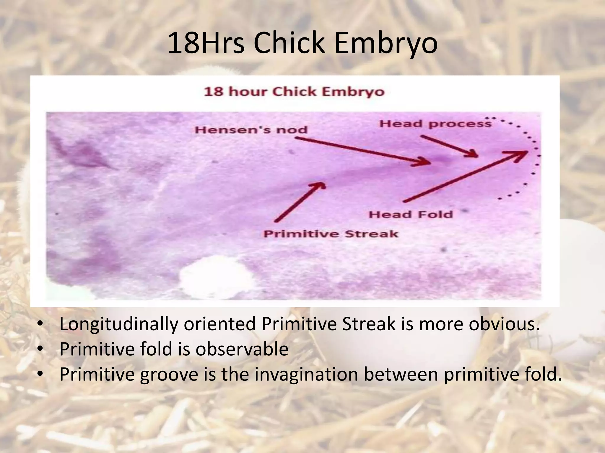
Developmental observations at 18 hrs and anatomical formations, including notochord and somite development.
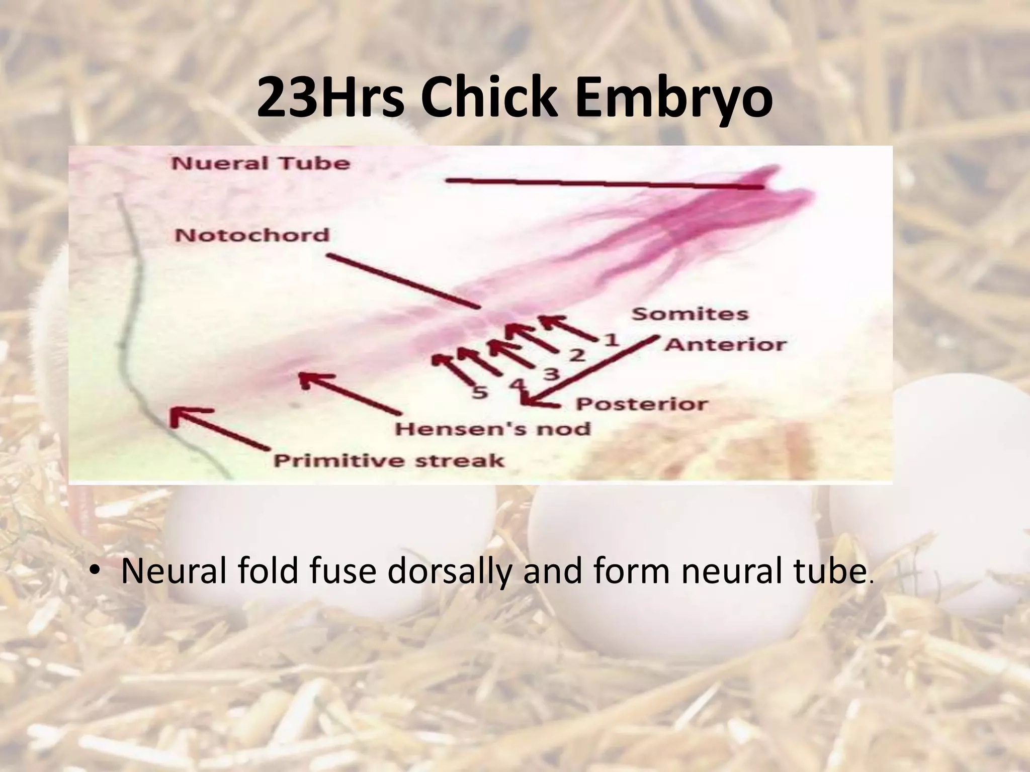
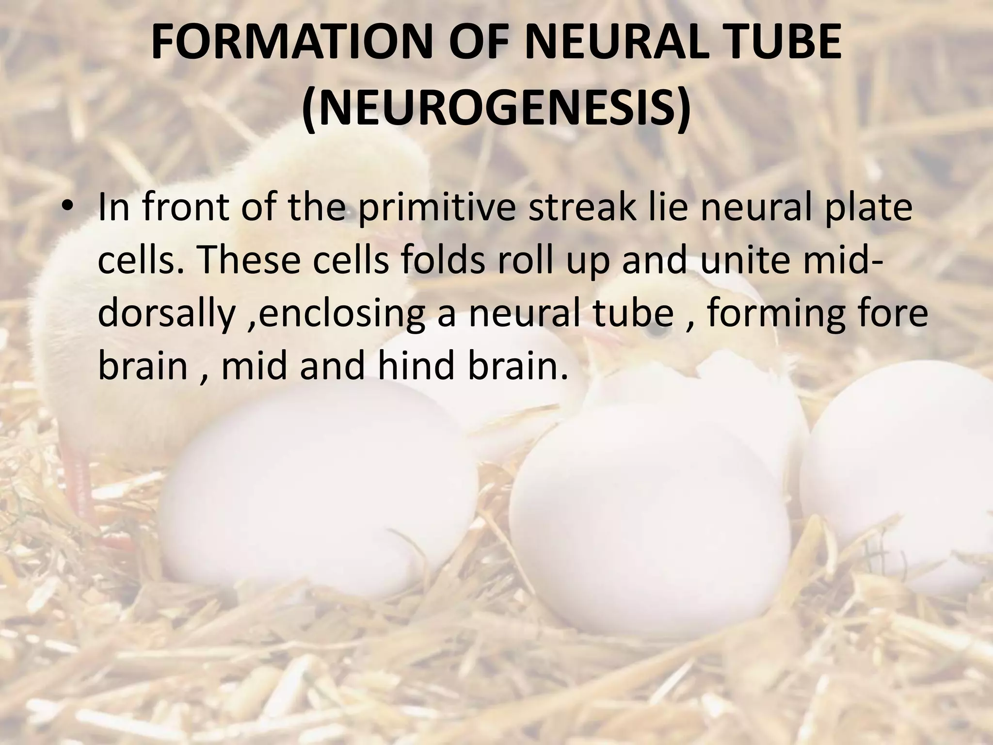
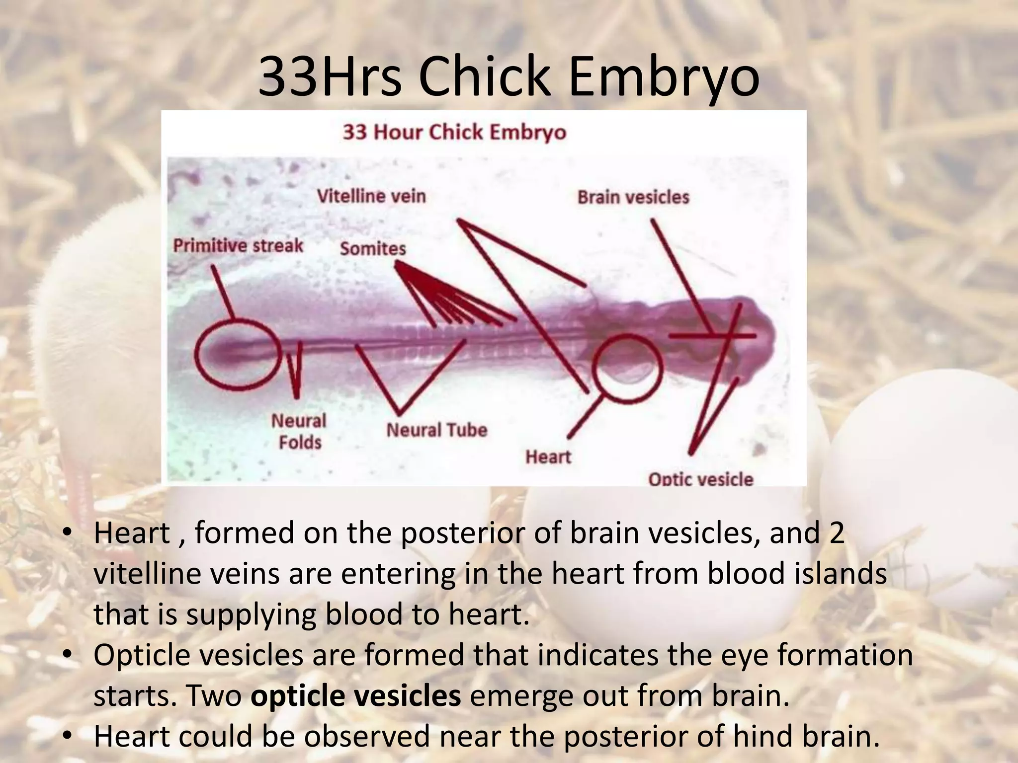
Neurogenesis in the chick embryos, detailing the formation of the neural tube and brain development.
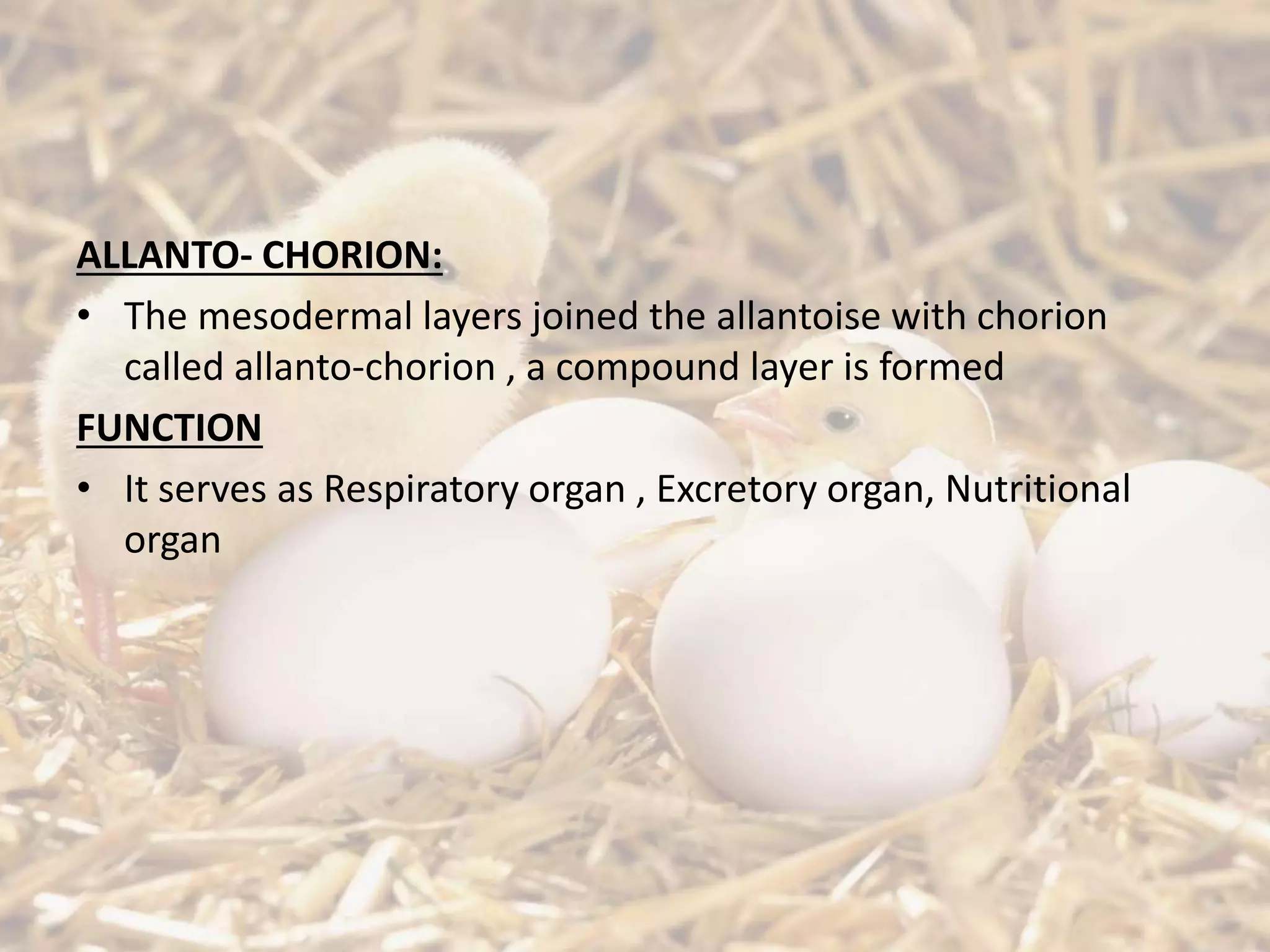
Details on organ formation, including the yolk sac and its function, along with descriptions of other membranes.
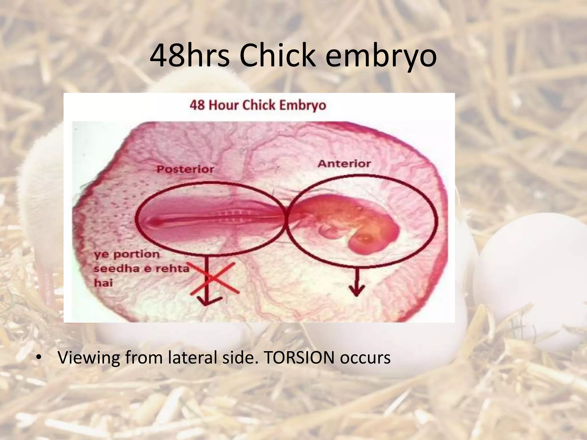
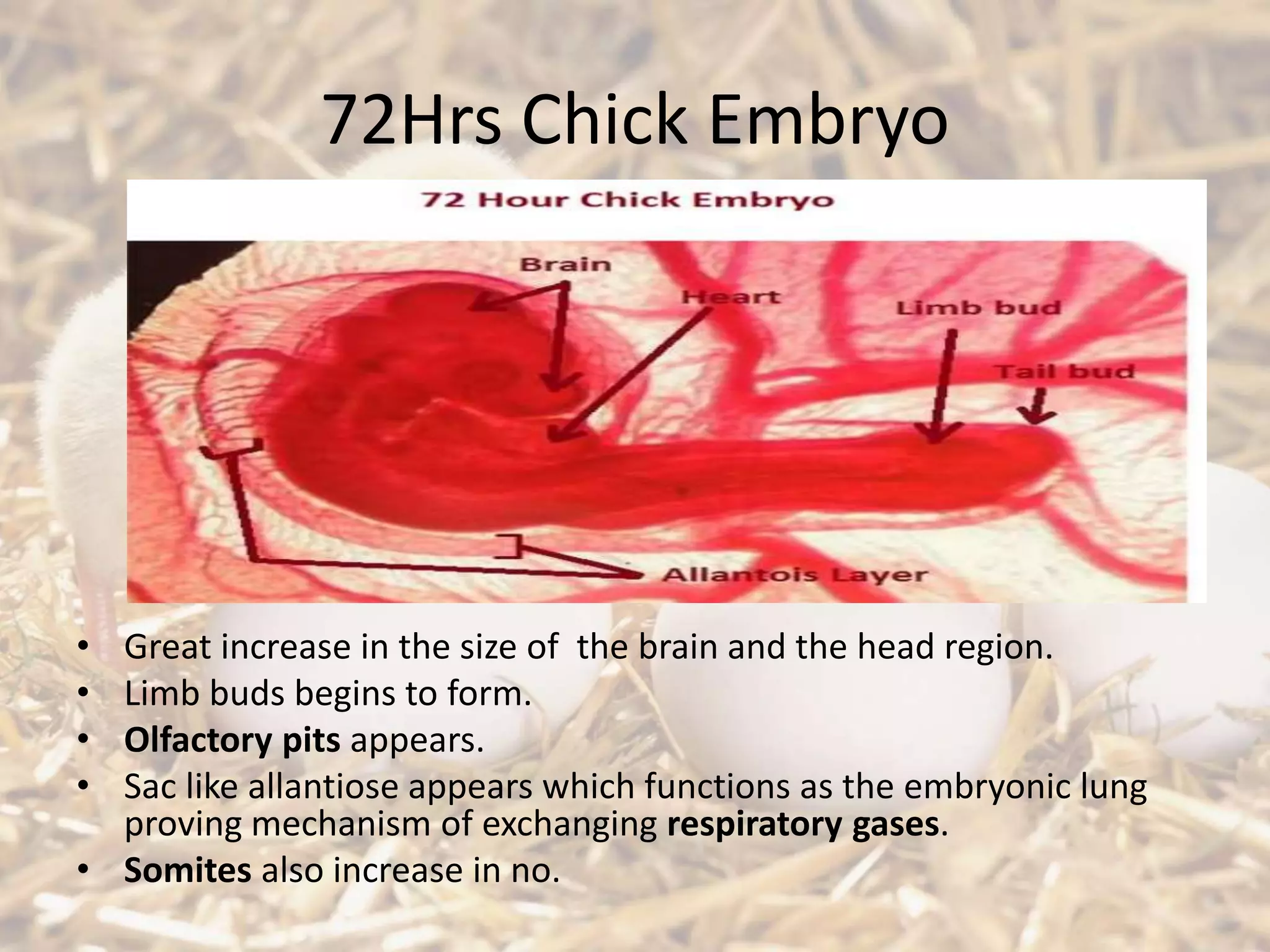
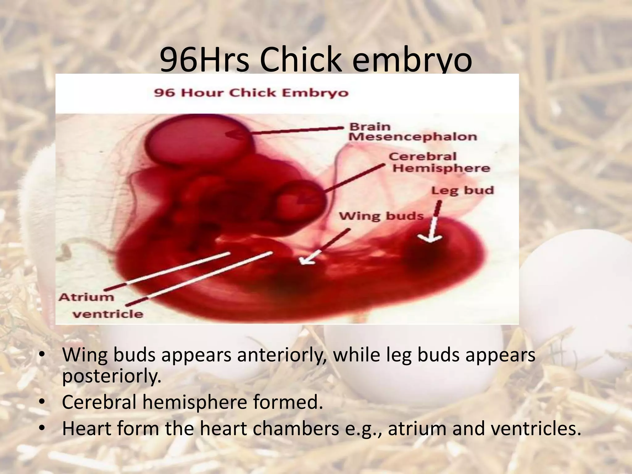
Final stages of chick embryo development including limb formation, brain enlargement, and respiratory organs.
Video concluding the presentation on chick embryonic development.


![Jaw suspension in vertebrates [autosaved]](https://cdn.slidesharecdn.com/ss_thumbnails/jawsuspensioninvertebratesautosaved-201219155254-thumbnail.jpg?width=640&height=640&fit=bounds)


