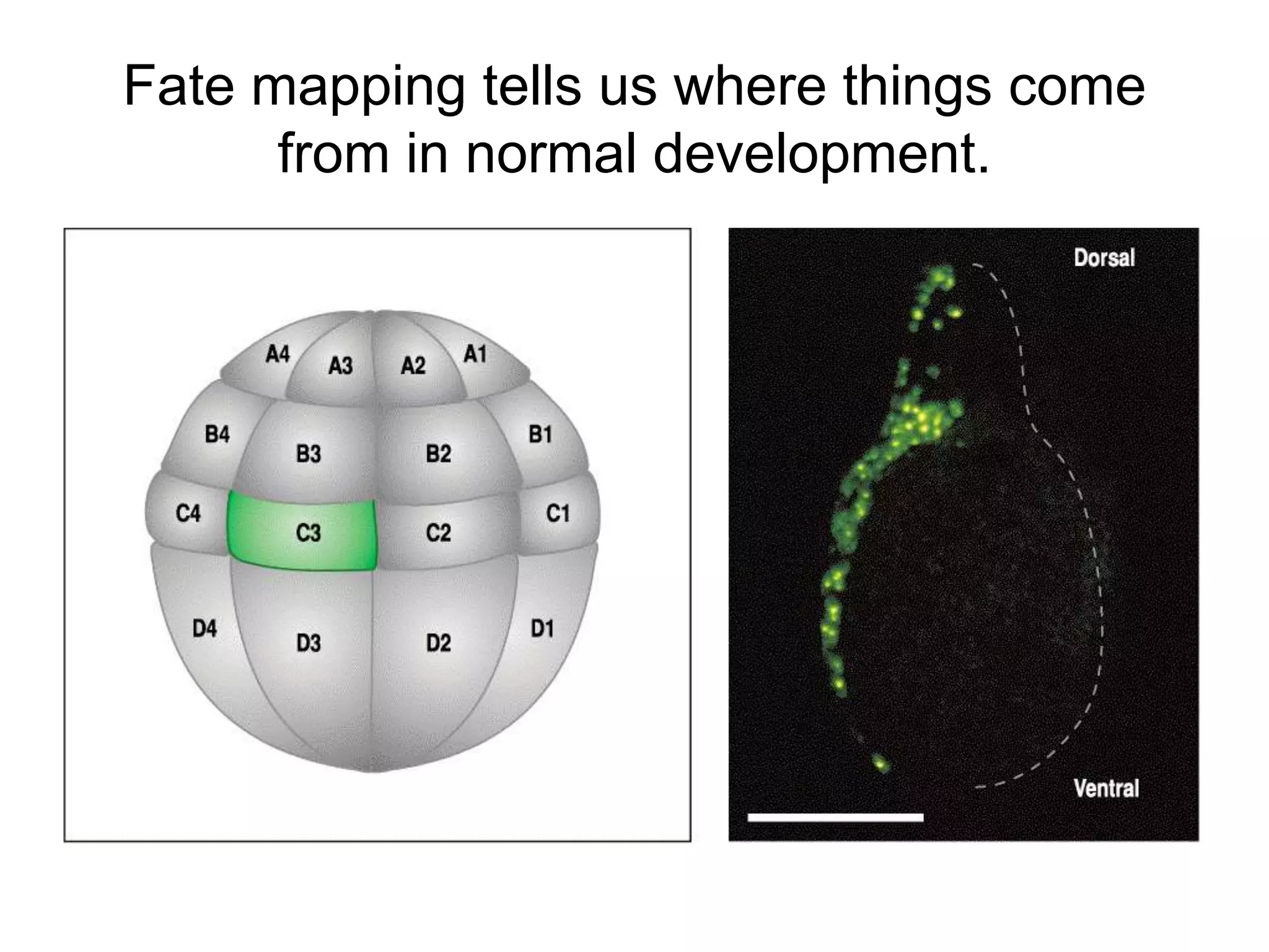
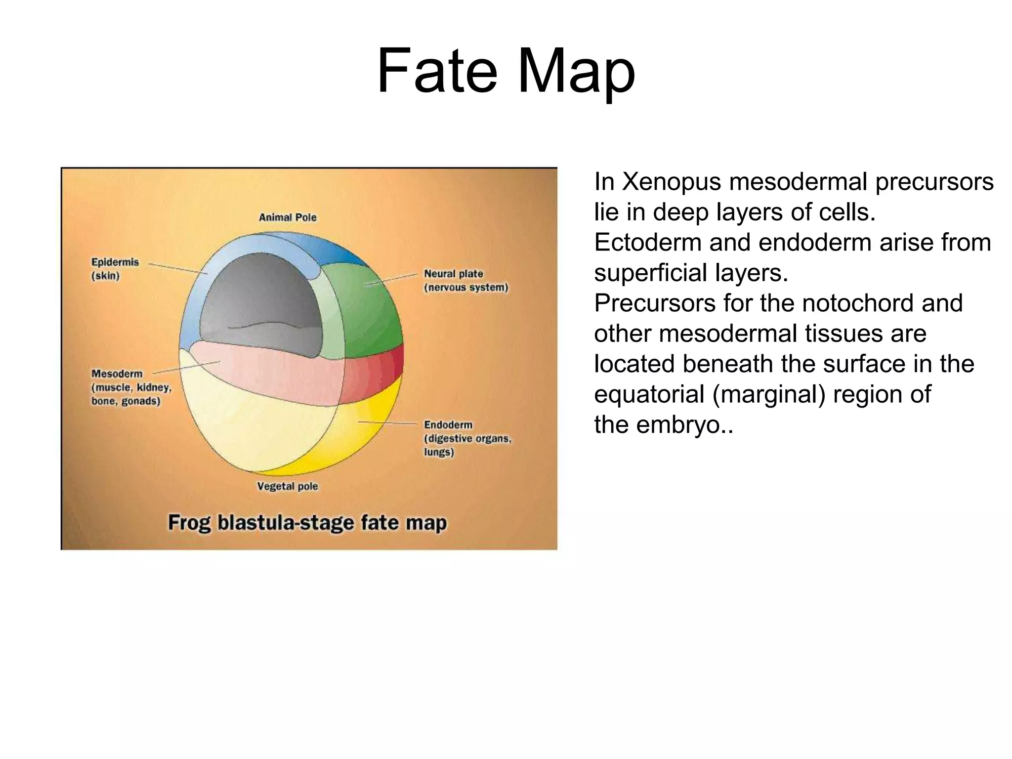
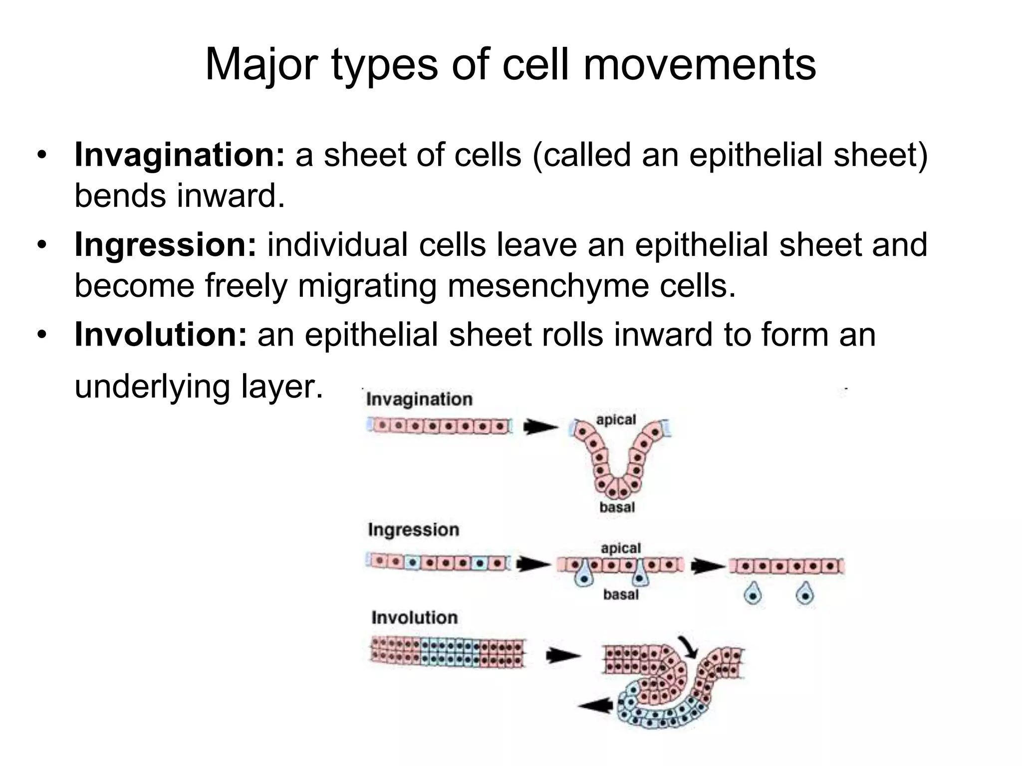
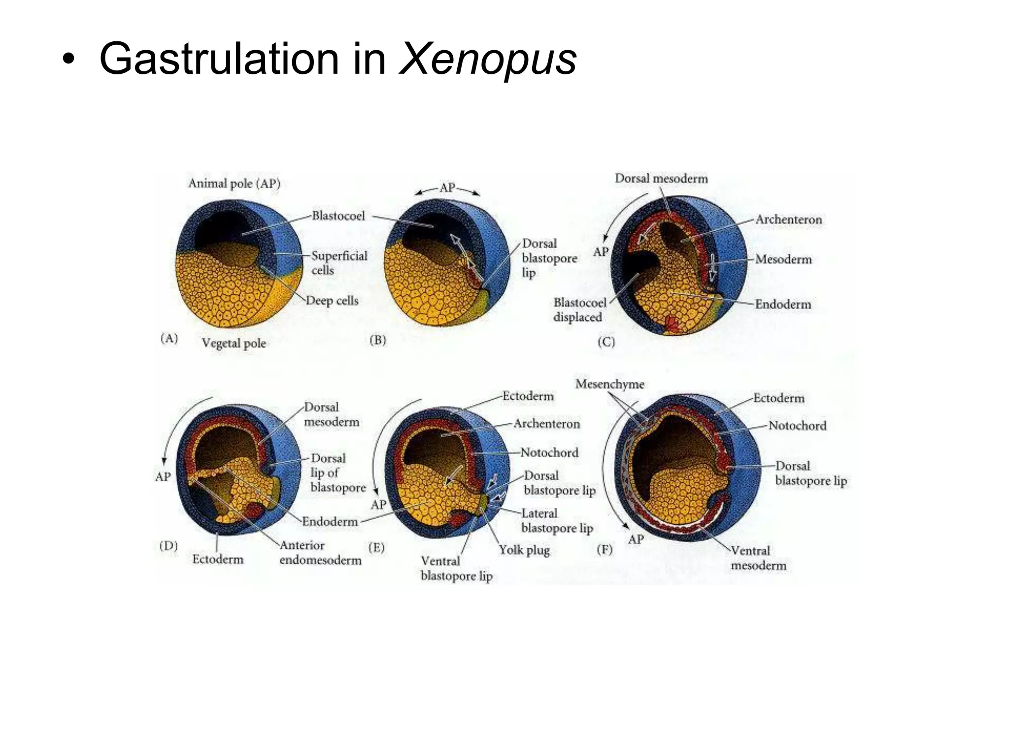
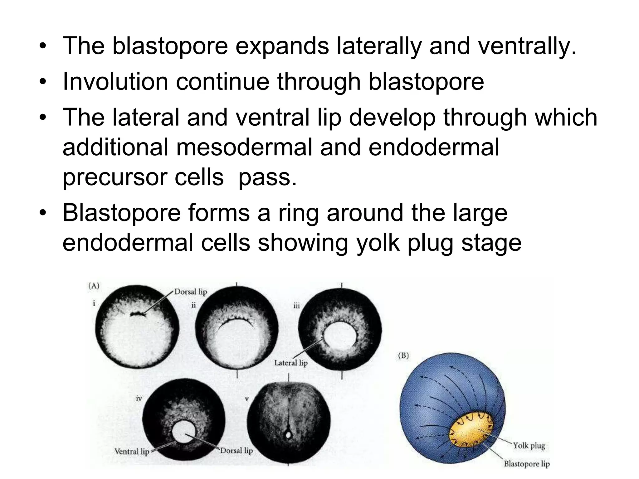
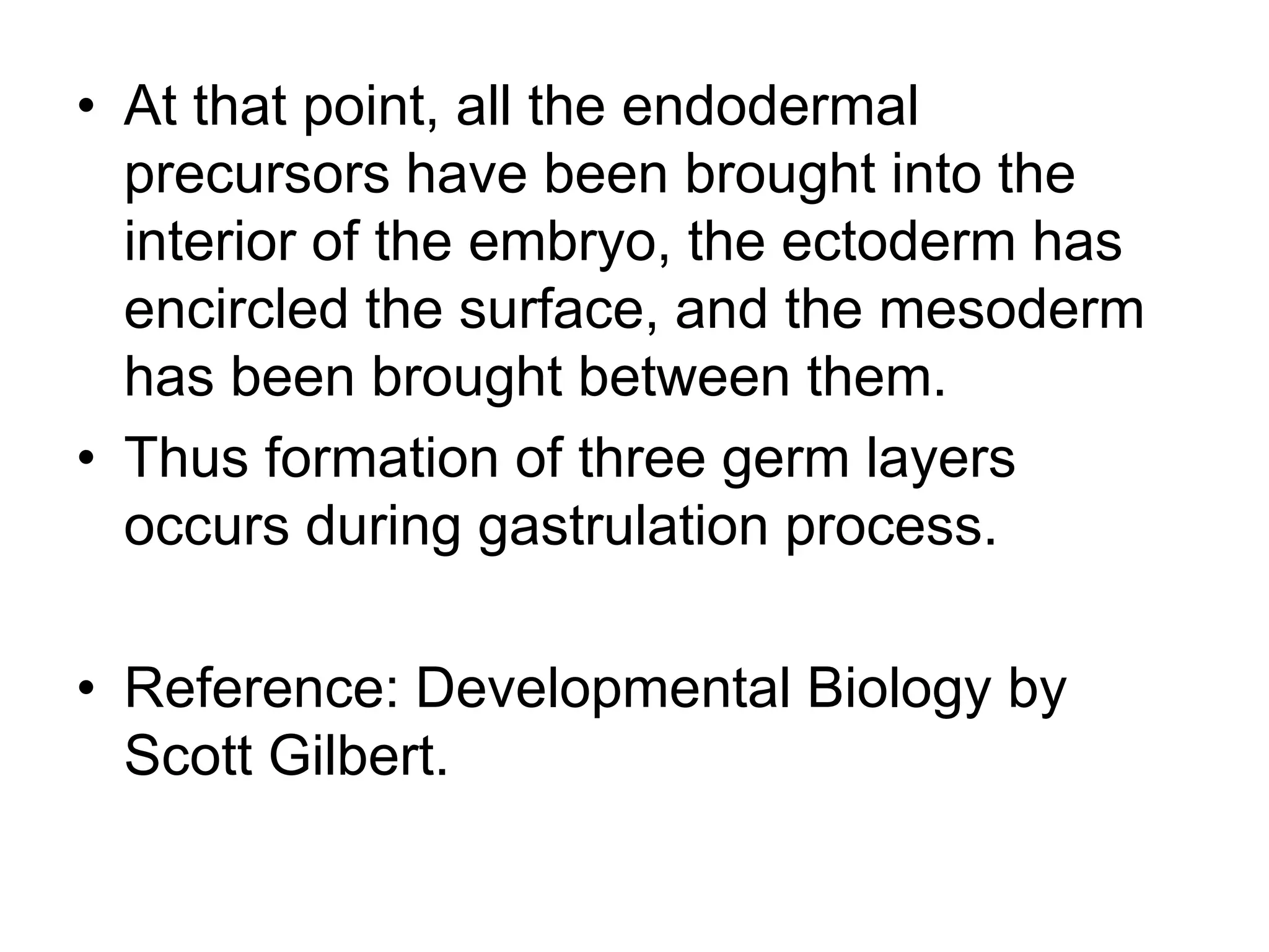
Gastrulation in Frog
Dr. Gauri Haval
Assistant professor
Abasaheb Garware College
• "It is not birth, marriage, or death, but gastrulation,
which is truly the most important time in your life."
Lewis Wolpert (1986)
• During gastrulation, cell movements result in a massive
reorganization of the embryo from a simple spherical ball
of cells, the blastula, into a multi-layered organism.
During gastrulation, many of the cells at or near the
surface of the embryo move to a new, more interior
location.
• The primary germ layers (endoderm, mesoderm, and
ectoderm) are formed and organized in their proper
locations during gastrulation.
Preparation of Fate Maps
• Tracing cell lineages. Groups of cells
studied by
1. Vital dyes
2. Radioactive labeling and fluorescent dyes.
3. Genetic marking.
Fate mapping tells us where things come
from in normal development.
Fate Map
In Xenopus mesodermal precursors
lie in deep layers of cells.
Ectoderm and endoderm arise from
superficial layers.
Precursors for the notochord and
other mesodermal tissues are
located beneath the surface in the
equatorial (marginal) region of
the embryo..
Process of Gastrulation
• The primary germ layers (endoderm, mesoderm, and
ectoderm) are formed and organized in their proper
locations during gastrulation. Endoderm, the most internal
germ layer, forms the lining of the gut and other internal
organs. Ectoderm, the most exterior germ layer, forms skin,
brain, the nervous system, and other external tissues.
Mesoderm, the the middle germ layer, forms muscle, the
skeletal system, and the circulatory system.
• Although the details of gastrulation differ between various
groups of animals, the cellular mechanisms involved in
gastrulation are common to all animals. Gastrulation
involves changes in cell motility, cell shape, and cell
adhesion.
Major types of cell movements
• Invagination: a sheet of cells (called an epithelial sheet)
bends inward.
• Ingression: individual cells leave an epithelial sheet and
become freely migrating mesenchyme cells.
• Involution: an epithelial sheet rolls inward to form an
underlying layer.
• Epiboly: a sheet of cells spreads by thinning.
• Intercalation: rows of cells move between one another,
creating an array of cells that is longer (in one or more
dimensions) but thinner.
• Convergent Extension: rows of cells intercalate, but the
intercalation is highly directional.
• Gastrulation
• Process of highly coordinated cell and
tissue movements
• Cells of blastula are dramatically arranged
• Cells take up new positions and new
neighbours
• Three germ layers are formed by
movements of the cells
• Gastrulation in frog embryos is initiated on
the future dorsal side of the embryo, just
below the equator in the region of the gray
crescent.
• In this region cells invaginate to form a slit
of blastopore
• In Xenopus laevis the change in shape of
cells is very essential for initiation of
gastrulation
• The cells in this region become bottle
shaped and invaginate inwards to form a
slit like blastopore.
Formation of blastopore lip
.
• The gastrulation in Xenopus begins at the
marginal zone. In this region the cells are
not yolky.
• The next phase is involution of marginal
zone cells and animal cells undergo
epiboly and converge at blastopore.
• When the migrating marginal cells reach
the dorsal lip of the blastopore, they turn
inward and travel alongthe inner surface of
the outer animal hemisphere cells. Thus,
the cells constituting the lip of the
blastopore are constantly changing.
• Gastrulation in Xenopus
• The first cells to compose the dorsal blastopore
lip are the bottle cells that invaginated to form
the leading edge of the archenteron. These cells
later become the pharyngeal cells of the foregut.
• The dorsal blastopore lip becomes composed of
cells that involute into the embryo to become the
prechordal plate (the precursor of the head
mesoderm).
• The next cells involuting into the embryo through
the dorsal blastopore lip are called the
chordamesoderm cells. These cells will form
the notochord.
• Blastocoel is displaced opposite to dorsal lip of
blastopore
• The blastopore expands laterally and ventrally.
• Involution continue through blastopore
• The lateral and ventral lip develop through which
additional mesodermal and endodermal
precursor cells pass.
• Blastopore forms a ring around the large
endodermal cells showing yolk plug stage
• At that point, all the endodermal
precursors have been brought into the
interior of the embryo, the ectoderm has
encircled the surface, and the mesoderm
has been brought between them.
• Thus formation of three germ layers
occurs during gastrulation process.
• Reference: Developmental Biology by
Scott Gilbert.

Gastrulation in frog

  • 1.
    Gastrulation in Frog Dr.Gauri Haval Assistant professor Abasaheb Garware College
  • 2.
    • "It isnot birth, marriage, or death, but gastrulation, which is truly the most important time in your life." Lewis Wolpert (1986) • During gastrulation, cell movements result in a massive reorganization of the embryo from a simple spherical ball of cells, the blastula, into a multi-layered organism. During gastrulation, many of the cells at or near the surface of the embryo move to a new, more interior location. • The primary germ layers (endoderm, mesoderm, and ectoderm) are formed and organized in their proper locations during gastrulation.
  • 3.
    Preparation of FateMaps • Tracing cell lineages. Groups of cells studied by 1. Vital dyes 2. Radioactive labeling and fluorescent dyes. 3. Genetic marking.
  • 4.
    Fate mapping tellsus where things come from in normal development.
  • 5.
    Fate Map In Xenopusmesodermal precursors lie in deep layers of cells. Ectoderm and endoderm arise from superficial layers. Precursors for the notochord and other mesodermal tissues are located beneath the surface in the equatorial (marginal) region of the embryo..
  • 6.
    Process of Gastrulation •The primary germ layers (endoderm, mesoderm, and ectoderm) are formed and organized in their proper locations during gastrulation. Endoderm, the most internal germ layer, forms the lining of the gut and other internal organs. Ectoderm, the most exterior germ layer, forms skin, brain, the nervous system, and other external tissues. Mesoderm, the the middle germ layer, forms muscle, the skeletal system, and the circulatory system. • Although the details of gastrulation differ between various groups of animals, the cellular mechanisms involved in gastrulation are common to all animals. Gastrulation involves changes in cell motility, cell shape, and cell adhesion.
  • 7.
    Major types ofcell movements • Invagination: a sheet of cells (called an epithelial sheet) bends inward. • Ingression: individual cells leave an epithelial sheet and become freely migrating mesenchyme cells. • Involution: an epithelial sheet rolls inward to form an underlying layer.
  • 8.
    • Epiboly: asheet of cells spreads by thinning. • Intercalation: rows of cells move between one another, creating an array of cells that is longer (in one or more dimensions) but thinner. • Convergent Extension: rows of cells intercalate, but the intercalation is highly directional.
  • 9.
    • Gastrulation • Processof highly coordinated cell and tissue movements • Cells of blastula are dramatically arranged • Cells take up new positions and new neighbours • Three germ layers are formed by movements of the cells
  • 10.
    • Gastrulation infrog embryos is initiated on the future dorsal side of the embryo, just below the equator in the region of the gray crescent. • In this region cells invaginate to form a slit of blastopore • In Xenopus laevis the change in shape of cells is very essential for initiation of gastrulation • The cells in this region become bottle shaped and invaginate inwards to form a slit like blastopore.
  • 11.
  • 12.
    • The gastrulationin Xenopus begins at the marginal zone. In this region the cells are not yolky. • The next phase is involution of marginal zone cells and animal cells undergo epiboly and converge at blastopore. • When the migrating marginal cells reach the dorsal lip of the blastopore, they turn inward and travel alongthe inner surface of the outer animal hemisphere cells. Thus, the cells constituting the lip of the blastopore are constantly changing.
  • 13.
  • 14.
    • The firstcells to compose the dorsal blastopore lip are the bottle cells that invaginated to form the leading edge of the archenteron. These cells later become the pharyngeal cells of the foregut. • The dorsal blastopore lip becomes composed of cells that involute into the embryo to become the prechordal plate (the precursor of the head mesoderm). • The next cells involuting into the embryo through the dorsal blastopore lip are called the chordamesoderm cells. These cells will form the notochord. • Blastocoel is displaced opposite to dorsal lip of blastopore
  • 15.
    • The blastoporeexpands laterally and ventrally. • Involution continue through blastopore • The lateral and ventral lip develop through which additional mesodermal and endodermal precursor cells pass. • Blastopore forms a ring around the large endodermal cells showing yolk plug stage
  • 16.
    • At thatpoint, all the endodermal precursors have been brought into the interior of the embryo, the ectoderm has encircled the surface, and the mesoderm has been brought between them. • Thus formation of three germ layers occurs during gastrulation process. • Reference: Developmental Biology by Scott Gilbert.