Downloaded 1,833 times
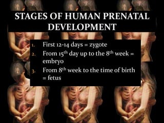
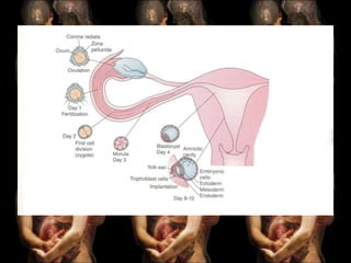
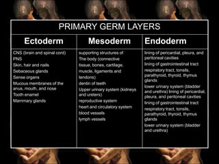
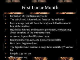


The document discusses human embryonic and fetal growth and development. It defines key terms and outlines the objectives of covering the stages of intrauterine development from zygote to fetus. It describes the three primary germ layers that develop into organ systems, and the changes that occur in each stage of embryonic and fetal growth. Finally, it provides detailed information about fetal growth and development in each lunar month of pregnancy.