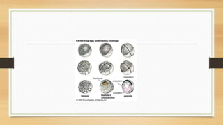
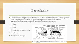
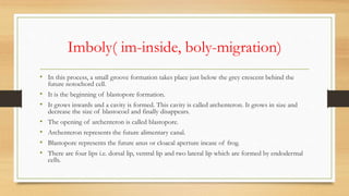
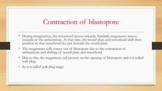
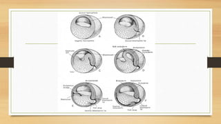
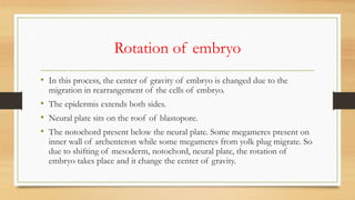
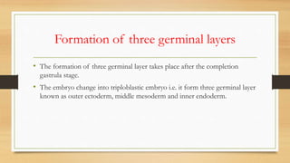
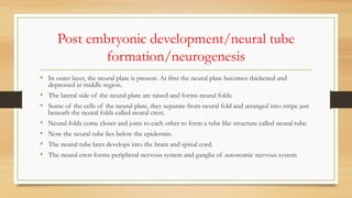
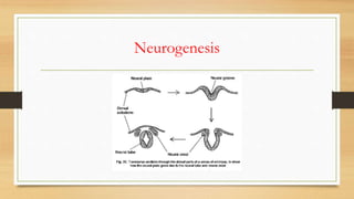
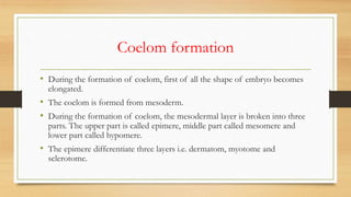
1. The development of frog consists of copulation, spawning, fertilization, cleavage, blastulation, gastrulation, and post-embryonic development.
2. During gastrulation, epiboly, imboly, contraction of the blastopore, and involution occur, forming the three germ layers - ectoderm, mesoderm, and endoderm.
3. Post-embryonic development includes neurogenesis forming the neural tube, notogenesis forming the notochord, and coelom formation separating the mesoderm into three layers.
2. The development of frog consists of:
• Copulation
• Spawning
• Fertilization
• Pre- embryonic development
• Cleavage
• Blastulation
• Gastrulation
• Post embryonic development
• Neurogenesis
• Notogenesis
• Coelom formation
3. Copulation
• Frogs lay their eggs in water in early spring.
• In pseudocopulation or mating, the male frog firmly clasps the body of the
female frog by his forelegs and enlarged thumb pads (nuptial pads).
• These nuptial pads help in clasping the body of female.
• This sexual behavior is called amplexus. As the eggs are extruded through
the cloaca of female, the male deposits sperm cells over them (insemination).
• Thus, fertilization is external, taking place in water.
4. Spawning
• The mesolecithal eggs of frog enclosed in a protective gelatinous albumen
are laid in water.
• The cluster or masses of eggs which remain stick together is called spawn.
• Generally a frog laid 3000-4000 eggs.
5. Fertilisation
• In frog, fertilization is external and occurs at once in water outside the body.
• In the fertilization process, sperm nucleus and female nucleus fuse together to
form zygote nucleus.
• The fusion of both male and female nuclei is called amphimixis.
• The upper half of the zygote or animal hemisphere is pigmented black due to
presence of melanin and it contains the cytoplasm and a nucleus, the lower vegetal
hemisphere is white and full of yolk.
• After fertilization, there is formation of grey crescent which represent the future
archenteron or alimentary canal.
7. Cleavage and Blastulation
• Cleavage or segmentation is holoblastic and equal. These are called bastomeres.
• A vertical furrow from the animal to the vegetal pole divides the zygote completely into two
equal-sized cells.
• A second vertical furrow at right angles to the first divides the zygote into four cells.
• The third cleavage is horizontal and above the equator which segments the zygote into
upper four smaller, black-colored cells in the animal pole and lower four larger, white-
colored cells.
• The cells formed by cleavage are blastomeres, the upper black blastomeres are called
micromeres, and lower white ones are macromeres.
9. • Further cleavages divide the micromeres more rapidly than the lower
macromeres whose division is hindered by yolk.
• The blastomeres’ mutual pressure flattens their surfaces in contact with each
other but free surfaces of each blastomere remain spherical.
• At this stage the whole embryo acquires a characteristic appearance like
mulberry shaped so it is called morula.
10. Blastulation
• Morula develops into the next embryonic stage called Blastula.
• As the cleavage proceeds ,the number of blastomeres of the morula increases and
blastomeres undergo rearrangement.
• The cells adhere to each other and they arrange themselves into true epithelium.
• This epithelium is called blastoderm.
• The fluid filled space or cavity called blastocoel appears in the center of blastoderm.
• This hollow ,spherical, and epithelial thick embryonic stage is called blastula and the
process of its formation is called blastulation.
11. • The blastocoel is consists of micromere cells from dorsal and lateral side
where as floor of blastocoel bears megameres.
• The arrangement of cells in blastula can determinate the future organs of
body called presumptive areas or layers.
12.
13. • The blastocoel contains different presumptive areas or layers. These layers are:
• Epidermis: arise from the first dorsal cell of micromeres present in the animal pole.
• Neural plate: forms from the adjacent cell to the dorsal cell of micromeres in the
animal pole.
• Notochord: it arise from the lateral cells of micromeres.
• Mesoderm: it also arises from the micromeres.
• Endoderm: it arises from the megameres.
14. Gastrulation
• Gastrulation is the process of formation of double or triple layered hollow gastrula
from single layered blastula. In gastrulation process, the movement and
differentiation of cell takes place. It consists of following stages:
• Epiboly
• Imboly
• Contraction of blastoporee
• Involution
• Rotation of embryo
15. Epiboly( epi- over, boly- move)
• During gastrulation process, the micromeres present on the animal pole
divides rapidly than megameres.
• So the micromeres migrate towards vegetal pores and covers the megameres.
• They roll over the megameres from animal poles to vegetal pole.
• So they cover all area except the yolk plug.
16. Imboly( im-inside, boly-migration)
• In this process, a small groove formation takes place just below the grey crescent behind the
future notochord cell.
• It is the beginning of blastopore formation.
• It grows inwards and a cavity is formed. This cavity is called archenteron. It grows in size and
decrease the size of blastocoel and finally disappears.
• The opening of archenteron is called blastopore.
• Archenteron represents the future alimentary canal.
• Blastopore represents the future anus or cloacal aperture incase of frog.
• There are four lips i.e. dorsal lip, ventral lip and two lateral lip which are formed by endodermal
cells.
17. Contraction of blastopore
• During invagination, the notochord moves inwards. Similarly megameres moves
inwards of the archenteron. At that time, the neural plate and notochord shift their
position so that notochord lies just beneath the neural plate.
• The megameres cells comes out of blastopore due to the contraction of
archenteron and shifting of neural plate and notochord.
• Due to this, the megameres cell present on the opening of blastopore and it is called
yolk plug.
• So it is called yolk plug stage.
18.
19. Involution
• During the invagination process, the mesoderm which is present just below
the lateral lip and above the ventral lip , they migrate inwards up to the
notochord.
• The mesoderm joins with notochord and forms chorda mesodermal layer.
So, involution is the inwards migration of the mesoderm above the ventral
lip and joining to notochord.
20. Rotation of embryo
• In this process, the center of gravity of embryo is changed due to the
migration in rearrangement of the cells of embryo.
• The epidermis extends both sides.
• Neural plate sits on the roof of blastopore.
• The notochord present below the neural plate. Some megameres present on
inner wall of archenteron while some megameres from yolk plug migrate. So
due to shifting of mesoderm, notochord, neural plate, the rotation of
embryo takes place and it change the center of gravity.
21. Formation of three germinal layers
• The formation of three germinal layer takes place after the completion
gastrula stage.
• The embryo change into triploblastic embryo i.e. it form three germinal layer
known as outer ectoderm, middle mesoderm and inner endoderm.
22. Post embryonic development/neural tube
formation/neurogenesis
• In outer layer, the neural plate is present. At first the neural plate becomes thickened and
depressed at middle region.
• The lateral side of the neural plate are raised and forms neural folds.
• Some of the cells of the neural plate, they separate from neural fold and arranged into stripe just
beneath the neural folds called neural crest.
• Neural folds come closer and joins to each other to form a tube like structure called neural tube.
• Now the neural tube lies below the epidermis.
• The neural tube later develops into the brain and spinal cord.
• The neural crest forms peripheral nervous system and ganglia of autonomic nervous system
24. Notochord formation/notogenesis
• In the notogenesis process, the notochordal or chorda-mesodermal cells are
responsible for notochord formation. These cells are present below the neural plate.
• At first, the notochord separated from the mesoderm from lateral side.
• These cells now forms the rod like structure just below the neural tube. After that
some cells of notochord becomes vacuolated in outer region. These vacuolated
cells later forms the meninges of brain and spinal chord.
• In late stage of embryo, this notochord is broken into small pieces called somites.
These somites later cover the neural tube and develops into vertebral column.
25. Coelom formation
• During the formation of coelom, first of all the shape of embryo becomes
elongated.
• The coelom is formed from mesoderm.
• During the formation of coelom, the mesodermal layer is broken into three
parts. The upper part is called epimere, middle part called mesomere and
lower part called hypomere.
• The epimere differentiate three layers i.e. dermatom, myotome and
sclerotome.
28. • Dermis forms the dermis of skin. The myotome forms the muscle and sclerotome
forms the skeleton around the notochord.
• The middle portion is modified into nephrostome.
• The hypodermal contains endodermal layer inside. In hypomere, first a small slit is
formed in the middle of hypomere. Due to this, the hypomere is divided into two
layers.
• The layer which is presents towards body wall called parietal layer and inner layer
called visceral layer.
• The gap between two layer called splanchnocoel.
29. • The formation of splanchnocoel indicates the beginning stage of coelom
formation.
• In later stage, the size of splanchnocoel increases, due to which the parietal
layer shift towards body wall and visceral layer moves towards gut. The space
between these layer called coelom.