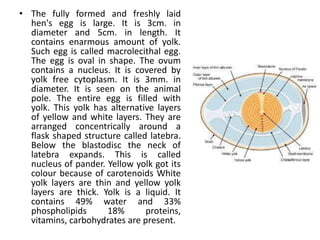
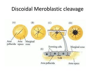
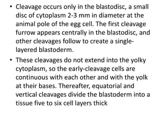
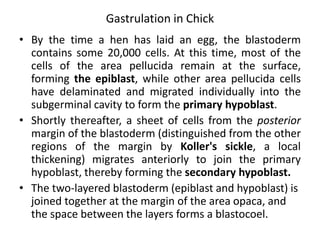
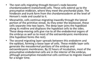
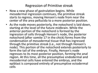
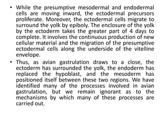
Development of Chick
Dr. Gauri Haval
• The fully formed and freshly laid
hen's egg is large. It is 3cm. in
diameter and 5cm. in length. It
contains enarmous amount of yolk.
Such egg is called macrolecithal egg.
The egg is oval in shape. The ovum
contains a nucleus. It is covered by
yolk free cytoplasm. It is 3mm. in
diameter. It is seen on the animal
pole. The entire egg is filled with
yolk. This yolk has alternative layers
of yellow and white layers. They are
arranged concentrically around a
flask shaped structure called latebra.
Below the blastodisc the neck of
latebra expands. This is called
nucleus of pander. Yellow yolk got its
colour because of carotenoids White
yolk layers are thin and yellow yolk
layers are thick. Yolk is a liquid. It
contains 49% water and 33%
phospholipids 18% proteins,
vitamins, carbohydrates are present.
• The entire ovum is covered by plasma membrane. It is called
plasmalemma. It is lipoprotein layer. This is ovum is covered by egg
membranes.
• Primary membranes: These membranes develop between oocyte
and follicle. The primary membranes are secreted by follicle cells. It
is called vitelline, membrane is come from two origins. Inner part is
produced by ovary. Outer part is from the falopian tube.
• Secondary membranes: Oviduct secretes secondary membranes.
Above vitelline membrane albumen is present. It is white in colour
and it contains water and proteins. The outer layer of albumen is
thin. It is called thin albumen. The middle layer of albumen is thick.
It is called thick albumen, or dense albumen. The inner most
albumen is very thick. It develops into chalazae. The chalazae are
called balancers. They keep the ovum in the centre.
• Shell membranes: Above the albumen two shell membranes are
present. Towards the broad end of egg, in between the shell
membranes an air space is present. This air space is formed when
egg is laid cooled from 60°C to lesser temperature.
• Shell: Above the shell membranes a shell is present, it is porous in
nature. It is calcareous. This porous shell is useful for exchange of
gases. In a freshly laid hen's egg shell is soft. Very soon it becomes
hard. L
• Fertilization of the chick egg occurs in the
oviduct, before the albumen and the shell are
secreted upon it. The egg is telolecithal (like
that of the fish), with a small disc of cytoplasm
sitting atop a large yolk. Like fish eggs, the
yolky eggs of birds undergo discoidal
meroblastic cleavage.
Discoidal Meroblastic cleavage
• Cleavage occurs only in the blastodisc, a small
disc of cytoplasm 2-3 mm in diameter at the
animal pole of the egg cell. The first cleavage
furrow appears centrally in the blastodisc, and
other cleavages follow to create a single-
layered blastoderm.
• These cleavages do not extend into the yolky
cytoplasm, so the early-cleavage cells are
continuous with each other and with the yolk
at their bases. Thereafter, equatorial and
vertical cleavages divide the blastoderm into a
tissue five to six cell layers thick
• Between the blastoderm and the yolk is a space
called the subgerminal cavity. This space is
created when the blastoderm cells absorb fluid
from the albumin ("egg white") and secrete it
between themselves and the yolk . At this stage,
the deep cells in the center of the blastoderm are
shed and die, leaving behind a one cell-thick area
pellucida. This part of the blastoderm forms most
of the actual embryo. The peripheral ring of
blastoderm cells that have not shed their deep
cells constitutes the area opaca.
• Between the area pellucida and the area opaca is
a thin layer of cells called the marginal zone (or
marginal belt). Some of the marginal zone cells
become very important in determining cell fate
during early chick development
Gastrulation in Chick
• By the time a hen has laid an egg, the blastoderm
contains some 20,000 cells. At this time, most of the
cells of the area pellucida remain at the surface,
forming the epiblast, while other area pellucida cells
have delaminated and migrated individually into the
subgerminal cavity to form the primary hypoblast.
• Shortly thereafter, a sheet of cells from the posterior
margin of the blastoderm (distinguished from the other
regions of the margin by Koller's sickle, a local
thickening) migrates anteriorly to join the primary
hypoblast, thereby forming the secondary hypoblast.
• The two-layered blastoderm (epiblast and hypoblast) is
joined together at the margin of the area opaca, and
the space between the layers forms a blastocoel.
• All three germ layers of the embryo proper (plus a
considerable amount of extraembryonic membrane)
are formed from the epiblastic cells.
• Fate map of chick embryo
Formation of Primitive streak
• The major structural characteristic
of avian, reptilian, and mammalian
gastrulation is the primitive streak.
This streak is first visible as a
thickening of the epiblast at the
posterior region of the embryo,
just anterior to Koller's sickle. This
thickening is caused by the
ingression of endodermal
precursors from the epiblast into
the blastocoel and by the migration
of cells from the lateral region of
the posterior epiblast toward the
center.
• As these cells enter the primitive
streak, the streak elongates toward
the future head region. At the
same time, the secondary
hypoblast cells continue to migrate
anteriorly from the posterior
margin of the blastoderm.
• The elongation of the primitive streak appears to
be coextensive with the anterior migration of
these secondary hypoblast cells. The streak
eventually extends 60 75% of the length of the
area pellucida.
• The primitive streak defines the axes of the
embryo. It extends from posterior to anterior;
migrating cells enter through its dorsal side and
move to its ventral side; and it separates the left
portion of the embryo from the right. Those
elements close to the streak will be the medial
(central) structures, while those farther from it
will be the distal (lateral) structures
• As cells converge to form the
primitive streak, a depression
forms within the streak. This
depression is called the primitive
groove, and it serves as an
opening through which migrating
cells pass into the blastocoel.
Thus, the primitive groove is
analogous to the amphibian
blastopore. At the anterior end of
the primitive streak is a regional
thickening of cells called the
primitive knot or Hensen's node.
The center of this node contains a
funnel-shaped depression
(sometimes called the primitive
pit) through which cells can pass
into the blastocoel. Hensen's
node is the functional equivalent
of the dorsal lip of the amphibian
blastopore (i.e., the organizer)
• As soon as the primitive streak has formed,
epiblast cells begin to migrate through it and into
the blastocoel.
• The primitive streak has a continually changing
cell population. Cells migrating through Hensen's
node pass down into the blastocoel and migrate
anteriorly, forming foregut, head mesoderm, and
notochord; cells passing through the lateral
portions of the primitive streak give rise to the
majority of endodermal and mesodermal tissues
Migration through Primitive streak and
formation of mesoderm and endoderm
• The first cells to migrate through Hensen's node are those
destined to become the pharyngial endoderm of the
foregut. Once inside the blastocoel, these endodermal cells
migrate anteriorly and eventually displace the hypoblast
cells, causing the hypoblast cells to be confined to a region
in the anterior portion of the area pellucida.
• The next cells entering the blastocoel through Hensen's
node also move anteriorly, but they do not move as far
ventrally as the presumptive foregut endodermal cells.
Rather, they remain between the endoderm and the
epiblast to form the head mesenchyme and the prechordal
plate mesoderm.
• These early-ingressing cells all move anteriorly, pushing up
the anterior midline region of the epiblast to form the head
process
• The next cells migrating through Hensen's node become
chordamesoderm (notochord) cells. These cells extend up to the
presumptive midbrain, where they meet the prechordal plate. The
hindbrain and trunk form from the chordamesoderm at the level of
Hensen's node and caudal to it.
• Meanwhile, cells continue migrating inwardly through the lateral
portion of the primitive streak. As they enter the blastocoel, these
cells separate into two layers. The deep layer joins the hypoblast
along its midline and displaces the hypoblast cells to the sides.
These deep-moving cells give rise to all the endodermal organs of
the embryo as well as to most of the extraembryonic membranes
(the hypoblast forms the rest).
• The second migrating layer spreads between this endoderm and
the epiblast, forming a loose layer of cells. These middle layer cells
generate the mesodermal portions of the embryo and
extraembryonic membranes. By 22 hours of incubation, most of the
presumptive endodermal cells are in the interior of the embryo,
although presumptive mesodermal cells continue to migrate inward
for a longer time.
Regression of Primitive streak
• Now a new phase of gastrulation begins. While
mesodermal ingression continues, the primitive streak
starts to regress, moving Hensen's node from near the
center of the area pellucida to a more posterior position.
• As the node moves posteriorly, the notochord is laid down,
starting at the level of the future midbrain. While the
anterior portion of the notochord is formed by the
ingression of cells through Hensen's node, the posterior
notochord (after somite 17 in the chick) forms from the
condensation of mesodermal tissue that has ingressed
through the primitive streak (i.e., not through Hensen's
node). This portion of the notochord extends posteriorly to
form the tail of the embryo. Finally, Hensen's node
regresses to its most posterior position, forming the anal
region. At this time, all the presumptive endodermal and
mesodermal cells have entered the embryo, and the
epiblast is composed entirely of presumptive ectodermal
cells
• While the presumptive mesodermal and endodermal
cells are moving inward, the ectodermal precursors
proliferate. Moreover, the ectodermal cells migrate to
surround the yolk by epiboly. The enclosure of the yolk
by the ectoderm takes the greater part of 4 days to
complete. It involves the continuous production of new
cellular material and the migration of the presumptive
ectodermal cells along the underside of the vitelline
envelope.
• Thus, as avian gastrulation draws to a close, the
ectoderm has surrounded the yolk, the endoderm has
replaced the hypoblast, and the mesoderm has
positioned itself between these two regions. We have
identified many of the processes involved in avian
gastrulation, but we remain ignorant as to the
mechanisms by which many of these processes are
carried out.

Development of chick

  • 1.
  • 2.
    • The fullyformed and freshly laid hen's egg is large. It is 3cm. in diameter and 5cm. in length. It contains enarmous amount of yolk. Such egg is called macrolecithal egg. The egg is oval in shape. The ovum contains a nucleus. It is covered by yolk free cytoplasm. It is 3mm. in diameter. It is seen on the animal pole. The entire egg is filled with yolk. This yolk has alternative layers of yellow and white layers. They are arranged concentrically around a flask shaped structure called latebra. Below the blastodisc the neck of latebra expands. This is called nucleus of pander. Yellow yolk got its colour because of carotenoids White yolk layers are thin and yellow yolk layers are thick. Yolk is a liquid. It contains 49% water and 33% phospholipids 18% proteins, vitamins, carbohydrates are present.
  • 3.
    • The entireovum is covered by plasma membrane. It is called plasmalemma. It is lipoprotein layer. This is ovum is covered by egg membranes. • Primary membranes: These membranes develop between oocyte and follicle. The primary membranes are secreted by follicle cells. It is called vitelline, membrane is come from two origins. Inner part is produced by ovary. Outer part is from the falopian tube. • Secondary membranes: Oviduct secretes secondary membranes. Above vitelline membrane albumen is present. It is white in colour and it contains water and proteins. The outer layer of albumen is thin. It is called thin albumen. The middle layer of albumen is thick. It is called thick albumen, or dense albumen. The inner most albumen is very thick. It develops into chalazae. The chalazae are called balancers. They keep the ovum in the centre. • Shell membranes: Above the albumen two shell membranes are present. Towards the broad end of egg, in between the shell membranes an air space is present. This air space is formed when egg is laid cooled from 60°C to lesser temperature. • Shell: Above the shell membranes a shell is present, it is porous in nature. It is calcareous. This porous shell is useful for exchange of gases. In a freshly laid hen's egg shell is soft. Very soon it becomes hard. L
  • 4.
    • Fertilization ofthe chick egg occurs in the oviduct, before the albumen and the shell are secreted upon it. The egg is telolecithal (like that of the fish), with a small disc of cytoplasm sitting atop a large yolk. Like fish eggs, the yolky eggs of birds undergo discoidal meroblastic cleavage.
  • 5.
  • 6.
    • Cleavage occursonly in the blastodisc, a small disc of cytoplasm 2-3 mm in diameter at the animal pole of the egg cell. The first cleavage furrow appears centrally in the blastodisc, and other cleavages follow to create a single- layered blastoderm. • These cleavages do not extend into the yolky cytoplasm, so the early-cleavage cells are continuous with each other and with the yolk at their bases. Thereafter, equatorial and vertical cleavages divide the blastoderm into a tissue five to six cell layers thick
  • 8.
    • Between theblastoderm and the yolk is a space called the subgerminal cavity. This space is created when the blastoderm cells absorb fluid from the albumin ("egg white") and secrete it between themselves and the yolk . At this stage, the deep cells in the center of the blastoderm are shed and die, leaving behind a one cell-thick area pellucida. This part of the blastoderm forms most of the actual embryo. The peripheral ring of blastoderm cells that have not shed their deep cells constitutes the area opaca. • Between the area pellucida and the area opaca is a thin layer of cells called the marginal zone (or marginal belt). Some of the marginal zone cells become very important in determining cell fate during early chick development
  • 9.
    Gastrulation in Chick •By the time a hen has laid an egg, the blastoderm contains some 20,000 cells. At this time, most of the cells of the area pellucida remain at the surface, forming the epiblast, while other area pellucida cells have delaminated and migrated individually into the subgerminal cavity to form the primary hypoblast. • Shortly thereafter, a sheet of cells from the posterior margin of the blastoderm (distinguished from the other regions of the margin by Koller's sickle, a local thickening) migrates anteriorly to join the primary hypoblast, thereby forming the secondary hypoblast. • The two-layered blastoderm (epiblast and hypoblast) is joined together at the margin of the area opaca, and the space between the layers forms a blastocoel.
  • 11.
    • All threegerm layers of the embryo proper (plus a considerable amount of extraembryonic membrane) are formed from the epiblastic cells. • Fate map of chick embryo
  • 12.
    Formation of Primitivestreak • The major structural characteristic of avian, reptilian, and mammalian gastrulation is the primitive streak. This streak is first visible as a thickening of the epiblast at the posterior region of the embryo, just anterior to Koller's sickle. This thickening is caused by the ingression of endodermal precursors from the epiblast into the blastocoel and by the migration of cells from the lateral region of the posterior epiblast toward the center. • As these cells enter the primitive streak, the streak elongates toward the future head region. At the same time, the secondary hypoblast cells continue to migrate anteriorly from the posterior margin of the blastoderm.
  • 13.
    • The elongationof the primitive streak appears to be coextensive with the anterior migration of these secondary hypoblast cells. The streak eventually extends 60 75% of the length of the area pellucida. • The primitive streak defines the axes of the embryo. It extends from posterior to anterior; migrating cells enter through its dorsal side and move to its ventral side; and it separates the left portion of the embryo from the right. Those elements close to the streak will be the medial (central) structures, while those farther from it will be the distal (lateral) structures
  • 14.
    • As cellsconverge to form the primitive streak, a depression forms within the streak. This depression is called the primitive groove, and it serves as an opening through which migrating cells pass into the blastocoel. Thus, the primitive groove is analogous to the amphibian blastopore. At the anterior end of the primitive streak is a regional thickening of cells called the primitive knot or Hensen's node. The center of this node contains a funnel-shaped depression (sometimes called the primitive pit) through which cells can pass into the blastocoel. Hensen's node is the functional equivalent of the dorsal lip of the amphibian blastopore (i.e., the organizer)
  • 15.
    • As soonas the primitive streak has formed, epiblast cells begin to migrate through it and into the blastocoel. • The primitive streak has a continually changing cell population. Cells migrating through Hensen's node pass down into the blastocoel and migrate anteriorly, forming foregut, head mesoderm, and notochord; cells passing through the lateral portions of the primitive streak give rise to the majority of endodermal and mesodermal tissues
  • 16.
    Migration through Primitivestreak and formation of mesoderm and endoderm • The first cells to migrate through Hensen's node are those destined to become the pharyngial endoderm of the foregut. Once inside the blastocoel, these endodermal cells migrate anteriorly and eventually displace the hypoblast cells, causing the hypoblast cells to be confined to a region in the anterior portion of the area pellucida. • The next cells entering the blastocoel through Hensen's node also move anteriorly, but they do not move as far ventrally as the presumptive foregut endodermal cells. Rather, they remain between the endoderm and the epiblast to form the head mesenchyme and the prechordal plate mesoderm. • These early-ingressing cells all move anteriorly, pushing up the anterior midline region of the epiblast to form the head process
  • 17.
    • The nextcells migrating through Hensen's node become chordamesoderm (notochord) cells. These cells extend up to the presumptive midbrain, where they meet the prechordal plate. The hindbrain and trunk form from the chordamesoderm at the level of Hensen's node and caudal to it. • Meanwhile, cells continue migrating inwardly through the lateral portion of the primitive streak. As they enter the blastocoel, these cells separate into two layers. The deep layer joins the hypoblast along its midline and displaces the hypoblast cells to the sides. These deep-moving cells give rise to all the endodermal organs of the embryo as well as to most of the extraembryonic membranes (the hypoblast forms the rest). • The second migrating layer spreads between this endoderm and the epiblast, forming a loose layer of cells. These middle layer cells generate the mesodermal portions of the embryo and extraembryonic membranes. By 22 hours of incubation, most of the presumptive endodermal cells are in the interior of the embryo, although presumptive mesodermal cells continue to migrate inward for a longer time.
  • 18.
    Regression of Primitivestreak • Now a new phase of gastrulation begins. While mesodermal ingression continues, the primitive streak starts to regress, moving Hensen's node from near the center of the area pellucida to a more posterior position. • As the node moves posteriorly, the notochord is laid down, starting at the level of the future midbrain. While the anterior portion of the notochord is formed by the ingression of cells through Hensen's node, the posterior notochord (after somite 17 in the chick) forms from the condensation of mesodermal tissue that has ingressed through the primitive streak (i.e., not through Hensen's node). This portion of the notochord extends posteriorly to form the tail of the embryo. Finally, Hensen's node regresses to its most posterior position, forming the anal region. At this time, all the presumptive endodermal and mesodermal cells have entered the embryo, and the epiblast is composed entirely of presumptive ectodermal cells
  • 19.
    • While thepresumptive mesodermal and endodermal cells are moving inward, the ectodermal precursors proliferate. Moreover, the ectodermal cells migrate to surround the yolk by epiboly. The enclosure of the yolk by the ectoderm takes the greater part of 4 days to complete. It involves the continuous production of new cellular material and the migration of the presumptive ectodermal cells along the underside of the vitelline envelope. • Thus, as avian gastrulation draws to a close, the ectoderm has surrounded the yolk, the endoderm has replaced the hypoblast, and the mesoderm has positioned itself between these two regions. We have identified many of the processes involved in avian gastrulation, but we remain ignorant as to the mechanisms by which many of these processes are carried out.