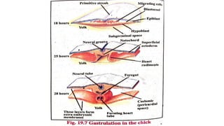
This is a slide for complete development in chick ,as chick is a vertebrate so with the help of the development in a chick we can we can understand development in vertebrates .
This topic explains the whole process of growth and development in animal the processes include
Fertilization and incubation
Cleavage
Morula
Blastula
Gastrulation
Notochord And Mesoderm Formation
Neurulation
2. EMBRYOLOGY
• The study of Growth and differentiation undergone by an organism in
the course of its development from a single fertilized egg into a highly
complex and an independent living being like his parents.
•DEVELOPMENT
• It is an ordered sequence of irreversible steps, with each step setting up the
necessary conditions for the next step.
• Since all animals are somehow related to each other due to evolution, there
are some similarities in their various forms of development
4. DEVELOPMENT OF CHICK
• PHYLUM: Chordata
• SUB-PHYLUM: Vertebrata
• CLASS: Aves
• The development of chick shows all the fundamental processes of
body formation which is common to all vertebrates.
5. Stages in the development of Chick:
1. Fertilization and incubation
2. Cleavage
3. Morula
4. Blastula
5. Gastrulation
6. Notochord formation
7. Neurulation
8. Mesoderm and coelom
formation
9. Organogenesis
6. 1. Fertilization and Incubation:
• The chick egg (oocyte), (called the YOLK) is surrounded by various
accessary coverings, which are secreted by female reproductive tract.
• Internal fertilization ( inside female reproductive tract)
• Fertilization takes place just as ovum is entering the oviduct.
• The shell is secreted as the egg is passing through the shell gland (
the uterus)
• The egg is laid after addition of albumin and coverings + shell.
7.
8. Incubation:
• The development inside the egg ceases unless the temperature of egg
is kept nearly up to the body temperature.
• The incubation temperature is between 36 – 38 º C.
• The same temperature is maintained when incubating eggs artificially
• Incubation time:
21 days
9. 2. Cleavage:
• Immediately after fertilization, the egg undergoes a series of mitotic
divisions, called cleavage.
• Discoidal cleavage:
• The process of cell division in chick embryo is confined to the small
disc of protoplasm lying on the surface of yolk at the animal pole. it
results in the formation of a disc of cells on surface of yolk.
(in a zygote there are two ends of the cell; the end with more yolk is
called vegetal pole and the other side which contains nucleus and
cytoplasm is called animal pole)
10. Cleavage (cont.)
• The cleavage furrow start in the clear cytoplasmic region.
• The first two cleavage planes are vertical
• while the third runs horizontally parallel to the surface and thus cuts
underneath the cytoplasm and separates it from the yolk.
• The successive cleavages become irregular and number of cells
increase.
14. Morula:
• Zygote → cleavage → morula
• Morula consists of a disc shaped mass of cells two or more layers in
thickness (blastoderm) lying close to the yolk.
• In the center of the blastoderm, the cells are smaller and completely
defined while those at the periphery are flattened are larger.
• Morula is a short-lived stage and soon changes into blastula.
15. Blastula:
• The blastula is characterized by the presence of a segmentation cavity
or blastocoel.
• The discoidal cap of cells above the blastocoel is called the
blastoderm.
• The marginal area of the blastoderm in which the cells remain
undetached from the yolk and are adherent to it is called the zone of
junction.
16.
17.
18. Gastrulation:
• The stage of development in which three germinal layers are formed.
i-e. ectoderm, mesoderm and endoderm.
• The gastrulation in chick involves the following changes:
1. The blastoderm splits into two layers, an upper layer called
epiblast and a lower layer of cells called hypoblast.
2. Epiblast is mainly presumptive (precursor of ) ectoderm and
mesoderm and hypoblast is presumptive endoderm.
3. The hypoblast cells grow outward over the surface of the yolk, then
downward around it to form the endodermal lining of a yolk sac.
The space between epiblast and hypoblast is called blastocoel.
19. 4. Area Pellucida and area opaqua:
• At this stage, the central cells of blastoderm can be separated from
the yolk. Under these central cells a pool of fluid
develops,(subgerminal space) raising them off the yolk and giving
the area a translucent appearance. This area is called area
pellucida. (allows light to pass)
• Area opaqua: The opaque area of blastoderm surrounding the
area pellucida is called area opaqua (peripheral region). Cells here
are unseparated from yolk . (doesnot allow light to pass, appears
darker)
Subgerminal space: the space between blastoderm and yolk. Filled
with a fluid.
20.
21. Migration of cells and formation of Primitive
streak:
• Primitive streak is a midline thickening on the surface of embryo.
• It is formed due to migration of cells medially (towards middle) and
caudally. This is the beginning of formation of three germ layers.
• It thickens and grows rapidly in length as more and more cells continue
to aggregate in the middle.
• The shape of the blastula changes from circular to pear shaped.
• Primitive node:
• The anterior end of the primitive streak is occupied by an aggregation
called the primitive node. The cells here are important because these are
notochordal cells. Rest of the cells of Primitive streak are mesodermal
cells.
22.
23.
24. • The formation of primitive streaks marks the anterior and posterior
ends and also the dorsal and ventral sides of the body.
• The cells from here begin to migrate into the blastocoel. That is from
surface to inside of embryo. Thus the primitive streaks is the blastopore
of the embryo and its margins are the dorsal and ventral lips of blastopore.
• Primitive Groove:
• The continuous migration of cells takes place between epiblast and
hypoblast and results in the formation of a groove (depression) along the
whole length of primitive streak. This is named as primitive groove.
• The margins of the primitive groove are called Primitive ridges.
25.
26. Formation of Henson’s node:
• At the cephalic (the side where head forms/ anterior) end of primitive
streak, closely packed cells form a thickening knows as Henson’s
node. ( an aggregation of cells)
• From Henson node an invagination will form which will lead to the
formation of notochord.
• The cells continue to migrate into the blastocoel. The first migrating
cells will form endoderm whereas the cells which migrate later will
form mesoderm. While the cells which remain on the surface will form
ectoderm. In this way the three germ layers are formed.
27.
28.
29.
30. Formation of Notochord:
• Notochord is a supportive structure formed in embryo on the dorsal
side. it also marks the main body axis.
• It forms shortly after the formation of primitive groove and primitive
streak.
• After the endoderm is well established, cells begin to push in from the
region of Henson’s node to form rod like notochord in the midline
beneath the ectoderm.
• In an 18 hours chick embryo notochord is one of the few prominent
features.
31.
32. • Beside notochord in an 18 hours embryo,
• The epidermis spreads into a coherent layer of cells. These cells
merge with the yolk on the periphery.
• The marginal area where the expanding germ layers merge with the
yolk is called the germ wall.
• Primitive Gut:
• The cavity between endoderm and yolk which was called the
gastrocoel, is now called the primitive gut (early gut because this
is where digestive system will form).
33.
34. Formation and differentiation of Mesoderm:
• The middle layer of the embryo is called mesoderm.
• It is formed due to migration of cells from primitive groove into
the blastocoel.
• The mesoderm differentiates as:
1. Dorsal mesoderm: it forms from Henson’s node and develops
into notochord and Somites.
• Somites are compact cell masses on both sides of notochord and
neural tube. These form skeleton and skeletal muscles.
35. 2. Lateral plate mesoderm: This layer is on the lateral sides of
embryo. It further splits into two layers.
• Somatic mesoderm: the layer towards ectoderm
• Splanchnic mesoderm: the layer towards endoderm.
• The space between somatic and splanchnic mesoderm is called
Coelom.
• The ectodermal cells lying just above the notochord develop into
neural plate ectoderm. This part of ectoderm forms nervous system.
36.
37.
38. Neurulation:
• Formation of neural tube in the embryo is called neurulation. This
neural tube forms the future nervous system of the embryo. The
embryo after formation of neural tube is called a neurula.
• The neural tube forms from presumptive neural ectoderm which
lies just above the notochord in the form of a band.
• It occurs in following steps:
1. Formation of neural plate:
• as gastrula elongates, the band thinkens to form the neural plate. In
18 hours embryo, it is a flat, thickened areaof ectoderm.
39. 2. Neural groove:
• In embryo Of 21-22 hours, the neural plate shows a longitudinal folding
i-e it invaginates longitudinally forming a neural groove in the mid
dorsal line. This is called neural groove. Its margins are called neural
folds.
• The anterior end of the groove is widest and it will form the future brain
whereas rest of the portion will form spinal cord.
• In 24 hours embryo the folding of neural plate is clearly visible. The
embryo is now called a neurula.
3. Neural tube formation:
• The neural fold grow towards each other and meet in the middle, fuse
and convert the neural groove into neural tube.
43. • The cavity enclosed by neural tube is called the neurocoel.
• At each end of the tube, small openings called anterior and posterior
neuropores are also seen. Which close later on.
• With the formation of neural tube there is formation of central nervous
system.
• The marginal area of the germ layers encloses the embryo by forming
the three extraembryonic membranes called amnion, chorion and
allantois. Which provide protection to embryo.