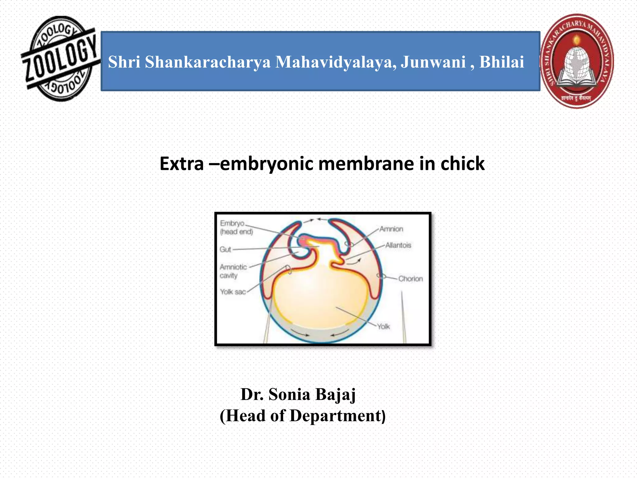
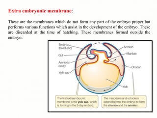
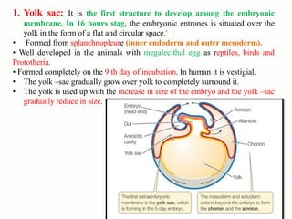
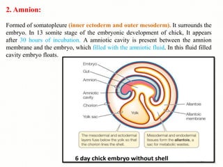
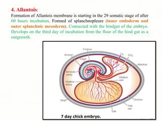
SlideShare a Scribd company logo
Shri Shankaracharya Mahavidyalaya, Junwani , Bhilai
Extra –embryonic membrane in chick
Dr. Sonia Bajaj
(Head of Department)
Extra embryonic membrane: These are the membranes which do not
form any part of the embryo proper but performs various functions which assist in
the development of the embryo. These are discarded at the time of hatching.
These membranes formed outside the embryo .
Amniotes: These are the vertebrates group whose eggs contain extra
embryonic membranes for protecting the embryo. They lay eggs on the land.
Therefore ,in chick and other amniotes the following extra embryonic membrane
develop in them for the protection, respiration and nutrition of the embryo.
Example: Reptiles, Birds and Mammals.
An amniotes: These are the vertebrates group whose eggs do not contain
extra embryonic membranes during embryonic development. They lay eggs in the
water. Example: Fish, Amphibian.
Extra embryonic membranes in chick
In chick, the presence of an enormous amount of yolk and embryonic life to
be spent within a shell is correlated with the development of extra-embryonic
membranes. Original blastoderm is a small disc, which spreads by peripheral
expansion and eventually covers the entire surface of the egg. But only the
most central region is directly connected with the formation of the embryo
proper. All the remainder of the blastoderm is extra-embryonic.
There are four types of extra embryonic membranes in birds-
1. Yolk sac.
2. Amnion.
3. Chorion.
4. Allantois
Extra embryonic membrane:
These are the membranes which do not form any part of the embryo proper but
performs various functions which assist in the development of the embryo. These
are discarded at the time of hatching. These membranes formed outside the
embryo.
1. Yolk sac: It is the first structure to develop among the embryonic
membrane. In 16 hours stag, the embryonic entrones is situated over the
yolk in the form of a flat and circular space.`
• Formed from splanchnopleure (inner endoderm and outer mesoderm).
• Well developed in the animals with megalecithal egg as reptiles, birds and
Prototheria.
• Formed completely on the 9 th day of incubation. In human it is vestigial.
• The yolk –sac gradually grow over yolk to completely surround it.
• The yolk is used up with the increase in size of the embryo and the yolk –sac
gradually reduce in size.
Functions of Yolk sac:
1.It surrounds the yolk. Its main function is in digestion. It
serve as extra embryonic gut.
2. It help in digestion of yolk and transfer the digested
material to the developing embryo.
3. First respiratory organ in the embryo.
4. Form yolk sac placenta in the marsupials.
Development of yolk sac-
In reptiles and birds, the somatopleure and splanchnopleure develop from the
periphery of the blast disc. These usually spread peripherally over the yolk mass.
Soon afterwards, the embryo undergoes series of folds, which appear all around
the body of the embryo. These folds are termed as the body folds. The extra
embryonic splanchnopleure (splanchnic mesoderm + endoderm) constantly
spreads over the yolk mass and eventually yolk sac encloses the mass of yolk in a
large measure. The yolk sac, however, not surrounds the yolk fully. A small
passage is left on the ventral side for the embryo to absorb the remains of
albumen at a later stage. Immediately with the formation of the yolk sac, the intra
embryonic splanchnopleure is subjected to fold resembling with the more
superficial body folds i.e., the intra embryonic folds. The intra embryonic folds
give rise to walled digestive tract, or gut, in the body of the embryo. The middle
of the embryonic gut remains open to the yolk beneath. At this level, yolk sac is
connected to the digestive tract by a constricted yolk stalk.
2. Amnion:
Formed of somatopleure (inner ectoderm and outer mesoderm). It surrounds the
embryo. In 13 somite stage of the embryonic development of chick, It appears
after 30 hours of incubation. A amniotic cavity is present between the amnion
membrane and the embryo, which filled with the amniotic fluid. In this fluid filled
cavity embryo floats.
Function of Amnion:
The fluid filled in the amniotic cavity forms a sort of pool for the embryo and
therefore, performs the following functions-
1. It prevents the embryo from drying.
2. Protection of the embryo from the mechanical injury and desiccation.
3. It protects the embryo from external shock.
4. It prevents the embryo from sticking to the shell or embryonic membranes.
5. It also helps in the absorption of the albumen.
6. The amniotic fluid allows freedom of motion to the embryo .
7. It is working as a barrier of outer organisms.
8. In most mammals of a part of the chorion forms finger-like out-growths called
chorionic villi. That penetrates into corresponding crypts or depressions in the wall
of the uterus. It is at this site where exchange of substances occurs found in the
inner cell mass begin to spread along the inner surface on the trophoblast and
surround the internal cavity of the blastocyte in the same way as the endodermal
cell in reptiles and birds surrounded the mass of uncleaved egg.
3. Chorion:
Formed of somatopleure (outer ectoderm and inner mesoderm). It forms the
outermost boundary. Space between amnion and chorion is called chorionic cavity
which further provides protection to the embryo.
Function of chorion:
1. It is outer layer, therefore, it is the place where exchange of substance occurs
between embryonic tissue in the maternal environment.
2. It acts as a respiratory organ and is in contact with the allantois.
3. In most mammals it develops villi which penetrate into the tissue of uterus wall
and increase the absorptive area of chorion.
4. Blood vessels are developing after the formation of allantochorion and this is
working as a lung. It takes O2, from atmosphere and removes CO2, to the
atmosphere.
5. It provides calcium for skeleton of developing embryo by transportation of Ca by
shell.
Development of Amnion & Chorion-
The amnion and chorion are developed jointly as upward projecting folds, the
amniotic folds of the extra embryonic somatopleure. The amniotic folds are
named according to their location. They are amniotic head fold and amniotic tail
fold. The amniotic fold first appears as a transverse fold in front of the head. It
is called amniotic head fold. It grows upwards and then bends backwards, over
the anterior end of the head and covers it as with a hood. Another fold develops
behind the embryo. It is termed as amniotic tail fold. All these folds finally
cover an embryo in two sheets of somatopleure. The inner somatopleuric sheet
becomes the amnion and the outer, the chorion.
The amnion consists of a layer of extra embryonic ectoderm on the inside and a
layer of extra embryonic somatic mesoderm on the outside whereas, the chorion
is made up of a layer of extra embryonic ectoderm on the outside and a layer of
extra embryonic somatic mesoderm on the inside. The cavity between the
amnion and the embryo is called the amniotic cavity. In between, the amnion
and the chorion is the chorionic cavity or extra embryonic coelom.
4. Allantois:
Formation of Allantois membrane is starting in the 29 somatic stage of after
60 hours incubation. Formed of splanchnopleure (inner endoderm and
outer splanchnic mesoderm). Connected with the hindgut of the embryo.
Develops on the third day of incubation from the floor of the hind gut as a
outgrowth.
Development of allantois -The allantois arises as a ventral outgrowth of the
splanchnopleure from the hindgut on the third day of incubation. It slowly enlarges
as holosac and expands inside exocoel. Its walls are formed of an outer splanchnic
mesoderm and inner endoderm. The proximal part of the allantois forms a slender
neck or the allantoic stalk with which it remains connected with the hindgut of the
embryo. The distal part of the allantois expands and penetrates between the amnion
and the yolk sac on one side and the chorion on the other side. By the middle of the
incubation period, the allantois spreads all around the egg underneath the chorion.
The mesoderm on the external surface of the allantois fuses with that of the chorion
forming a conjoined chorio-allantoic membrane.
Function of allantois:
The allontois is highly muscular from the beginning, As a result of the
formation of the allanto-chorion the blood vessels become dispersed over the
inner surface of the porous shell. Thus, it performs the following functions –
1. It acts as a respiratory organ. It is working as a lung and provides
atmospheric O2, to the embryo by porous shell. It is working of the
exchange of gases because it is situated near the porous shell.
2. It collects the embryonic villi and working as a excretory organ (urinary
bladder) of embryo.
3. Also helps in digestion and nutrition from albumen and calcium of the shell.
4. It helps in removal of waste products, e.g, CO2, urea, etc. of metabolism
from the embryo.
5.
Importance of foetal membranes:
• The presence of amnion, allantois and chorion is an adaptation
for terrestrial life of animals. Their presence enables the embryo
and adult to live on land.
• The presence of yolk-sac is for different purposes. It acts as a
digestive and absorptive surface through which the embryo
gets the yolk.
References-
Modern text book – R.L.Kotpal
Jantu Vigyan- S.M. Sexsena
Jantu Vigyan- Dr.H.N. Baijal

More Related Content

What's hot

Cleavage, Types of cleavage
Cleavage, Types of cleavage Cleavage, Types of cleavage
Cleavage, Types of cleavage
SoniaBajaj10
 
Origin and Organization of Coelom_MS.pptx
Origin and Organization of Coelom_MS.pptxOrigin and Organization of Coelom_MS.pptx
Origin and Organization of Coelom_MS.pptx
dean137192
 
succession of kidneys in a vertebrate series
succession of kidneys in a vertebrate seriessuccession of kidneys in a vertebrate series
succession of kidneys in a vertebrate series
Naman Sharma
 
Regeneration, Types of Regeneration,Invertebrates&Vertebrates sp. ,Mechanism,...
Regeneration, Types of Regeneration,Invertebrates&Vertebrates sp. ,Mechanism,...Regeneration, Types of Regeneration,Invertebrates&Vertebrates sp. ,Mechanism,...
Regeneration, Types of Regeneration,Invertebrates&Vertebrates sp. ,Mechanism,...
SoniaBajaj10
 
embryonic-induction.pdf
embryonic-induction.pdfembryonic-induction.pdf
embryonic-induction.pdf
souryajyotidash
 
Fetal membranes in chick
Fetal membranes in chickFetal membranes in chick
Fetal membranes in chick
Sandip Rathod
 
Integument.pdf
Integument.pdfIntegument.pdf
Integument.pdf
Mohd Asif Kanth
 
Skull of rabbit
Skull of rabbit Skull of rabbit
Skull of rabbit
Shivani Rawat
 
Evolution of Urinogenital Ducts
Evolution of Urinogenital DuctsEvolution of Urinogenital Ducts
Evolution of Urinogenital Ducts
D. B. S. College Kanpur
 
Extra embryonic membranes
Extra embryonic membranesExtra embryonic membranes
Extra embryonic membranes
jyoti malik
 
Biochemistry of fertilization.
Biochemistry of fertilization.Biochemistry of fertilization.
Biochemistry of fertilization.
KrishnaSahu94
 
Lateral line system
Lateral line systemLateral line system
Lateral line system
Govt.college,Nagda, ujjain.M.P
 
Placenta structure and Classification
Placenta structure and ClassificationPlacenta structure and Classification
Placenta structure and Classification
Dr. Dinesh C. Sharma
 
Regeneration
RegenerationRegeneration
Regeneration
Syed Muhammad Khan
 
Comparative anatomy of aortic arches
Comparative anatomy of aortic archesComparative anatomy of aortic arches
Comparative anatomy of aortic arches
Shahla Yasmin
 
Gastrulation in frog embryo
Gastrulation in frog embryoGastrulation in frog embryo
Gastrulation in frog embryo
NARTHANAA SENAVARAYAN
 
Gastrulation chick
Gastrulation chickGastrulation chick
Gastrulation chick
jyoti malik
 
Regeneration in vertebrates
Regeneration in vertebratesRegeneration in vertebrates
Regeneration in vertebrates
Kashif Manzoor
 
FATE Map
FATE MapFATE Map
Development of frog/ embryogenesis
Development of frog/ embryogenesisDevelopment of frog/ embryogenesis
Development of frog/ embryogenesis
Dambar Khatri
 

What's hot (20)

Cleavage, Types of cleavage
Cleavage, Types of cleavage Cleavage, Types of cleavage
Cleavage, Types of cleavage
 
Origin and Organization of Coelom_MS.pptx
Origin and Organization of Coelom_MS.pptxOrigin and Organization of Coelom_MS.pptx
Origin and Organization of Coelom_MS.pptx
 
succession of kidneys in a vertebrate series
succession of kidneys in a vertebrate seriessuccession of kidneys in a vertebrate series
succession of kidneys in a vertebrate series
 
Regeneration, Types of Regeneration,Invertebrates&Vertebrates sp. ,Mechanism,...
Regeneration, Types of Regeneration,Invertebrates&Vertebrates sp. ,Mechanism,...Regeneration, Types of Regeneration,Invertebrates&Vertebrates sp. ,Mechanism,...
Regeneration, Types of Regeneration,Invertebrates&Vertebrates sp. ,Mechanism,...
 
embryonic-induction.pdf
embryonic-induction.pdfembryonic-induction.pdf
embryonic-induction.pdf
 
Fetal membranes in chick
Fetal membranes in chickFetal membranes in chick
Fetal membranes in chick
 
Integument.pdf
Integument.pdfIntegument.pdf
Integument.pdf
 
Skull of rabbit
Skull of rabbit Skull of rabbit
Skull of rabbit
 
Evolution of Urinogenital Ducts
Evolution of Urinogenital DuctsEvolution of Urinogenital Ducts
Evolution of Urinogenital Ducts
 
Extra embryonic membranes
Extra embryonic membranesExtra embryonic membranes
Extra embryonic membranes
 
Biochemistry of fertilization.
Biochemistry of fertilization.Biochemistry of fertilization.
Biochemistry of fertilization.
 
Lateral line system
Lateral line systemLateral line system
Lateral line system
 
Placenta structure and Classification
Placenta structure and ClassificationPlacenta structure and Classification
Placenta structure and Classification
 
Regeneration
RegenerationRegeneration
Regeneration
 
Comparative anatomy of aortic arches
Comparative anatomy of aortic archesComparative anatomy of aortic arches
Comparative anatomy of aortic arches
 
Gastrulation in frog embryo
Gastrulation in frog embryoGastrulation in frog embryo
Gastrulation in frog embryo
 
Gastrulation chick
Gastrulation chickGastrulation chick
Gastrulation chick
 
Regeneration in vertebrates
Regeneration in vertebratesRegeneration in vertebrates
Regeneration in vertebrates
 
FATE Map
FATE MapFATE Map
FATE Map
 
Development of frog/ embryogenesis
Development of frog/ embryogenesisDevelopment of frog/ embryogenesis
Development of frog/ embryogenesis
 

Similar to Extra embryonic membrane in chick, Types, Developments, Functions

Implantation, Embryonic membrane and Twinning
Implantation, Embryonic membrane and TwinningImplantation, Embryonic membrane and Twinning
Implantation, Embryonic membrane and Twinning
Amani Riyadh
 
Extra embryonic structures
Extra embryonic structuresExtra embryonic structures
Extra embryonic structures
Najam ul Sehar Aiman
 
General Embryology
General EmbryologyGeneral Embryology
General Embryology
Eneutron
 
embryologymo-150405023631-conversion-gate01.pdf
embryologymo-150405023631-conversion-gate01.pdfembryologymo-150405023631-conversion-gate01.pdf
embryologymo-150405023631-conversion-gate01.pdf
Ullas Chandra Sahoo
 
Implantation and placentation , and overview
Implantation and placentation , and overviewImplantation and placentation , and overview
Implantation and placentation , and overview
Pranjal Gupta
 
Embryonic and post embryonic development in insect
Embryonic and post embryonic development in insectEmbryonic and post embryonic development in insect
Embryonic and post embryonic development in insect
Indian Agricultural Research Institute, Pusa Campus, New Delhi
 
Obg ppt
Obg pptObg ppt
Obg ppt
naz khan
 
BIOUnnes_Oogenesis
BIOUnnes_OogenesisBIOUnnes_Oogenesis
BIOUnnes_Oogenesis
Nur Aini
 
FORMATION OF EMBRYO and FETAL DEVELOPMENT.pdf
FORMATION OF EMBRYO and FETAL DEVELOPMENT.pdfFORMATION OF EMBRYO and FETAL DEVELOPMENT.pdf
FORMATION OF EMBRYO and FETAL DEVELOPMENT.pdf
Dolisha Warbi
 
Basics of Human Embryology including dental applications
Basics of Human Embryology including dental applicationsBasics of Human Embryology including dental applications
Basics of Human Embryology including dental applications
sadafcmw1
 
Embryology of face
Embryology of faceEmbryology of face
Embryology of facesukhjit kaur
 
Embryology part 2
Embryology part 2Embryology part 2
Embryology part 2
Amirrasa Kateb
 
Development of face, paranasal sinus.
Development of face, paranasal sinus.Development of face, paranasal sinus.
Development of face, paranasal sinus.
ashwin harekal
 
357( lectures-1) 2013
357( lectures-1) 2013357( lectures-1) 2013
357( lectures-1) 2013
GD_357
 
2nd week-implantation.ppt
2nd week-implantation.ppt2nd week-implantation.ppt
2nd week-implantation.ppt
ShrutiYadav335673
 
Extraembryonic structures in reptiles and birds
Extraembryonic structures in reptiles and birdsExtraembryonic structures in reptiles and birds
Extraembryonic structures in reptiles and birds
BilawalGhafoor2
 
Embryology{extra embryonic membrane}(1)
Embryology{extra embryonic membrane}(1)Embryology{extra embryonic membrane}(1)
Embryology{extra embryonic membrane}(1)
Madurai kamaraj university
 
Developmental Biology
Developmental BiologyDevelopmental Biology

Similar to Extra embryonic membrane in chick, Types, Developments, Functions (20)

Implantation, Embryonic membrane and Twinning
Implantation, Embryonic membrane and TwinningImplantation, Embryonic membrane and Twinning
Implantation, Embryonic membrane and Twinning
 
Extra embryonic structures
Extra embryonic structuresExtra embryonic structures
Extra embryonic structures
 
General Embryology
General EmbryologyGeneral Embryology
General Embryology
 
Embryology
Embryology Embryology
Embryology
 
embryologymo-150405023631-conversion-gate01.pdf
embryologymo-150405023631-conversion-gate01.pdfembryologymo-150405023631-conversion-gate01.pdf
embryologymo-150405023631-conversion-gate01.pdf
 
Implantation and placentation , and overview
Implantation and placentation , and overviewImplantation and placentation , and overview
Implantation and placentation , and overview
 
Embryonic and post embryonic development in insect
Embryonic and post embryonic development in insectEmbryonic and post embryonic development in insect
Embryonic and post embryonic development in insect
 
Obg ppt
Obg pptObg ppt
Obg ppt
 
BIOUnnes_Oogenesis
BIOUnnes_OogenesisBIOUnnes_Oogenesis
BIOUnnes_Oogenesis
 
FORMATION OF EMBRYO and FETAL DEVELOPMENT.pdf
FORMATION OF EMBRYO and FETAL DEVELOPMENT.pdfFORMATION OF EMBRYO and FETAL DEVELOPMENT.pdf
FORMATION OF EMBRYO and FETAL DEVELOPMENT.pdf
 
Basics of Human Embryology including dental applications
Basics of Human Embryology including dental applicationsBasics of Human Embryology including dental applications
Basics of Human Embryology including dental applications
 
Embryology of face
Embryology of faceEmbryology of face
Embryology of face
 
Germ Layers Ppdf
Germ Layers PpdfGerm Layers Ppdf
Germ Layers Ppdf
 
Embryology part 2
Embryology part 2Embryology part 2
Embryology part 2
 
Development of face, paranasal sinus.
Development of face, paranasal sinus.Development of face, paranasal sinus.
Development of face, paranasal sinus.
 
357( lectures-1) 2013
357( lectures-1) 2013357( lectures-1) 2013
357( lectures-1) 2013
 
2nd week-implantation.ppt
2nd week-implantation.ppt2nd week-implantation.ppt
2nd week-implantation.ppt
 
Extraembryonic structures in reptiles and birds
Extraembryonic structures in reptiles and birdsExtraembryonic structures in reptiles and birds
Extraembryonic structures in reptiles and birds
 
Embryology{extra embryonic membrane}(1)
Embryology{extra embryonic membrane}(1)Embryology{extra embryonic membrane}(1)
Embryology{extra embryonic membrane}(1)
 
Developmental Biology
Developmental BiologyDevelopmental Biology
Developmental Biology
 

More from SoniaBajaj10

Probability.pptx, Types of Probability, UG
Probability.pptx, Types of Probability, UGProbability.pptx, Types of Probability, UG
Probability.pptx, Types of Probability, UG
SoniaBajaj10
 
Measures of Central Tendency.pptx for UG
Measures of Central Tendency.pptx for UGMeasures of Central Tendency.pptx for UG
Measures of Central Tendency.pptx for UG
SoniaBajaj10
 
Primitive,diffused & advance Nervous system in Non chordata.pptx
Primitive,diffused & advance Nervous system in Non chordata.pptxPrimitive,diffused & advance Nervous system in Non chordata.pptx
Primitive,diffused & advance Nervous system in Non chordata.pptx
SoniaBajaj10
 
Comparative anatomy & Physiology of Excretion in Invertebrates.pptx
Comparative anatomy & Physiology of  Excretion in Invertebrates.pptxComparative anatomy & Physiology of  Excretion in Invertebrates.pptx
Comparative anatomy & Physiology of Excretion in Invertebrates.pptx
SoniaBajaj10
 
Comparative Anatomy & Physiology of Respiration in Non chordata.pptx
Comparative Anatomy & Physiology of Respiration in  Non chordata.pptxComparative Anatomy & Physiology of Respiration in  Non chordata.pptx
Comparative Anatomy & Physiology of Respiration in Non chordata.pptx
SoniaBajaj10
 
Primitive,diffused & advance Nervous system in Non chordata.pptx
Primitive,diffused & advance Nervous system in Non chordata.pptxPrimitive,diffused & advance Nervous system in Non chordata.pptx
Primitive,diffused & advance Nervous system in Non chordata.pptx
SoniaBajaj10
 
Locomotry organ in non chordata.pptx
Locomotry organ in non chordata.pptxLocomotry organ in non chordata.pptx
Locomotry organ in non chordata.pptx
SoniaBajaj10
 
Pathogenic nemadodes & disease.pptx
Pathogenic nemadodes & disease.pptxPathogenic nemadodes & disease.pptx
Pathogenic nemadodes & disease.pptx
SoniaBajaj10
 
Animal Behaviour
Animal BehaviourAnimal Behaviour
Animal Behaviour
SoniaBajaj10
 
External part of Body (oral & ab oral Surface) , Pedicellaria & water vascula...
External part of Body (oral & ab oral Surface) , Pedicellaria & water vascula...External part of Body (oral & ab oral Surface) , Pedicellaria & water vascula...
External part of Body (oral & ab oral Surface) , Pedicellaria & water vascula...
SoniaBajaj10
 
Respiratory system of pila
Respiratory system of pilaRespiratory system of pila
Respiratory system of pila
SoniaBajaj10
 
Digestive system of pila
Digestive system of pilaDigestive system of pila
Digestive system of pila
SoniaBajaj10
 
Placentation in mammal,classification of placenta and function
Placentation  in mammal,classification of placenta and function Placentation  in mammal,classification of placenta and function
Placentation in mammal,classification of placenta and function
SoniaBajaj10
 
Development of chick embryo- structure of egg,cleavages,fate map and primitiv...
Development of chick embryo- structure of egg,cleavages,fate map and primitiv...Development of chick embryo- structure of egg,cleavages,fate map and primitiv...
Development of chick embryo- structure of egg,cleavages,fate map and primitiv...
SoniaBajaj10
 
Diffrentiation,Cell diffrentiation,Types of differentiation,Mechanism,Factors...
Diffrentiation,Cell diffrentiation,Types of differentiation,Mechanism,Factors...Diffrentiation,Cell diffrentiation,Types of differentiation,Mechanism,Factors...
Diffrentiation,Cell diffrentiation,Types of differentiation,Mechanism,Factors...
SoniaBajaj10
 
Prawn respiratory system,Types of gills,structure of gill plates,working of ...
Prawn  respiratory system,Types of gills,structure of gill plates,working of ...Prawn  respiratory system,Types of gills,structure of gill plates,working of ...
Prawn respiratory system,Types of gills,structure of gill plates,working of ...
SoniaBajaj10
 
Prawn digestive system,parts of Alimentary canal,structure of hastate plate,
Prawn  digestive system,parts of Alimentary canal,structure of hastate plate,Prawn  digestive system,parts of Alimentary canal,structure of hastate plate,
Prawn digestive system,parts of Alimentary canal,structure of hastate plate,
SoniaBajaj10
 
Reproductive system of earthworm-Male reproductive part and female reproducti...
Reproductive system of earthworm-Male reproductive part and female reproducti...Reproductive system of earthworm-Male reproductive part and female reproducti...
Reproductive system of earthworm-Male reproductive part and female reproducti...
SoniaBajaj10
 
Excretory system of earthworm , Three types of nephridia, structure of septa...
Excretory system of earthworm ,  Three types of nephridia, structure of septa...Excretory system of earthworm ,  Three types of nephridia, structure of septa...
Excretory system of earthworm , Three types of nephridia, structure of septa...
SoniaBajaj10
 
Blood circulatory system of earthworm
Blood circulatory system of earthwormBlood circulatory system of earthworm
Blood circulatory system of earthworm
SoniaBajaj10
 

More from SoniaBajaj10 (20)

Probability.pptx, Types of Probability, UG
Probability.pptx, Types of Probability, UGProbability.pptx, Types of Probability, UG
Probability.pptx, Types of Probability, UG
 
Measures of Central Tendency.pptx for UG
Measures of Central Tendency.pptx for UGMeasures of Central Tendency.pptx for UG
Measures of Central Tendency.pptx for UG
 
Primitive,diffused & advance Nervous system in Non chordata.pptx
Primitive,diffused & advance Nervous system in Non chordata.pptxPrimitive,diffused & advance Nervous system in Non chordata.pptx
Primitive,diffused & advance Nervous system in Non chordata.pptx
 
Comparative anatomy & Physiology of Excretion in Invertebrates.pptx
Comparative anatomy & Physiology of  Excretion in Invertebrates.pptxComparative anatomy & Physiology of  Excretion in Invertebrates.pptx
Comparative anatomy & Physiology of Excretion in Invertebrates.pptx
 
Comparative Anatomy & Physiology of Respiration in Non chordata.pptx
Comparative Anatomy & Physiology of Respiration in  Non chordata.pptxComparative Anatomy & Physiology of Respiration in  Non chordata.pptx
Comparative Anatomy & Physiology of Respiration in Non chordata.pptx
 
Primitive,diffused & advance Nervous system in Non chordata.pptx
Primitive,diffused & advance Nervous system in Non chordata.pptxPrimitive,diffused & advance Nervous system in Non chordata.pptx
Primitive,diffused & advance Nervous system in Non chordata.pptx
 
Locomotry organ in non chordata.pptx
Locomotry organ in non chordata.pptxLocomotry organ in non chordata.pptx
Locomotry organ in non chordata.pptx
 
Pathogenic nemadodes & disease.pptx
Pathogenic nemadodes & disease.pptxPathogenic nemadodes & disease.pptx
Pathogenic nemadodes & disease.pptx
 
Animal Behaviour
Animal BehaviourAnimal Behaviour
Animal Behaviour
 
External part of Body (oral & ab oral Surface) , Pedicellaria & water vascula...
External part of Body (oral & ab oral Surface) , Pedicellaria & water vascula...External part of Body (oral & ab oral Surface) , Pedicellaria & water vascula...
External part of Body (oral & ab oral Surface) , Pedicellaria & water vascula...
 
Respiratory system of pila
Respiratory system of pilaRespiratory system of pila
Respiratory system of pila
 
Digestive system of pila
Digestive system of pilaDigestive system of pila
Digestive system of pila
 
Placentation in mammal,classification of placenta and function
Placentation  in mammal,classification of placenta and function Placentation  in mammal,classification of placenta and function
Placentation in mammal,classification of placenta and function
 
Development of chick embryo- structure of egg,cleavages,fate map and primitiv...
Development of chick embryo- structure of egg,cleavages,fate map and primitiv...Development of chick embryo- structure of egg,cleavages,fate map and primitiv...
Development of chick embryo- structure of egg,cleavages,fate map and primitiv...
 
Diffrentiation,Cell diffrentiation,Types of differentiation,Mechanism,Factors...
Diffrentiation,Cell diffrentiation,Types of differentiation,Mechanism,Factors...Diffrentiation,Cell diffrentiation,Types of differentiation,Mechanism,Factors...
Diffrentiation,Cell diffrentiation,Types of differentiation,Mechanism,Factors...
 
Prawn respiratory system,Types of gills,structure of gill plates,working of ...
Prawn  respiratory system,Types of gills,structure of gill plates,working of ...Prawn  respiratory system,Types of gills,structure of gill plates,working of ...
Prawn respiratory system,Types of gills,structure of gill plates,working of ...
 
Prawn digestive system,parts of Alimentary canal,structure of hastate plate,
Prawn  digestive system,parts of Alimentary canal,structure of hastate plate,Prawn  digestive system,parts of Alimentary canal,structure of hastate plate,
Prawn digestive system,parts of Alimentary canal,structure of hastate plate,
 
Reproductive system of earthworm-Male reproductive part and female reproducti...
Reproductive system of earthworm-Male reproductive part and female reproducti...Reproductive system of earthworm-Male reproductive part and female reproducti...
Reproductive system of earthworm-Male reproductive part and female reproducti...
 
Excretory system of earthworm , Three types of nephridia, structure of septa...
Excretory system of earthworm ,  Three types of nephridia, structure of septa...Excretory system of earthworm ,  Three types of nephridia, structure of septa...
Excretory system of earthworm , Three types of nephridia, structure of septa...
 
Blood circulatory system of earthworm
Blood circulatory system of earthwormBlood circulatory system of earthworm
Blood circulatory system of earthworm
 

Recently uploaded

Circulatory system_ Laplace law. Ohms law.reynaults law,baro-chemo-receptors-...
Circulatory system_ Laplace law. Ohms law.reynaults law,baro-chemo-receptors-...Circulatory system_ Laplace law. Ohms law.reynaults law,baro-chemo-receptors-...
Circulatory system_ Laplace law. Ohms law.reynaults law,baro-chemo-receptors-...
muralinath2
 
Lab report on liquid viscosity of glycerin
Lab report on liquid viscosity of glycerinLab report on liquid viscosity of glycerin
Lab report on liquid viscosity of glycerin
ossaicprecious19
 
Hemostasis_importance& clinical significance.pptx
Hemostasis_importance& clinical significance.pptxHemostasis_importance& clinical significance.pptx
Hemostasis_importance& clinical significance.pptx
muralinath2
 
NuGOweek 2024 Ghent - programme - final version
NuGOweek 2024 Ghent - programme - final versionNuGOweek 2024 Ghent - programme - final version
NuGOweek 2024 Ghent - programme - final version
pablovgd
 
RNA INTERFERENCE: UNRAVELING GENETIC SILENCING
RNA INTERFERENCE: UNRAVELING GENETIC SILENCINGRNA INTERFERENCE: UNRAVELING GENETIC SILENCING
RNA INTERFERENCE: UNRAVELING GENETIC SILENCING
AADYARAJPANDEY1
 
Body fluids_tonicity_dehydration_hypovolemia_hypervolemia.pptx
Body fluids_tonicity_dehydration_hypovolemia_hypervolemia.pptxBody fluids_tonicity_dehydration_hypovolemia_hypervolemia.pptx
Body fluids_tonicity_dehydration_hypovolemia_hypervolemia.pptx
muralinath2
 
Lateral Ventricles.pdf very easy good diagrams comprehensive
Lateral Ventricles.pdf very easy good diagrams comprehensiveLateral Ventricles.pdf very easy good diagrams comprehensive
Lateral Ventricles.pdf very easy good diagrams comprehensive
silvermistyshot
 
EY - Supply Chain Services 2018_template.pptx
EY - Supply Chain Services 2018_template.pptxEY - Supply Chain Services 2018_template.pptx
EY - Supply Chain Services 2018_template.pptx
AlguinaldoKong
 
GBSN- Microbiology (Lab 3) Gram Staining
GBSN- Microbiology (Lab 3) Gram StainingGBSN- Microbiology (Lab 3) Gram Staining
GBSN- Microbiology (Lab 3) Gram Staining
Areesha Ahmad
 
(May 29th, 2024) Advancements in Intravital Microscopy- Insights for Preclini...
(May 29th, 2024) Advancements in Intravital Microscopy- Insights for Preclini...(May 29th, 2024) Advancements in Intravital Microscopy- Insights for Preclini...
(May 29th, 2024) Advancements in Intravital Microscopy- Insights for Preclini...
Scintica Instrumentation
 
filosofia boliviana introducción jsjdjd.pptx
filosofia boliviana introducción jsjdjd.pptxfilosofia boliviana introducción jsjdjd.pptx
filosofia boliviana introducción jsjdjd.pptx
IvanMallco1
 
Observation of Io’s Resurfacing via Plume Deposition Using Ground-based Adapt...
Observation of Io’s Resurfacing via Plume Deposition Using Ground-based Adapt...Observation of Io’s Resurfacing via Plume Deposition Using Ground-based Adapt...
Observation of Io’s Resurfacing via Plume Deposition Using Ground-based Adapt...
Sérgio Sacani
 
Seminar of U.V. Spectroscopy by SAMIR PANDA
 Seminar of U.V. Spectroscopy by SAMIR PANDA Seminar of U.V. Spectroscopy by SAMIR PANDA
Seminar of U.V. Spectroscopy by SAMIR PANDA
SAMIR PANDA
 
role of pramana in research.pptx in science
role of pramana in research.pptx in sciencerole of pramana in research.pptx in science
role of pramana in research.pptx in science
sonaliswain16
 
Astronomy Update- Curiosity’s exploration of Mars _ Local Briefs _ leadertele...
Astronomy Update- Curiosity’s exploration of Mars _ Local Briefs _ leadertele...Astronomy Update- Curiosity’s exploration of Mars _ Local Briefs _ leadertele...
Astronomy Update- Curiosity’s exploration of Mars _ Local Briefs _ leadertele...
NathanBaughman3
 
extra-chromosomal-inheritance[1].pptx.pdfpdf
extra-chromosomal-inheritance[1].pptx.pdfpdfextra-chromosomal-inheritance[1].pptx.pdfpdf
extra-chromosomal-inheritance[1].pptx.pdfpdf
DiyaBiswas10
 
Mammalian Pineal Body Structure and Also Functions
Mammalian Pineal Body Structure and Also FunctionsMammalian Pineal Body Structure and Also Functions
Mammalian Pineal Body Structure and Also Functions
YOGESH DOGRA
 
Richard's entangled aventures in wonderland
Richard's entangled aventures in wonderlandRichard's entangled aventures in wonderland
Richard's entangled aventures in wonderland
Richard Gill
 
Leaf Initiation, Growth and Differentiation.pdf
Leaf Initiation, Growth and Differentiation.pdfLeaf Initiation, Growth and Differentiation.pdf
Leaf Initiation, Growth and Differentiation.pdf
RenuJangid3
 
Hemoglobin metabolism_pathophysiology.pptx
Hemoglobin metabolism_pathophysiology.pptxHemoglobin metabolism_pathophysiology.pptx
Hemoglobin metabolism_pathophysiology.pptx
muralinath2
 

Recently uploaded (20)

Circulatory system_ Laplace law. Ohms law.reynaults law,baro-chemo-receptors-...
Circulatory system_ Laplace law. Ohms law.reynaults law,baro-chemo-receptors-...Circulatory system_ Laplace law. Ohms law.reynaults law,baro-chemo-receptors-...
Circulatory system_ Laplace law. Ohms law.reynaults law,baro-chemo-receptors-...
 
Lab report on liquid viscosity of glycerin
Lab report on liquid viscosity of glycerinLab report on liquid viscosity of glycerin
Lab report on liquid viscosity of glycerin
 
Hemostasis_importance& clinical significance.pptx
Hemostasis_importance& clinical significance.pptxHemostasis_importance& clinical significance.pptx
Hemostasis_importance& clinical significance.pptx
 
NuGOweek 2024 Ghent - programme - final version
NuGOweek 2024 Ghent - programme - final versionNuGOweek 2024 Ghent - programme - final version
NuGOweek 2024 Ghent - programme - final version
 
RNA INTERFERENCE: UNRAVELING GENETIC SILENCING
RNA INTERFERENCE: UNRAVELING GENETIC SILENCINGRNA INTERFERENCE: UNRAVELING GENETIC SILENCING
RNA INTERFERENCE: UNRAVELING GENETIC SILENCING
 
Body fluids_tonicity_dehydration_hypovolemia_hypervolemia.pptx
Body fluids_tonicity_dehydration_hypovolemia_hypervolemia.pptxBody fluids_tonicity_dehydration_hypovolemia_hypervolemia.pptx
Body fluids_tonicity_dehydration_hypovolemia_hypervolemia.pptx
 
Lateral Ventricles.pdf very easy good diagrams comprehensive
Lateral Ventricles.pdf very easy good diagrams comprehensiveLateral Ventricles.pdf very easy good diagrams comprehensive
Lateral Ventricles.pdf very easy good diagrams comprehensive
 
EY - Supply Chain Services 2018_template.pptx
EY - Supply Chain Services 2018_template.pptxEY - Supply Chain Services 2018_template.pptx
EY - Supply Chain Services 2018_template.pptx
 
GBSN- Microbiology (Lab 3) Gram Staining
GBSN- Microbiology (Lab 3) Gram StainingGBSN- Microbiology (Lab 3) Gram Staining
GBSN- Microbiology (Lab 3) Gram Staining
 
(May 29th, 2024) Advancements in Intravital Microscopy- Insights for Preclini...
(May 29th, 2024) Advancements in Intravital Microscopy- Insights for Preclini...(May 29th, 2024) Advancements in Intravital Microscopy- Insights for Preclini...
(May 29th, 2024) Advancements in Intravital Microscopy- Insights for Preclini...
 
filosofia boliviana introducción jsjdjd.pptx
filosofia boliviana introducción jsjdjd.pptxfilosofia boliviana introducción jsjdjd.pptx
filosofia boliviana introducción jsjdjd.pptx
 
Observation of Io’s Resurfacing via Plume Deposition Using Ground-based Adapt...
Observation of Io’s Resurfacing via Plume Deposition Using Ground-based Adapt...Observation of Io’s Resurfacing via Plume Deposition Using Ground-based Adapt...
Observation of Io’s Resurfacing via Plume Deposition Using Ground-based Adapt...
 
Seminar of U.V. Spectroscopy by SAMIR PANDA
 Seminar of U.V. Spectroscopy by SAMIR PANDA Seminar of U.V. Spectroscopy by SAMIR PANDA
Seminar of U.V. Spectroscopy by SAMIR PANDA
 
role of pramana in research.pptx in science
role of pramana in research.pptx in sciencerole of pramana in research.pptx in science
role of pramana in research.pptx in science
 
Astronomy Update- Curiosity’s exploration of Mars _ Local Briefs _ leadertele...
Astronomy Update- Curiosity’s exploration of Mars _ Local Briefs _ leadertele...Astronomy Update- Curiosity’s exploration of Mars _ Local Briefs _ leadertele...
Astronomy Update- Curiosity’s exploration of Mars _ Local Briefs _ leadertele...
 
extra-chromosomal-inheritance[1].pptx.pdfpdf
extra-chromosomal-inheritance[1].pptx.pdfpdfextra-chromosomal-inheritance[1].pptx.pdfpdf
extra-chromosomal-inheritance[1].pptx.pdfpdf
 
Mammalian Pineal Body Structure and Also Functions
Mammalian Pineal Body Structure and Also FunctionsMammalian Pineal Body Structure and Also Functions
Mammalian Pineal Body Structure and Also Functions
 
Richard's entangled aventures in wonderland
Richard's entangled aventures in wonderlandRichard's entangled aventures in wonderland
Richard's entangled aventures in wonderland
 
Leaf Initiation, Growth and Differentiation.pdf
Leaf Initiation, Growth and Differentiation.pdfLeaf Initiation, Growth and Differentiation.pdf
Leaf Initiation, Growth and Differentiation.pdf
 
Hemoglobin metabolism_pathophysiology.pptx
Hemoglobin metabolism_pathophysiology.pptxHemoglobin metabolism_pathophysiology.pptx
Hemoglobin metabolism_pathophysiology.pptx
 

Extra embryonic membrane in chick, Types, Developments, Functions

  • 1. Shri Shankaracharya Mahavidyalaya, Junwani , Bhilai Extra –embryonic membrane in chick Dr. Sonia Bajaj (Head of Department)
  • 2. Extra embryonic membrane: These are the membranes which do not form any part of the embryo proper but performs various functions which assist in the development of the embryo. These are discarded at the time of hatching. These membranes formed outside the embryo . Amniotes: These are the vertebrates group whose eggs contain extra embryonic membranes for protecting the embryo. They lay eggs on the land. Therefore ,in chick and other amniotes the following extra embryonic membrane develop in them for the protection, respiration and nutrition of the embryo. Example: Reptiles, Birds and Mammals. An amniotes: These are the vertebrates group whose eggs do not contain extra embryonic membranes during embryonic development. They lay eggs in the water. Example: Fish, Amphibian.
  • 3. Extra embryonic membranes in chick In chick, the presence of an enormous amount of yolk and embryonic life to be spent within a shell is correlated with the development of extra-embryonic membranes. Original blastoderm is a small disc, which spreads by peripheral expansion and eventually covers the entire surface of the egg. But only the most central region is directly connected with the formation of the embryo proper. All the remainder of the blastoderm is extra-embryonic. There are four types of extra embryonic membranes in birds- 1. Yolk sac. 2. Amnion. 3. Chorion. 4. Allantois
  • 4. Extra embryonic membrane: These are the membranes which do not form any part of the embryo proper but performs various functions which assist in the development of the embryo. These are discarded at the time of hatching. These membranes formed outside the embryo.
  • 5. 1. Yolk sac: It is the first structure to develop among the embryonic membrane. In 16 hours stag, the embryonic entrones is situated over the yolk in the form of a flat and circular space.` • Formed from splanchnopleure (inner endoderm and outer mesoderm). • Well developed in the animals with megalecithal egg as reptiles, birds and Prototheria. • Formed completely on the 9 th day of incubation. In human it is vestigial. • The yolk –sac gradually grow over yolk to completely surround it. • The yolk is used up with the increase in size of the embryo and the yolk –sac gradually reduce in size.
  • 6. Functions of Yolk sac: 1.It surrounds the yolk. Its main function is in digestion. It serve as extra embryonic gut. 2. It help in digestion of yolk and transfer the digested material to the developing embryo. 3. First respiratory organ in the embryo. 4. Form yolk sac placenta in the marsupials.
  • 7. Development of yolk sac- In reptiles and birds, the somatopleure and splanchnopleure develop from the periphery of the blast disc. These usually spread peripherally over the yolk mass. Soon afterwards, the embryo undergoes series of folds, which appear all around the body of the embryo. These folds are termed as the body folds. The extra embryonic splanchnopleure (splanchnic mesoderm + endoderm) constantly spreads over the yolk mass and eventually yolk sac encloses the mass of yolk in a large measure. The yolk sac, however, not surrounds the yolk fully. A small passage is left on the ventral side for the embryo to absorb the remains of albumen at a later stage. Immediately with the formation of the yolk sac, the intra embryonic splanchnopleure is subjected to fold resembling with the more superficial body folds i.e., the intra embryonic folds. The intra embryonic folds give rise to walled digestive tract, or gut, in the body of the embryo. The middle of the embryonic gut remains open to the yolk beneath. At this level, yolk sac is connected to the digestive tract by a constricted yolk stalk.
  • 8. 2. Amnion: Formed of somatopleure (inner ectoderm and outer mesoderm). It surrounds the embryo. In 13 somite stage of the embryonic development of chick, It appears after 30 hours of incubation. A amniotic cavity is present between the amnion membrane and the embryo, which filled with the amniotic fluid. In this fluid filled cavity embryo floats.
  • 9. Function of Amnion: The fluid filled in the amniotic cavity forms a sort of pool for the embryo and therefore, performs the following functions- 1. It prevents the embryo from drying. 2. Protection of the embryo from the mechanical injury and desiccation. 3. It protects the embryo from external shock. 4. It prevents the embryo from sticking to the shell or embryonic membranes. 5. It also helps in the absorption of the albumen. 6. The amniotic fluid allows freedom of motion to the embryo . 7. It is working as a barrier of outer organisms. 8. In most mammals of a part of the chorion forms finger-like out-growths called chorionic villi. That penetrates into corresponding crypts or depressions in the wall of the uterus. It is at this site where exchange of substances occurs found in the inner cell mass begin to spread along the inner surface on the trophoblast and surround the internal cavity of the blastocyte in the same way as the endodermal cell in reptiles and birds surrounded the mass of uncleaved egg.
  • 10. 3. Chorion: Formed of somatopleure (outer ectoderm and inner mesoderm). It forms the outermost boundary. Space between amnion and chorion is called chorionic cavity which further provides protection to the embryo. Function of chorion: 1. It is outer layer, therefore, it is the place where exchange of substance occurs between embryonic tissue in the maternal environment. 2. It acts as a respiratory organ and is in contact with the allantois. 3. In most mammals it develops villi which penetrate into the tissue of uterus wall and increase the absorptive area of chorion. 4. Blood vessels are developing after the formation of allantochorion and this is working as a lung. It takes O2, from atmosphere and removes CO2, to the atmosphere. 5. It provides calcium for skeleton of developing embryo by transportation of Ca by shell.
  • 11. Development of Amnion & Chorion- The amnion and chorion are developed jointly as upward projecting folds, the amniotic folds of the extra embryonic somatopleure. The amniotic folds are named according to their location. They are amniotic head fold and amniotic tail fold. The amniotic fold first appears as a transverse fold in front of the head. It is called amniotic head fold. It grows upwards and then bends backwards, over the anterior end of the head and covers it as with a hood. Another fold develops behind the embryo. It is termed as amniotic tail fold. All these folds finally cover an embryo in two sheets of somatopleure. The inner somatopleuric sheet becomes the amnion and the outer, the chorion. The amnion consists of a layer of extra embryonic ectoderm on the inside and a layer of extra embryonic somatic mesoderm on the outside whereas, the chorion is made up of a layer of extra embryonic ectoderm on the outside and a layer of extra embryonic somatic mesoderm on the inside. The cavity between the amnion and the embryo is called the amniotic cavity. In between, the amnion and the chorion is the chorionic cavity or extra embryonic coelom.
  • 12. 4. Allantois: Formation of Allantois membrane is starting in the 29 somatic stage of after 60 hours incubation. Formed of splanchnopleure (inner endoderm and outer splanchnic mesoderm). Connected with the hindgut of the embryo. Develops on the third day of incubation from the floor of the hind gut as a outgrowth.
  • 13. Development of allantois -The allantois arises as a ventral outgrowth of the splanchnopleure from the hindgut on the third day of incubation. It slowly enlarges as holosac and expands inside exocoel. Its walls are formed of an outer splanchnic mesoderm and inner endoderm. The proximal part of the allantois forms a slender neck or the allantoic stalk with which it remains connected with the hindgut of the embryo. The distal part of the allantois expands and penetrates between the amnion and the yolk sac on one side and the chorion on the other side. By the middle of the incubation period, the allantois spreads all around the egg underneath the chorion. The mesoderm on the external surface of the allantois fuses with that of the chorion forming a conjoined chorio-allantoic membrane.
  • 14. Function of allantois: The allontois is highly muscular from the beginning, As a result of the formation of the allanto-chorion the blood vessels become dispersed over the inner surface of the porous shell. Thus, it performs the following functions – 1. It acts as a respiratory organ. It is working as a lung and provides atmospheric O2, to the embryo by porous shell. It is working of the exchange of gases because it is situated near the porous shell. 2. It collects the embryonic villi and working as a excretory organ (urinary bladder) of embryo. 3. Also helps in digestion and nutrition from albumen and calcium of the shell. 4. It helps in removal of waste products, e.g, CO2, urea, etc. of metabolism from the embryo. 5.
  • 15. Importance of foetal membranes: • The presence of amnion, allantois and chorion is an adaptation for terrestrial life of animals. Their presence enables the embryo and adult to live on land. • The presence of yolk-sac is for different purposes. It acts as a digestive and absorptive surface through which the embryo gets the yolk.
  • 16. References- Modern text book – R.L.Kotpal Jantu Vigyan- S.M. Sexsena Jantu Vigyan- Dr.H.N. Baijal