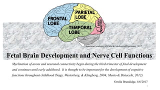
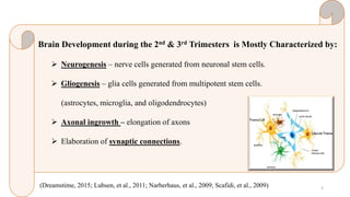
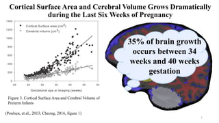
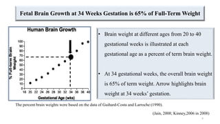
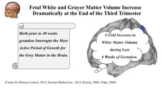
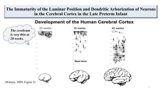
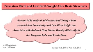
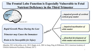
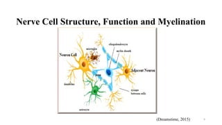
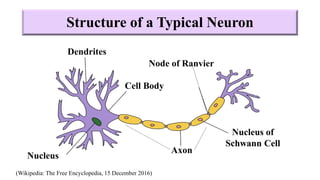
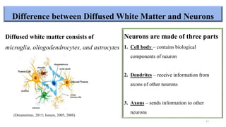
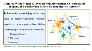
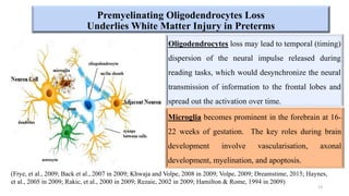
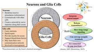
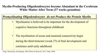
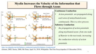
Fetal Brain Development and Nerve Cell Functions
Myelination of axons and neuronal connectivity begin during the third trimester of fetal development
and continues until early adulthood. It is thought to be important for the development of cognitive
functions throughout childhood (Nagy, Westerberg, & Klingberg, 2004; Mento & Bisiacchi, 2012).
1
American Academy of Child & Adolescent Psychiatry, 2017
Ozella Brundidge, 4/6/2017
Brain Development during the 2nd & 3rd Trimesters is Mostly Characterized by:
 Neurogenesis – nerve cells generated from neuronal stem cells.
 Gliogenesis – glia cells generated from multipotent stem cells.
(astrocytes, microglia, and oligodendrocytes)
 Axonal ingrowth – elongation of axons
 Elaboration of synaptic connections.
(Dreamstime, 2015; Lubsen, et al., 2011; Narberhaus, et al., 2009; Scafidi, et al., 2009) 2
Cortical Surface Area and Cerebral Volume Grows Dramatically
during the Last Six Weeks of Pregnancy
3
(Poulsen, et al., 2013; Cheong, 2016, figure 1)
35% of brain growth
occurs between 34
weeks and 40 weeks
gestation
Figure 3. Cortical Surface Area and Cerebral Volume of
Preterm Infants
Fetal Brain Growth at 34 Weeks Gestation is 65% of Full-Term Weight
4
• Brain weight at different ages from 20 to 40
gestational weeks is illustrated at each
gestational age as a percent of term brain weight.
• At 34 gestational weeks, the overall brain weight
is 65% of term weight. Arrow highlights brain
weight at 34 weeks’ gestation.
(Jain, 2008; Kinney,2006 in 2008)
The percent brain weights were based on the data of Guihard-Costa and Larroche (1990).
(Center for Disease Control, 2015; Nuclear Medical Inc., 2013; Kinney, 2006; Volpe, 2006)
Birth prior to 40 weeks
gestation Interrupts the Most
Active Period of Growth for
the Grey Matter in the Brain.
5-Fold Increase in
White Matter Volume
during Last
6 Weeks of Gestation.
Fetal White and Grayer Matter Volume Increase
Dramatically at the End of the Third Trimester
The Immaturity of the Laminar Position and Dendritic Arborization of Neurons
in the Cerebral Cortex in the Late Preterm Infant
6
(Kinney, 2009, Figure 2)
The cerebrum
is very thin at
20 weeks.
Premature Birth and Low Birth Weight Alter Brain Structures
7
n=137 participants
Age 13-22 years (Spencer et al., 2008 in Park, et al., 2014)
A recent MRI study of Adolescents and Young Adults
revealed that Prematurity and Low Birth Weight are
Associated with Reduced Gray Matter Density Bilaterally in
the Temporal Lobe and Cerebellum.
Rapid growth phase during the last trimester may cause the immature brain to be susceptible for
injury, leading to impaired growth of cerebral cortical gray matter and myelination of white
matter, disturbed development of white matter microstructure
8
(Baschat, 2011 in Reveillon, et al., 2013; Huppi, et al., 2001 in Jiang, Ping, & Wilkinson, 2012; American Academy of
Child & Adolescent Psychiatry, February 2017)
…impaired growth of cerebral
cortical gray matter
…impaired myelination of
white matter
…disturbed development of
white matter microstructure
The Frontal Lobe Function is Especially Vulnerable to Fetal
Nutrient Deficiency in the Third Trimester
Rapid Growth Phase During the Last
Trimester may Cause the Immature
Brain to be Susceptible for Injury
AACAP, 2017
Nerve Cell Structure, Function and Myelination
9(Dreamstime, 2015)
Structure of a Typical Neuron
Dendrites
Cell Body
Nucleus
Axon
Nucleus of
Schwann Cell
Node of Ranvier
(Wikipedia: The Free Encyclopedia, 15 December 2016)
Difference between Diffused White Matter and Neurons
Diffused white matter consists of
microglia, oliogodendrocytes, and astrocytes
Neurons are made of three parts
1. Cell body – contains biological
components of neuron
2. Dendrites – receive information from
axons of other neurons
3. Axons – sends information to other
neurons(Dreamstime, 2015; Jensen, 2005, 2008)
11
Diffused White Matter is Involved with Myelination, Cytostructural
Support, and Possibly has its own Communication Network*
(Dreamstime, 2015; Lubsen, Vohr, Myers, Hampson, Lacadie, Schneider, Katz, et al., 2011)
12
Diffuse white matter injury is the major
cause of neurodevelopmental conditions
experienced by some preterm born children.
The three types of diffuse white matter are:
1. Oligodendrocyte
2. Microglia
3. Astrocyte
Oligodendrocytes loss may lead to temporal (timing)
dispersion of the neural impulse released during
reading tasks, which would desynchronize the neural
transmission of information to the frontal lobes and
spread out the activation over time.
(Frye, et al., 2009; Back et al., 2007 in 2009; Khwaja and Volpe, 2008 in 2009; Volpe, 2009; Dreamstime, 2015; Haynes,
et al., 2005 in 2009; Rakic, et al., 2000 in 2009; Rezaie, 2002 in 2009; Hamilton & Rome, 1994 in 2009)
Premyelinating Oligodendrocytes Loss
Underlies White Matter Injury in Preterms
13
Microglia becomes prominent in the forebrain at 16-
22 weeks of gestation. The key roles during brain
development involve vascularisation, axonal
development, myelination, and apoptosis.
Neurons and Glia Cells
Neurons
Glia Cells
Both
Release
transmitters*
Have long-range
signaling
Can enwrap synaptic
terminals
Are connected
by gap junctions
(Jensen, 2005; Dreamstime, 2015)
Neurons:
• Dendrites receive
stimulation (information)
• Communicate with other
cells
• Create a network by nerve
impulses along their axons
Glia cells:
• Carry nutrients
• Speed repair
• Provide myelin for axons
• Support blood-brain barrier
• May form their own
communication network
• Involved in neurogenesis
*Neurotransmitters are the brain’s chemical messengers.
14
Integrate
neural output
Myelin is an Essential Protein for Brain Development
 Myelin is a fatty white substance that coats, supports, and
insulates axons.
 It increases efficiency by allowing electrical impulses to
travel up to 12 times faster.
 Myelin may be involved with the formation of habits*.
(Jensen, 2005; Sousa, 2008)*Non-declarative/Implicit memory
15
Myelin-Producing Oligodendrocytes become Abundant in the Cerebrum
White Matter After Term (37 weeks gestation)
• Myelination is believed to be important for the development of
cognitive functions throughout childhood.
• The myelination of axons and neuronal connectivity begin
during the third trimester (week 27) of fetal development and
continues until early adulthood.
Premyelinating Oligodendrocytes do not Produce the Protein Myelin
(Nagy, Westerberg, & Klingberg, 2004; Mento & Bisiacchi, 2012; Volpe, 2009)
Premyelinating Oligodendrocytes Underlies White Matter Injury and
Alters the Time it Takes for Information to Reach Target Regions
A decrease in the total number of
neurons firing synchronously result in:
• the release of neurotransmitters at
fewer synapses at any particular time
• an upregulation of post-synaptic
receptors could compensate for this
phenomenon.
(Frye, et al., 2009; Back et al., 2007 in 2009; Khwaja and Volpe, 2008 in 2009)
17
Sensory Information Travels through Myelinated Axons
(A2 nervesystemscolstons 2013, December 11 slide 9)
Information Travels through Myelinated Neurons at a Greater Velocity
(A2 nervesystemscolstons 2013, December 11 slide 8)
• Peripheral neurons have an insulating sheath around the axon called myelin which is formed by
Schwann cells.
• Myelin sheathing allows these neurons to conduct nerve impulses faster than in non-myelinated
neurons
Information is Transported through the Brain
Myelin sheathing has
bare sections of axons
called Nodes of Ranvier.
Action potentials (AP)
jump from node to node
(A2 nervesystemscolstons 2013, December 11, slide 7)
Myelin Increases the Velocity of the Information that
Flows through Axons
Continuous Conduction
• Action potentials are propagated along
each area of unmyelinated axons
continuously. This is a slow process.
Saltatory Conduction
• the propagation of action potentials
along myelinated axons from one node
of Ranvier to the next node, increasing
the conduction velocity of action
potentials.
(Jensen, 2005; Sousa, 2008; Ms Sime, April 16, 2016; Wikipedia: The Free Encyclopedia (15 December 2016).
21
https://en.wikipedia.org/wiki/Saltatory_conduction#/media/File:Saltatory_Conduction.gif
End of
Fetal Brain Development and Cellular Functions
22
American Academy of Child & Adolescent Psychiatry, 2017

Fetal brain development and cellular functions

  • 1.
    Fetal Brain Developmentand Nerve Cell Functions Myelination of axons and neuronal connectivity begin during the third trimester of fetal development and continues until early adulthood. It is thought to be important for the development of cognitive functions throughout childhood (Nagy, Westerberg, & Klingberg, 2004; Mento & Bisiacchi, 2012). 1 American Academy of Child & Adolescent Psychiatry, 2017 Ozella Brundidge, 4/6/2017
  • 2.
    Brain Development duringthe 2nd & 3rd Trimesters is Mostly Characterized by:  Neurogenesis – nerve cells generated from neuronal stem cells.  Gliogenesis – glia cells generated from multipotent stem cells. (astrocytes, microglia, and oligodendrocytes)  Axonal ingrowth – elongation of axons  Elaboration of synaptic connections. (Dreamstime, 2015; Lubsen, et al., 2011; Narberhaus, et al., 2009; Scafidi, et al., 2009) 2
  • 3.
    Cortical Surface Areaand Cerebral Volume Grows Dramatically during the Last Six Weeks of Pregnancy 3 (Poulsen, et al., 2013; Cheong, 2016, figure 1) 35% of brain growth occurs between 34 weeks and 40 weeks gestation Figure 3. Cortical Surface Area and Cerebral Volume of Preterm Infants
  • 4.
    Fetal Brain Growthat 34 Weeks Gestation is 65% of Full-Term Weight 4 • Brain weight at different ages from 20 to 40 gestational weeks is illustrated at each gestational age as a percent of term brain weight. • At 34 gestational weeks, the overall brain weight is 65% of term weight. Arrow highlights brain weight at 34 weeks’ gestation. (Jain, 2008; Kinney,2006 in 2008) The percent brain weights were based on the data of Guihard-Costa and Larroche (1990).
  • 5.
    (Center for DiseaseControl, 2015; Nuclear Medical Inc., 2013; Kinney, 2006; Volpe, 2006) Birth prior to 40 weeks gestation Interrupts the Most Active Period of Growth for the Grey Matter in the Brain. 5-Fold Increase in White Matter Volume during Last 6 Weeks of Gestation. Fetal White and Grayer Matter Volume Increase Dramatically at the End of the Third Trimester
  • 6.
    The Immaturity ofthe Laminar Position and Dendritic Arborization of Neurons in the Cerebral Cortex in the Late Preterm Infant 6 (Kinney, 2009, Figure 2) The cerebrum is very thin at 20 weeks.
  • 7.
    Premature Birth andLow Birth Weight Alter Brain Structures 7 n=137 participants Age 13-22 years (Spencer et al., 2008 in Park, et al., 2014) A recent MRI study of Adolescents and Young Adults revealed that Prematurity and Low Birth Weight are Associated with Reduced Gray Matter Density Bilaterally in the Temporal Lobe and Cerebellum.
  • 8.
    Rapid growth phaseduring the last trimester may cause the immature brain to be susceptible for injury, leading to impaired growth of cerebral cortical gray matter and myelination of white matter, disturbed development of white matter microstructure 8 (Baschat, 2011 in Reveillon, et al., 2013; Huppi, et al., 2001 in Jiang, Ping, & Wilkinson, 2012; American Academy of Child & Adolescent Psychiatry, February 2017) …impaired growth of cerebral cortical gray matter …impaired myelination of white matter …disturbed development of white matter microstructure The Frontal Lobe Function is Especially Vulnerable to Fetal Nutrient Deficiency in the Third Trimester Rapid Growth Phase During the Last Trimester may Cause the Immature Brain to be Susceptible for Injury AACAP, 2017
  • 9.
    Nerve Cell Structure,Function and Myelination 9(Dreamstime, 2015)
  • 10.
    Structure of aTypical Neuron Dendrites Cell Body Nucleus Axon Nucleus of Schwann Cell Node of Ranvier (Wikipedia: The Free Encyclopedia, 15 December 2016)
  • 11.
    Difference between DiffusedWhite Matter and Neurons Diffused white matter consists of microglia, oliogodendrocytes, and astrocytes Neurons are made of three parts 1. Cell body – contains biological components of neuron 2. Dendrites – receive information from axons of other neurons 3. Axons – sends information to other neurons(Dreamstime, 2015; Jensen, 2005, 2008) 11
  • 12.
    Diffused White Matteris Involved with Myelination, Cytostructural Support, and Possibly has its own Communication Network* (Dreamstime, 2015; Lubsen, Vohr, Myers, Hampson, Lacadie, Schneider, Katz, et al., 2011) 12 Diffuse white matter injury is the major cause of neurodevelopmental conditions experienced by some preterm born children. The three types of diffuse white matter are: 1. Oligodendrocyte 2. Microglia 3. Astrocyte
  • 13.
    Oligodendrocytes loss maylead to temporal (timing) dispersion of the neural impulse released during reading tasks, which would desynchronize the neural transmission of information to the frontal lobes and spread out the activation over time. (Frye, et al., 2009; Back et al., 2007 in 2009; Khwaja and Volpe, 2008 in 2009; Volpe, 2009; Dreamstime, 2015; Haynes, et al., 2005 in 2009; Rakic, et al., 2000 in 2009; Rezaie, 2002 in 2009; Hamilton & Rome, 1994 in 2009) Premyelinating Oligodendrocytes Loss Underlies White Matter Injury in Preterms 13 Microglia becomes prominent in the forebrain at 16- 22 weeks of gestation. The key roles during brain development involve vascularisation, axonal development, myelination, and apoptosis.
  • 14.
    Neurons and GliaCells Neurons Glia Cells Both Release transmitters* Have long-range signaling Can enwrap synaptic terminals Are connected by gap junctions (Jensen, 2005; Dreamstime, 2015) Neurons: • Dendrites receive stimulation (information) • Communicate with other cells • Create a network by nerve impulses along their axons Glia cells: • Carry nutrients • Speed repair • Provide myelin for axons • Support blood-brain barrier • May form their own communication network • Involved in neurogenesis *Neurotransmitters are the brain’s chemical messengers. 14 Integrate neural output
  • 15.
    Myelin is anEssential Protein for Brain Development  Myelin is a fatty white substance that coats, supports, and insulates axons.  It increases efficiency by allowing electrical impulses to travel up to 12 times faster.  Myelin may be involved with the formation of habits*. (Jensen, 2005; Sousa, 2008)*Non-declarative/Implicit memory 15
  • 16.
    Myelin-Producing Oligodendrocytes becomeAbundant in the Cerebrum White Matter After Term (37 weeks gestation) • Myelination is believed to be important for the development of cognitive functions throughout childhood. • The myelination of axons and neuronal connectivity begin during the third trimester (week 27) of fetal development and continues until early adulthood. Premyelinating Oligodendrocytes do not Produce the Protein Myelin (Nagy, Westerberg, & Klingberg, 2004; Mento & Bisiacchi, 2012; Volpe, 2009)
  • 17.
    Premyelinating Oligodendrocytes UnderliesWhite Matter Injury and Alters the Time it Takes for Information to Reach Target Regions A decrease in the total number of neurons firing synchronously result in: • the release of neurotransmitters at fewer synapses at any particular time • an upregulation of post-synaptic receptors could compensate for this phenomenon. (Frye, et al., 2009; Back et al., 2007 in 2009; Khwaja and Volpe, 2008 in 2009) 17
  • 18.
    Sensory Information Travelsthrough Myelinated Axons (A2 nervesystemscolstons 2013, December 11 slide 9)
  • 19.
    Information Travels throughMyelinated Neurons at a Greater Velocity (A2 nervesystemscolstons 2013, December 11 slide 8) • Peripheral neurons have an insulating sheath around the axon called myelin which is formed by Schwann cells. • Myelin sheathing allows these neurons to conduct nerve impulses faster than in non-myelinated neurons
  • 20.
    Information is Transportedthrough the Brain Myelin sheathing has bare sections of axons called Nodes of Ranvier. Action potentials (AP) jump from node to node (A2 nervesystemscolstons 2013, December 11, slide 7)
  • 21.
    Myelin Increases theVelocity of the Information that Flows through Axons Continuous Conduction • Action potentials are propagated along each area of unmyelinated axons continuously. This is a slow process. Saltatory Conduction • the propagation of action potentials along myelinated axons from one node of Ranvier to the next node, increasing the conduction velocity of action potentials. (Jensen, 2005; Sousa, 2008; Ms Sime, April 16, 2016; Wikipedia: The Free Encyclopedia (15 December 2016). 21 https://en.wikipedia.org/wiki/Saltatory_conduction#/media/File:Saltatory_Conduction.gif
  • 22.
    End of Fetal BrainDevelopment and Cellular Functions 22 American Academy of Child & Adolescent Psychiatry, 2017

Editor's Notes

  • #2 Myelination of white matter and neuronal connectivity are two important events that also contribute to general brain development. They start in the last trimester of gestation and proceed rapidly for several years postnatally following an initial process of massive transient connections and the successive pruning of exuberant axons in accordance with an economic principle (Innocenti and Price, 2005). Interestingly, by analysing the cellular composition of both grey and white matter in several species of primates, including humans, Herculano-Houzel et al. (2010) demonstrated that connectivity decreases in larger cerebral cortices as a slowly diminishing fraction of neurons project myelinated axons into the white matter. This differential structural maturation has been suggested to form the basis for new cognitive skills emerging during the first year of life. Nowadays, neonatal MRI (and derived software and hardware applications) is one of the best tools for addressing these issues (Giovanni Mento∗, Patrizia Silvia Bisiacchi, 2012)
  • #7 Figure 2 The immaturity of the laminar position and dendritic arborization of neurons, as demonstrated by Golgi drawings, in the cerebral cortex in the late preterm infant at 35 gestational weeks is striking in comparison to neurons at midgestation (20 weeks) and at term (40 weeks). The drawings by Chan and Armstrong are reprinted by permission.12 Kinney HC, Armstrong DL: Perinatal neuropathology, in Graham DI, Lantos PE (eds): Greenfield’s Neuropathology (ed 7). London, Arnold, 2002, pp 557-559
  • #8 Park, H. Y., Maitra, K., Achon, E., Loyola, & Rincon, M. (2014). Effects of early intervention on mental or neuromusculoskeletal and movement-related functions children born low birthweight or preterm: A meta-analysis. The American Journal of Occupational Therapy, 63(3), 268-276. http://dx.doi.org/10.5014/ajot.2014.010371 Spencer, M. D., Moorhead, T. W. J., Gibson, R. J., McIntosh, A.M., Sussmann, J. E.D., Owens, D.G. C., . . . Johnstone, E. C. (2008). Low birthweight and preterm birth in youngpeople with special educational needs: A magnetic resonance imaging analysis. BMC Medicine, 6, 1–11. http://dx.doi.org/10.1186/1741-7015-6-1
  • #9 American Academy of Child & Adolescent Psychiatry (February 2017). ADHD & the Brain. https://www.aacap.org/AACAP/Families_and_Youth/Facts_for_Families/FFF-Guide/ADHD_and_the_Brain.aspx
  • #13 Microglia have key roles during brain development, involving apoptosis, vascularisation, axonal development, and myelination.126–129 Astrocytes are high in interferon, which is a protein that signals damage.
  • #14 Apoptosis is cell death
  • #15 Microglia key roles during brain development 1) apoptosis, 2) vascularization, 3) axonal development, 4) myelination (126. Haynes, et al., 2005 Axonal development in the cerebral white matter of the human fetus and infant; 127. Rakie, et al., 2000 Programmed cell death in the developing human telencephalon; 128. Razaie, et al., 2005 Differientiation, ramification and distribution of microglia within the central nervous system examined; 129. Hamilton, et al., 1994 Stimulation of in vitro stimulation by microglia (all in Volpe, 2009)
  • #17  Immature oligodendroglial progenitors often lack processes, seem not to have the capacity for full differentiation to mature myelin-producing cells. Cerebral white matter axons (ie, projection, commissural, and association fibres) are in a phase of rapid growth during the premature period Herculano-Houzel et al. (2010) demonstrated that connectivity decreases in larger cerebral cortices as a slowly diminishing fraction of neurons project myelinated axons into the white matter. This differential structural maturation has been suggested to form the basis for new cognitive skills emerging during the first year of life.
  • #18 Possibly laying the foundation for the development of alternative pathways
  • #19 A2 nervesystemscolstons (2013, December 11). Saltatory Conduction in Myelinated Axons Myelin. Retrieved from https://image.slidesharecdn.com/a2nervesystemscolstons-131211062501-phpapp01/95/a2-nervesystemscolstons-9-638.jpg?cb=1386743163
  • #22 Wikipedia: The Free Encyclopedia (15 December 2016). Saltatory conduction. https://en.wikipedia.org/wiki/Saltatory_conduction Ms Sime (2016, April 16). Myelinated vs unmyelinated axons. Retrieved from https://alsiraatbiology14.wordpress.com/2015/04/16/myelinated-vs-unmyelinated-axons/
  • #23 Myelination of white matter and neuronal connectivity are two important events that also contribute to general brain development. They start in the last trimester of gestation and proceed rapidly for several years postnatally following an initial process of massive transient connections and the successive pruning of exuberant axons in accordance with an economic principle (Innocenti and Price, 2005). Interestingly, by analysing the cellular composition of both grey and white matter in several species of primates, including humans, Herculano-Houzel et al. (2010) demonstrated that connectivity decreases in larger cerebral cortices as a slowly diminishing fraction of neurons project myelinated axons into the white matter. This differential structural maturation has been suggested to form the basis for new cognitive skills emerging during the first year of life. Nowadays, neonatal MRI (and derived software and hardware applications) is one of the best tools for addressing these issues (Giovanni Mento∗, Patrizia Silvia Bisiacchi, 2012)