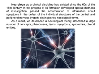
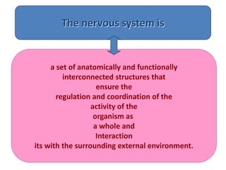
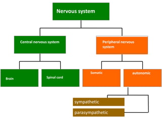
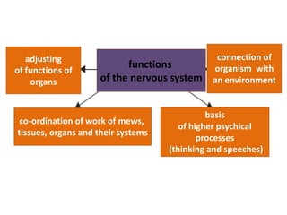
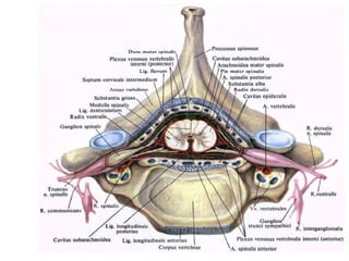
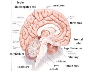
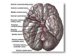
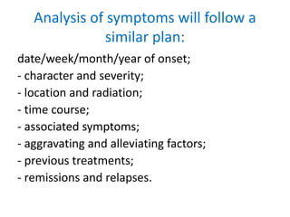
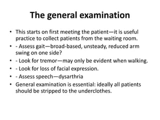
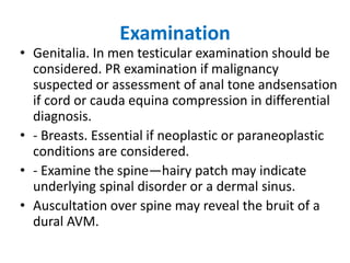
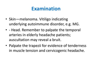
Introduction to neurology
Neurology as a clinical discipline has existed since the 60s of the
18th century. In the process of its formation developed special methods
of investigation, passed the accumulation of information about
symptoms in the defeat of the individual structures of the central and
peripheral nervous system, distinguished nosological forms.
As a result, we developed a neurological theory, described a large
number of concepts, phenomena, terms, symptoms, syndromes, clinical
entities.
The nervous system is
a set of anatomically and functionally
interconnected structures that
ensure the
regulation and coordination of the
activity of the
organism as
a whole and
Interaction
its with the surrounding external environment.
Brain
Central nervous system
Somatic autonomic
Peripheral nervous
system
Nervous system
sympathetic
parasympathetic
Spinal cord
Ontogenesis of the brain
• Development of the human nervous system starts on
day 15 post-conception (p/c) when a primitive streak of
specialised neuroectoderm forms on the dorsal surface
of the embryo.
• Dorsal induction is responsible for the formation and
closure of the neural tube as well as the three primary
vesicles at the rostral end of the neural tube.
• Ventral induction leads to formation of the cerebral
hemispheres, eye vesicles, olfactory bulbs, pituitary
glands and part of the face while dorsal induction
includes primary and secondary neurulation.
Ontogenesis
• Primary neurulation
begins with formation of
the neural plate and tube,
ending when the neural
tube is separated from
the surface ectoderm by
the intervening
mesenchyme.
• Formation of the neural
plate starts on day 17 p/c
and is complete by day 18
p/c when the edges of
the neural plate begin to
elevate, folding over to
form the neural tube.
Ontogenesis of the brain
The nervous system develops from
the ectoderm (neurons and glia) and
mesoderm (vessels and membranes
of the brain) .
By 3 weeks – nervous plate.
By the end of 4 weeks – three brain
bladders.
12 weeks –common features of the
brain.
By week 20, the process of
myelination begins, which indicates
functional maturity.
First, myelination occurs in the
sensitive part of the nervous system,
then in the coordination part.
Immediately after birth, there is
myelination of the frontal lobes of
the brain, the pyramidal tract.
• Nervous tissue is the basis of the structure and activity of the nervous system. The
main properties of this tissue are excitability and conductivity. It consists of two
types of cells: nerve cells - neurons and auxiliary satellite cells (glial cells), as well
as Schwann cells. Between the cells of the nervous tissue, intercellular spaces are
well developed, filled with a fat - like intercellular substance- glia (neuroglia). Glia
and satellite cells perform auxiliary functions for neurons: supporting, protective,
trophic, and metabolic
co-ordination of work of mews,
tissues, organs and their systems
functions
of the nervous system
adjusting
of functions of
organs
connection of
organism with
an environment
basis
of higher psychical
processes
(thinking and speeches)
A typical neuron structure
dendrite axon terminal
branch
cell body
intercept
of Ranvie
kernel
axon
Schwann cell
myelin sheath
dendrite
axon
kernel
soma
synapse
Classification of neurons
By body shape: pyramidal, pear-shaped, etc.
By body size: small, medium, large
By the number of processes: unipolar,bipolar,
pseudo-unipolar,
multipolar
By functional significance:receptor, effector,
associative
nervous tissue
The main component of the nervous system
which is built
Neurons
nerve cells
Neuroglia
perceived annoyance,
becomes excited
and transmit nerve impulses
cells that in the space between
neurons
perform reference,
demarcation,
trophic, secretory,
protective functions
Microglia cells
Nerve fibers are
processes of nerve cells, covered with a glial membrane and conducting a nerve
impulse
myelinated myelin-free
there is myelin in the glial membrane myelin is absent
Electric Chemical
Синапсы
Synapses are places of
connectionnerve cells where
the nerve impulse is
transmitted.
This can happen directly
(electrical transmission), but
most often it is carried out
indirectly, using chemical
molecules -
neurotransmitters:
•Acetylcholine,
•Norepinephrine
•Aspartate
•Glutamate
•Gamma-aminobutyric acid
•Dopamine
•Serotonin
•Histamine
•Arginine
The Structure of the Nervous System
The Spinal Cord
• is encased in the bony vertebral
column
• is attached to the brain stem
• is the major conduit of
information from the skin,
joints, and muscles of the body
to the brain, and vice versa
• A transection of the spinal cord
results in anesthesia (lack of
feeling) in the skin and paralysis
of the muscles in parts of the
body caudal to the cut.
• 31 segments:
• 8 neck (C)
• 12 breast (T)
• 5 lumbar (L)
• 5 sacral (S)
• and 1 coccyx.
Gray matter of the spinal cord (nuclei of the posterior,
lateral and anterior horns)
White matter of the spinal cord (pathways)
White matter of the spinal cord
(pathways)
• Fasciculus gracilis et cuneatus
White matter (pathways) of the lateral cord
Tr. cortico-spinalis lateralis
Tr. spinocerebellaris anterior et posterior
Tr. spino-thalamicus lateralis
Tr. rubro-spinalis
White matter (pathways) of the anterior cord
Tr. spino-thalamicus anterior
Tr. vestibulo-spinalis
Tr. tecto-spinalis
Tr. reticulo-spinalis
Оболочки СМ
• имеет три оболочки: и
наружная – твердая
• средняя - паутинная
• внутренняя - мягкая
(сосудистая), Между твердой
оболочкой и надкостницей
позвоночного канала -
эпидуральное пространство,
между твердой и паутинной -
субдуральное пространство.
• От мягкой (сосудистой)
оболочки паутинную оболочку
отделяет подпаутинное
(субарахноидальное)
пространство, содержащее
спинномозговую жидкость
(до 150 мл)
brain
an elongated slit cerebrum
thalamus
cerebellum
frontal
lobe
hypothalamus
pituitary
brain axis
midbrain
pons
medulla
spinal cord
Кровоснабжение головного мозга
Neurologic disorders
• Neurologic disorders are globally among the
most important causes of human illness and
mortality.
• Neurologic diseases are notable for more
frequently causing disability than death.
• Current estimates of the burden of
neurological disorders indicate that they have
a huge impact on human health.
Neurologic disorders
• Primary neurologic disorders affecting the
nervous system only
• Secondary neurologic disorders, in which
injury or dysfunction of the nervous system
occurs as a result of a disease that primarily
affects another organ system
• Primary neurologic disorders (Alzheimer
disease, migraine, and multiple sclerosis)
• Secondary neurologic disorders include
seizures secondary to infection with malaria,
paraneoplastic syndromes associated with
primary systemic cancer, and peripheral
neuropathy occurring in the setting of
nutritional deficiencies
Neurologist
• A doctor who has specialisation in neurology is known as a
neurologist. The neurologist treats disorders that affect the
brain, spinal cord, and nerves, such as:
• Demyelinating diseases of the central nervous system, such
as multiple sclerosis
• Cerebrovascular disease, such as stroke
• Headache disorders
• Infections of the brain and peripheral nervous system
• Neurodegenerative disorders, such as Alzheimer’s disease,
Parkinson’s disease, and Amyotrophic Lateral Sclerosis (Lou
Gehrig’s disease)
• Movement disorders, such as Parkinson’s disease
• Seizure disorders, such as epilepsy
• Spinal cord disorders
• Speech and language disorders.
NEUROLOGICAL EXAMINATION
• During this examination, the health history of the patient is
reviewed by neurologist with special attention to the current
condition.
• The patient then takes a neurological exam. Typically, the exam tests
mental status, function of the cranial nerves (including vision), strength,
coordination, reflexes, and sensation.
• This information endorse the neurologist determine whether the
problem exists in the nervous system and the clinical localization.
• Localization of the pathology is the key process by which
neurologists develop their differential diagnosis.
NEUROLOGICAL EXAMINATION
Components of the neurological examination
include assessment of the patient’s cognitive
function, cranial nerves, motor strength,
sensation, reflexes, coordination, and gait. In few
examples, neurologists may order additional
diagnostic tests as part of the evaluation.
Principles of neurological history taking
The usual approach to a clinical problem is to ask the
following:
- Where is the lesion, e.g. brain, spinal cord, anterior
horn cell, peripheral nerve, neuromuscular
junction, muscle?
- What is the aetiology, e.g. vascular, degenerative,
toxic, infective genetic, inflammatory, neoplastic,
functional?
- What is the differential diagnosis?
- Is treatment possible?
- What is the prognosis?
A detailed history usually will yield more information than the
neurological examination and ancillary tests
• Family members and eyewitness
accounts are essential, e.g in patients
with dementia and blackouts.
• Obtain a history by telephone if
necessary.
• A review of the case notes if available is
very useful.
Analysis of symptoms will follow a
similar plan:
date/week/month/year of onset;
- character and severity;
- location and radiation;
- time course;
- associated symptoms;
- aggravating and alleviating factors;
- previous treatments;
- remissions and relapses.
Past medical history
• Do not always accept the patient’s diagnostic
terms—enquire into specific symptoms, e.g.
‘migraine’, ‘seizure’, ‘stroke’.
Family history
• Draw a family tree. Document specific
illnesses and cause of death if known.
• In certain communities enquire about
consanguinity.
Social history
This should include:
• - alcohol;
• - smoking;
• - recreational drug use;
• - risk factors for HIV;
• - detailed travel history;
• - dietary habits.
The general examination
• This starts on first meeting the patient—it is useful
practice to collect patients from the waiting room.
• - Assess gait—broad-based, unsteady, reduced arm
swing on one side?
• - Look for tremor—may only be evident when walking.
• - Look for loss of facial expression.
• - Assess speech—dysarthria
• General examination is essential: ideally all patients
should be stripped to the underclothes.
Examination
• Cardiovascular system. Pulse, heart sounds,
blood pressure (lying down and standing after
3 minutes if any suggestion of autonomic
involvement).
• - Respiratory system. Diaphragmatic
movement. May need to measure forced vital
capacity (FVC).
• - Gastrointestinal system. Palpate for
hepatosplenomegaly or abdominal masses.
Examination
• Genitalia. In men testicular examination should be
considered. PR examination if malignancy
suspected or assessment of anal tone andsensation
if cord or cauda equina compression in differential
diagnosis.
• - Breasts. Essential if neoplastic or paraneoplastic
conditions are considered.
• - Examine the spine—hairy patch may indicate
underlying spinal disorder or a dermal sinus.
• Auscultation over spine may reveal the bruit of a
dural AVM.
Examination
• Skin—melanoma. Vitiligo indicating
underlying autoimmune disorder, e.g. MG.
• - Head. Remember to palpate the temporal
arteries in elderly headache patients;
auscultation may reveal a bruit.
• Palpate the trapezii for evidence of tenderness
in muscle tension and cervicogenic headache.
Methods for studying brain functions:
• Сlinical method - study of
cortical damage disorders
electrical stimulation of various
areas of the cortex the method of
conditioned reflexes EEG
registration of biopotentials of
the brain
• Echoencephaloscopy rapid
removal of individual sections
of the cortex.
• Carotid artery Doppler
• Transcranial Doppler performed using
a high-frequency probe over the thin
portion of the temporal bone using it
as an acoustic window.
Nerve conduction studies (NCS) and needle
electromyography (EMG):
- Study of sensory and motor peripheral nerve
disorders, e.g. neuropathies, radiculopathies,
dorsal root ganglionopathies, and anterior
horn cell disorders.
- Neuromuscular junction disorders.
- Skeletal muscle disorders.
- Cranial nerve disorders, e.g. facial nerve,
trigeminal nerve.
Evoked potentials (EPs).
- Study sensory and motor pathways in the
peripheral and central nervous systems.
- Useful in investigation of multiple sclerosis,
other spinal cord and brainstem disorders, and
cranial neuropathies.
- Monitoring of spinal cord function during
surgery for scoliosis and of the facial nerve
during acoustic neuroma surgery.
Neuro Radiological Examinations
• Computerized tomography (CT)
• Magnetic resonance angiography (MRA)
• Magnetic resonance imaging (MRI)
• Magnetic resonance spectroscopy (MRS)
• Functional MRI (fMRI)
• Catheter angiography
Thank you

introduction to neurology..pptx

  • 1.
  • 2.
    Neurology as aclinical discipline has existed since the 60s of the 18th century. In the process of its formation developed special methods of investigation, passed the accumulation of information about symptoms in the defeat of the individual structures of the central and peripheral nervous system, distinguished nosological forms. As a result, we developed a neurological theory, described a large number of concepts, phenomena, terms, symptoms, syndromes, clinical entities.
  • 3.
    The nervous systemis a set of anatomically and functionally interconnected structures that ensure the regulation and coordination of the activity of the organism as a whole and Interaction its with the surrounding external environment.
  • 4.
    Brain Central nervous system Somaticautonomic Peripheral nervous system Nervous system sympathetic parasympathetic Spinal cord
  • 5.
    Ontogenesis of thebrain • Development of the human nervous system starts on day 15 post-conception (p/c) when a primitive streak of specialised neuroectoderm forms on the dorsal surface of the embryo. • Dorsal induction is responsible for the formation and closure of the neural tube as well as the three primary vesicles at the rostral end of the neural tube. • Ventral induction leads to formation of the cerebral hemispheres, eye vesicles, olfactory bulbs, pituitary glands and part of the face while dorsal induction includes primary and secondary neurulation.
  • 6.
    Ontogenesis • Primary neurulation beginswith formation of the neural plate and tube, ending when the neural tube is separated from the surface ectoderm by the intervening mesenchyme. • Formation of the neural plate starts on day 17 p/c and is complete by day 18 p/c when the edges of the neural plate begin to elevate, folding over to form the neural tube.
  • 7.
    Ontogenesis of thebrain The nervous system develops from the ectoderm (neurons and glia) and mesoderm (vessels and membranes of the brain) . By 3 weeks – nervous plate. By the end of 4 weeks – three brain bladders. 12 weeks –common features of the brain. By week 20, the process of myelination begins, which indicates functional maturity. First, myelination occurs in the sensitive part of the nervous system, then in the coordination part. Immediately after birth, there is myelination of the frontal lobes of the brain, the pyramidal tract.
  • 8.
    • Nervous tissueis the basis of the structure and activity of the nervous system. The main properties of this tissue are excitability and conductivity. It consists of two types of cells: nerve cells - neurons and auxiliary satellite cells (glial cells), as well as Schwann cells. Between the cells of the nervous tissue, intercellular spaces are well developed, filled with a fat - like intercellular substance- glia (neuroglia). Glia and satellite cells perform auxiliary functions for neurons: supporting, protective, trophic, and metabolic
  • 9.
    co-ordination of workof mews, tissues, organs and their systems functions of the nervous system adjusting of functions of organs connection of organism with an environment basis of higher psychical processes (thinking and speeches)
  • 10.
    A typical neuronstructure dendrite axon terminal branch cell body intercept of Ranvie kernel axon Schwann cell myelin sheath
  • 11.
  • 12.
    Classification of neurons Bybody shape: pyramidal, pear-shaped, etc. By body size: small, medium, large By the number of processes: unipolar,bipolar, pseudo-unipolar, multipolar By functional significance:receptor, effector, associative
  • 13.
    nervous tissue The maincomponent of the nervous system which is built Neurons nerve cells Neuroglia perceived annoyance, becomes excited and transmit nerve impulses cells that in the space between neurons perform reference, demarcation, trophic, secretory, protective functions
  • 14.
  • 15.
    Nerve fibers are processesof nerve cells, covered with a glial membrane and conducting a nerve impulse myelinated myelin-free there is myelin in the glial membrane myelin is absent
  • 16.
    Electric Chemical Синапсы Synapses areplaces of connectionnerve cells where the nerve impulse is transmitted. This can happen directly (electrical transmission), but most often it is carried out indirectly, using chemical molecules - neurotransmitters: •Acetylcholine, •Norepinephrine •Aspartate •Glutamate •Gamma-aminobutyric acid •Dopamine •Serotonin •Histamine •Arginine
  • 18.
    The Structure ofthe Nervous System The Spinal Cord • is encased in the bony vertebral column • is attached to the brain stem • is the major conduit of information from the skin, joints, and muscles of the body to the brain, and vice versa • A transection of the spinal cord results in anesthesia (lack of feeling) in the skin and paralysis of the muscles in parts of the body caudal to the cut.
  • 19.
    • 31 segments: •8 neck (C) • 12 breast (T) • 5 lumbar (L) • 5 sacral (S) • and 1 coccyx.
  • 20.
    Gray matter ofthe spinal cord (nuclei of the posterior, lateral and anterior horns)
  • 21.
    White matter ofthe spinal cord (pathways)
  • 22.
    White matter ofthe spinal cord (pathways) • Fasciculus gracilis et cuneatus
  • 23.
    White matter (pathways)of the lateral cord Tr. cortico-spinalis lateralis Tr. spinocerebellaris anterior et posterior Tr. spino-thalamicus lateralis Tr. rubro-spinalis
  • 24.
    White matter (pathways)of the anterior cord White matter (pathways) of the anterior cord Tr. spino-thalamicus anterior Tr. vestibulo-spinalis Tr. tecto-spinalis Tr. reticulo-spinalis
  • 25.
    Оболочки СМ • имееттри оболочки: и наружная – твердая • средняя - паутинная • внутренняя - мягкая (сосудистая), Между твердой оболочкой и надкостницей позвоночного канала - эпидуральное пространство, между твердой и паутинной - субдуральное пространство. • От мягкой (сосудистой) оболочки паутинную оболочку отделяет подпаутинное (субарахноидальное) пространство, содержащее спинномозговую жидкость (до 150 мл)
  • 26.
    brain an elongated slitcerebrum thalamus cerebellum frontal lobe hypothalamus pituitary brain axis midbrain pons medulla spinal cord
  • 27.
  • 30.
    Neurologic disorders • Neurologicdisorders are globally among the most important causes of human illness and mortality. • Neurologic diseases are notable for more frequently causing disability than death. • Current estimates of the burden of neurological disorders indicate that they have a huge impact on human health.
  • 31.
    Neurologic disorders • Primaryneurologic disorders affecting the nervous system only • Secondary neurologic disorders, in which injury or dysfunction of the nervous system occurs as a result of a disease that primarily affects another organ system
  • 32.
    • Primary neurologicdisorders (Alzheimer disease, migraine, and multiple sclerosis) • Secondary neurologic disorders include seizures secondary to infection with malaria, paraneoplastic syndromes associated with primary systemic cancer, and peripheral neuropathy occurring in the setting of nutritional deficiencies
  • 33.
    Neurologist • A doctorwho has specialisation in neurology is known as a neurologist. The neurologist treats disorders that affect the brain, spinal cord, and nerves, such as: • Demyelinating diseases of the central nervous system, such as multiple sclerosis • Cerebrovascular disease, such as stroke • Headache disorders • Infections of the brain and peripheral nervous system • Neurodegenerative disorders, such as Alzheimer’s disease, Parkinson’s disease, and Amyotrophic Lateral Sclerosis (Lou Gehrig’s disease) • Movement disorders, such as Parkinson’s disease • Seizure disorders, such as epilepsy • Spinal cord disorders • Speech and language disorders.
  • 34.
    NEUROLOGICAL EXAMINATION • Duringthis examination, the health history of the patient is reviewed by neurologist with special attention to the current condition. • The patient then takes a neurological exam. Typically, the exam tests mental status, function of the cranial nerves (including vision), strength, coordination, reflexes, and sensation. • This information endorse the neurologist determine whether the problem exists in the nervous system and the clinical localization. • Localization of the pathology is the key process by which neurologists develop their differential diagnosis.
  • 35.
    NEUROLOGICAL EXAMINATION Components ofthe neurological examination include assessment of the patient’s cognitive function, cranial nerves, motor strength, sensation, reflexes, coordination, and gait. In few examples, neurologists may order additional diagnostic tests as part of the evaluation.
  • 36.
    Principles of neurologicalhistory taking The usual approach to a clinical problem is to ask the following: - Where is the lesion, e.g. brain, spinal cord, anterior horn cell, peripheral nerve, neuromuscular junction, muscle? - What is the aetiology, e.g. vascular, degenerative, toxic, infective genetic, inflammatory, neoplastic, functional? - What is the differential diagnosis? - Is treatment possible? - What is the prognosis?
  • 37.
    A detailed historyusually will yield more information than the neurological examination and ancillary tests • Family members and eyewitness accounts are essential, e.g in patients with dementia and blackouts. • Obtain a history by telephone if necessary. • A review of the case notes if available is very useful.
  • 38.
    Analysis of symptomswill follow a similar plan: date/week/month/year of onset; - character and severity; - location and radiation; - time course; - associated symptoms; - aggravating and alleviating factors; - previous treatments; - remissions and relapses.
  • 39.
    Past medical history •Do not always accept the patient’s diagnostic terms—enquire into specific symptoms, e.g. ‘migraine’, ‘seizure’, ‘stroke’. Family history • Draw a family tree. Document specific illnesses and cause of death if known. • In certain communities enquire about consanguinity.
  • 40.
    Social history This shouldinclude: • - alcohol; • - smoking; • - recreational drug use; • - risk factors for HIV; • - detailed travel history; • - dietary habits.
  • 41.
    The general examination •This starts on first meeting the patient—it is useful practice to collect patients from the waiting room. • - Assess gait—broad-based, unsteady, reduced arm swing on one side? • - Look for tremor—may only be evident when walking. • - Look for loss of facial expression. • - Assess speech—dysarthria • General examination is essential: ideally all patients should be stripped to the underclothes.
  • 42.
    Examination • Cardiovascular system.Pulse, heart sounds, blood pressure (lying down and standing after 3 minutes if any suggestion of autonomic involvement). • - Respiratory system. Diaphragmatic movement. May need to measure forced vital capacity (FVC). • - Gastrointestinal system. Palpate for hepatosplenomegaly or abdominal masses.
  • 43.
    Examination • Genitalia. Inmen testicular examination should be considered. PR examination if malignancy suspected or assessment of anal tone andsensation if cord or cauda equina compression in differential diagnosis. • - Breasts. Essential if neoplastic or paraneoplastic conditions are considered. • - Examine the spine—hairy patch may indicate underlying spinal disorder or a dermal sinus. • Auscultation over spine may reveal the bruit of a dural AVM.
  • 44.
    Examination • Skin—melanoma. Vitiligoindicating underlying autoimmune disorder, e.g. MG. • - Head. Remember to palpate the temporal arteries in elderly headache patients; auscultation may reveal a bruit. • Palpate the trapezii for evidence of tenderness in muscle tension and cervicogenic headache.
  • 45.
    Methods for studyingbrain functions: • Сlinical method - study of cortical damage disorders electrical stimulation of various areas of the cortex the method of conditioned reflexes EEG registration of biopotentials of the brain • Echoencephaloscopy rapid removal of individual sections of the cortex. • Carotid artery Doppler • Transcranial Doppler performed using a high-frequency probe over the thin portion of the temporal bone using it as an acoustic window.
  • 46.
    Nerve conduction studies(NCS) and needle electromyography (EMG): - Study of sensory and motor peripheral nerve disorders, e.g. neuropathies, radiculopathies, dorsal root ganglionopathies, and anterior horn cell disorders. - Neuromuscular junction disorders. - Skeletal muscle disorders. - Cranial nerve disorders, e.g. facial nerve, trigeminal nerve.
  • 47.
    Evoked potentials (EPs). -Study sensory and motor pathways in the peripheral and central nervous systems. - Useful in investigation of multiple sclerosis, other spinal cord and brainstem disorders, and cranial neuropathies. - Monitoring of spinal cord function during surgery for scoliosis and of the facial nerve during acoustic neuroma surgery.
  • 48.
    Neuro Radiological Examinations •Computerized tomography (CT) • Magnetic resonance angiography (MRA) • Magnetic resonance imaging (MRI) • Magnetic resonance spectroscopy (MRS) • Functional MRI (fMRI) • Catheter angiography
  • 49.