Download to read offline
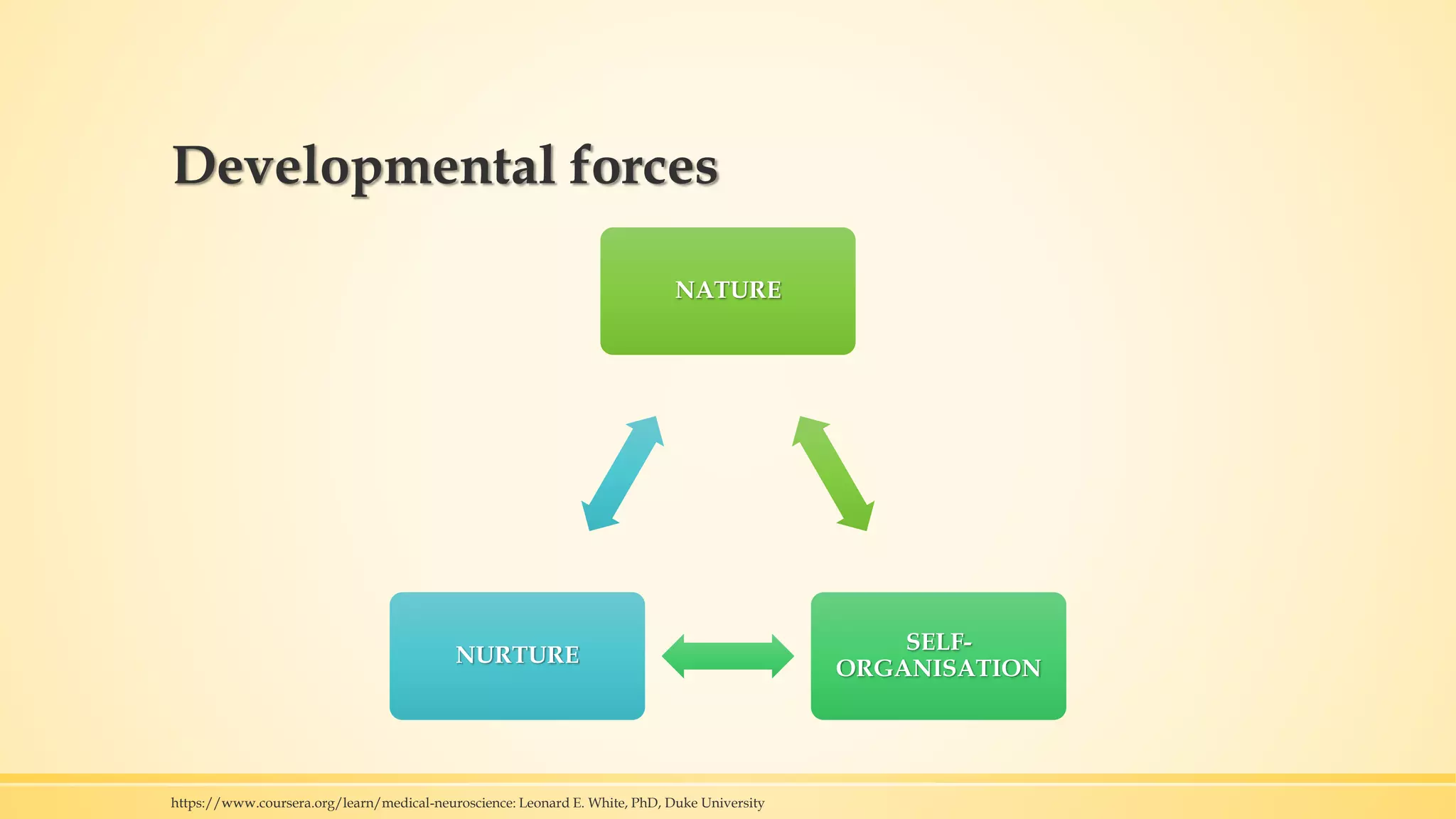
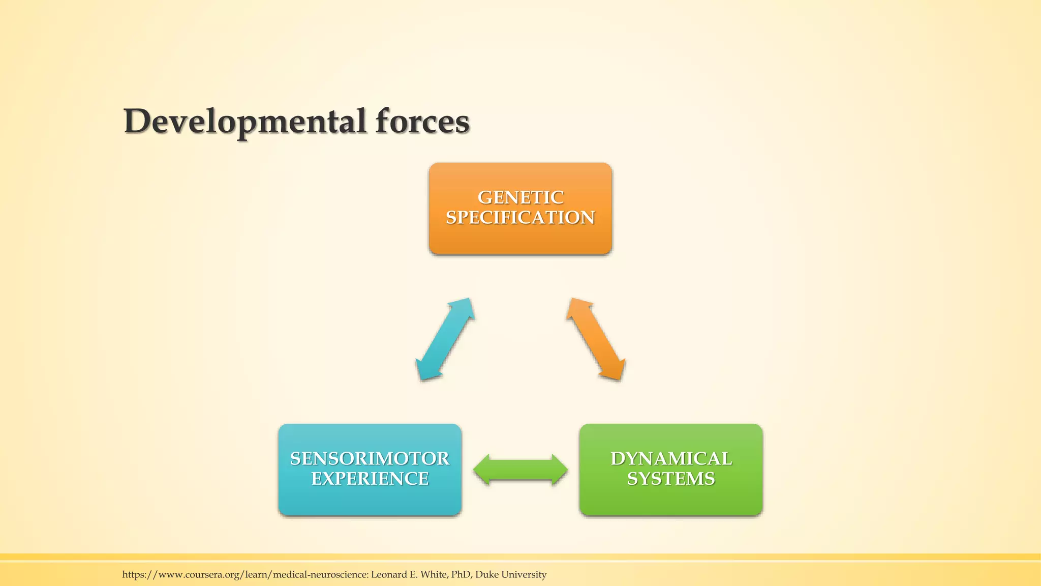
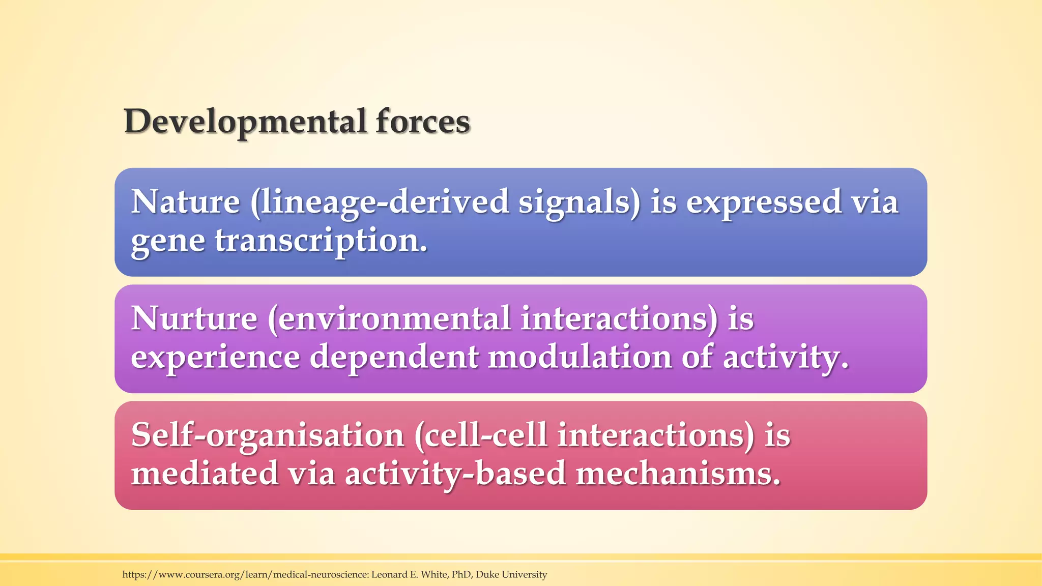
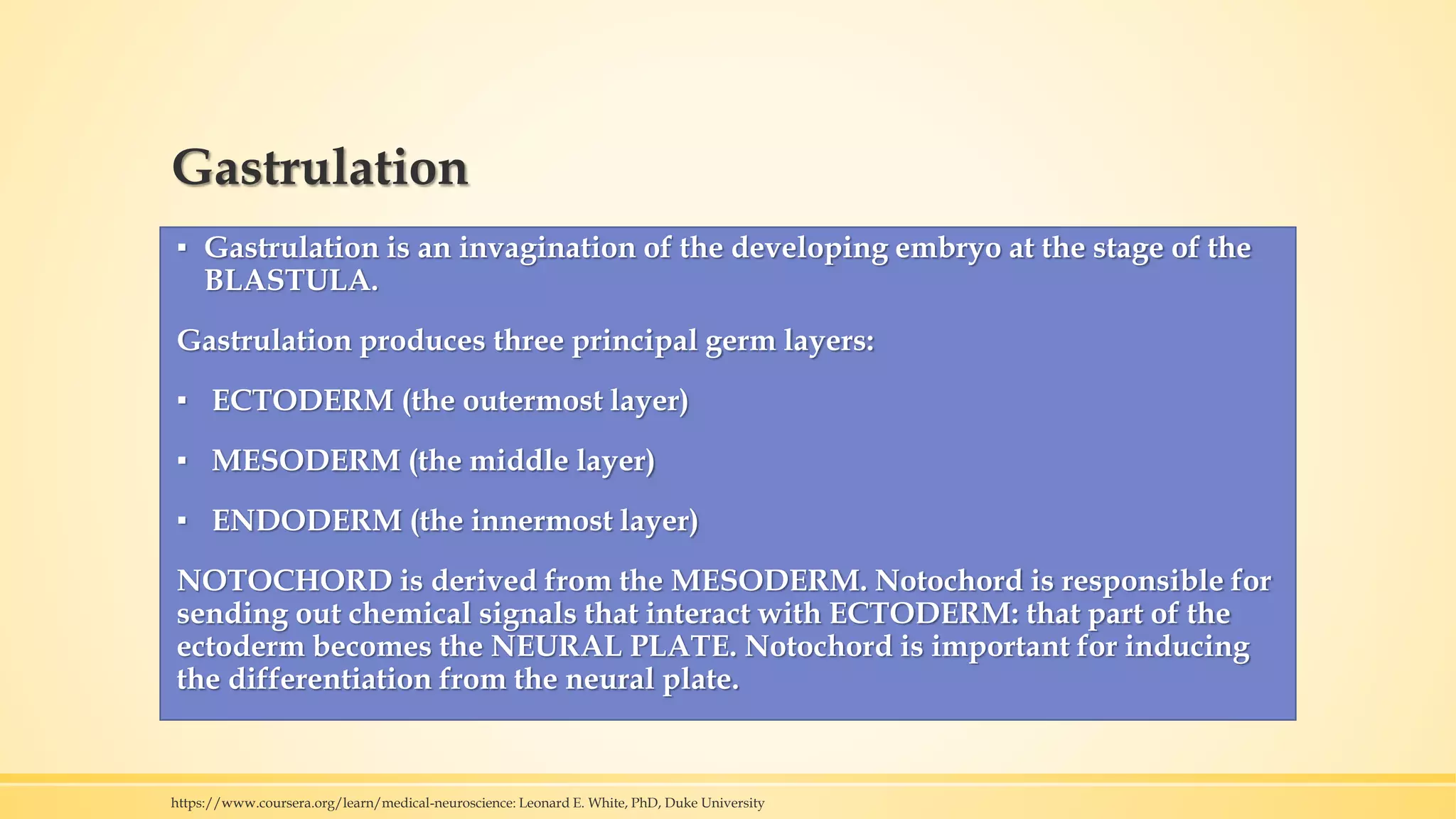
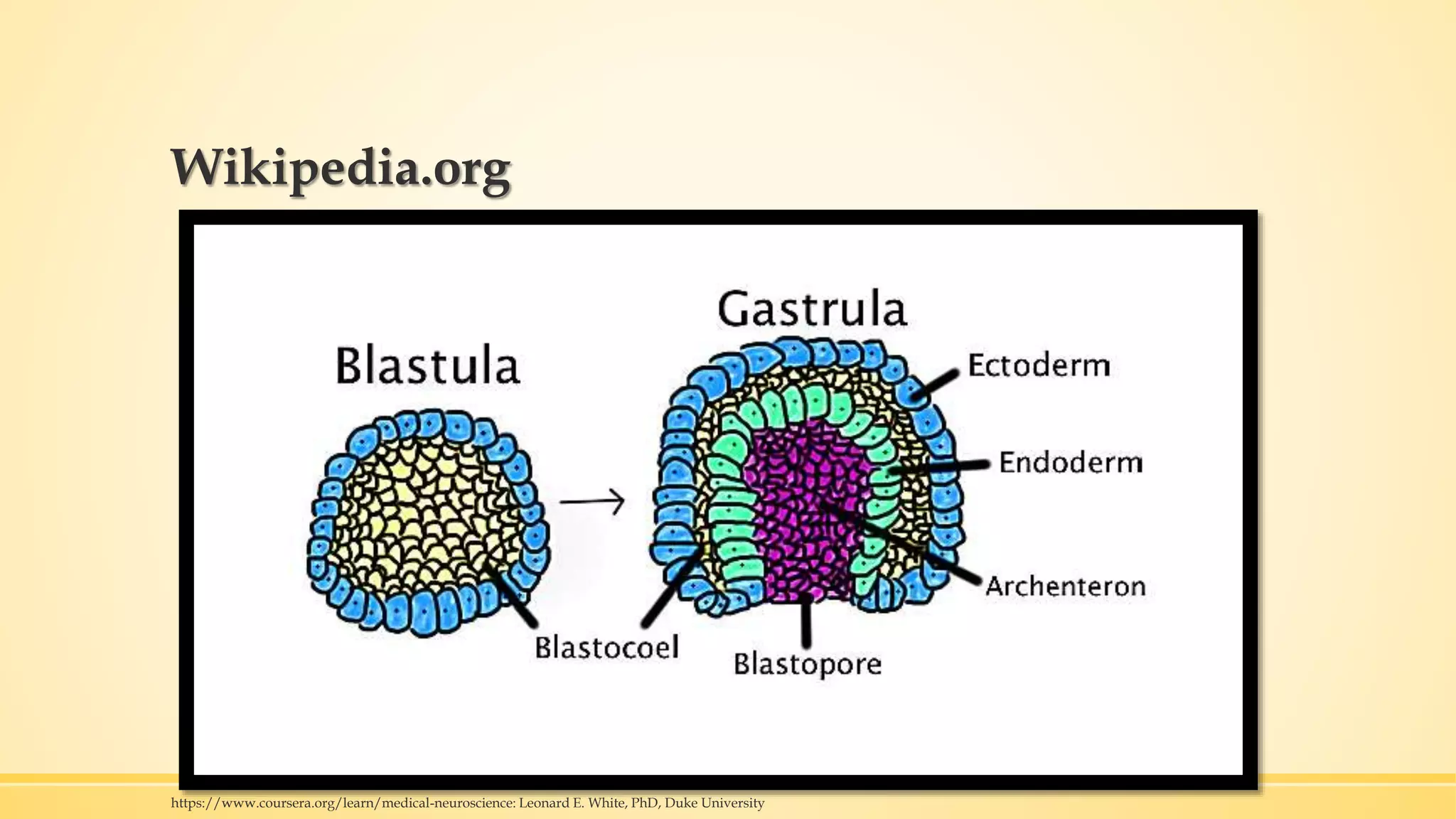
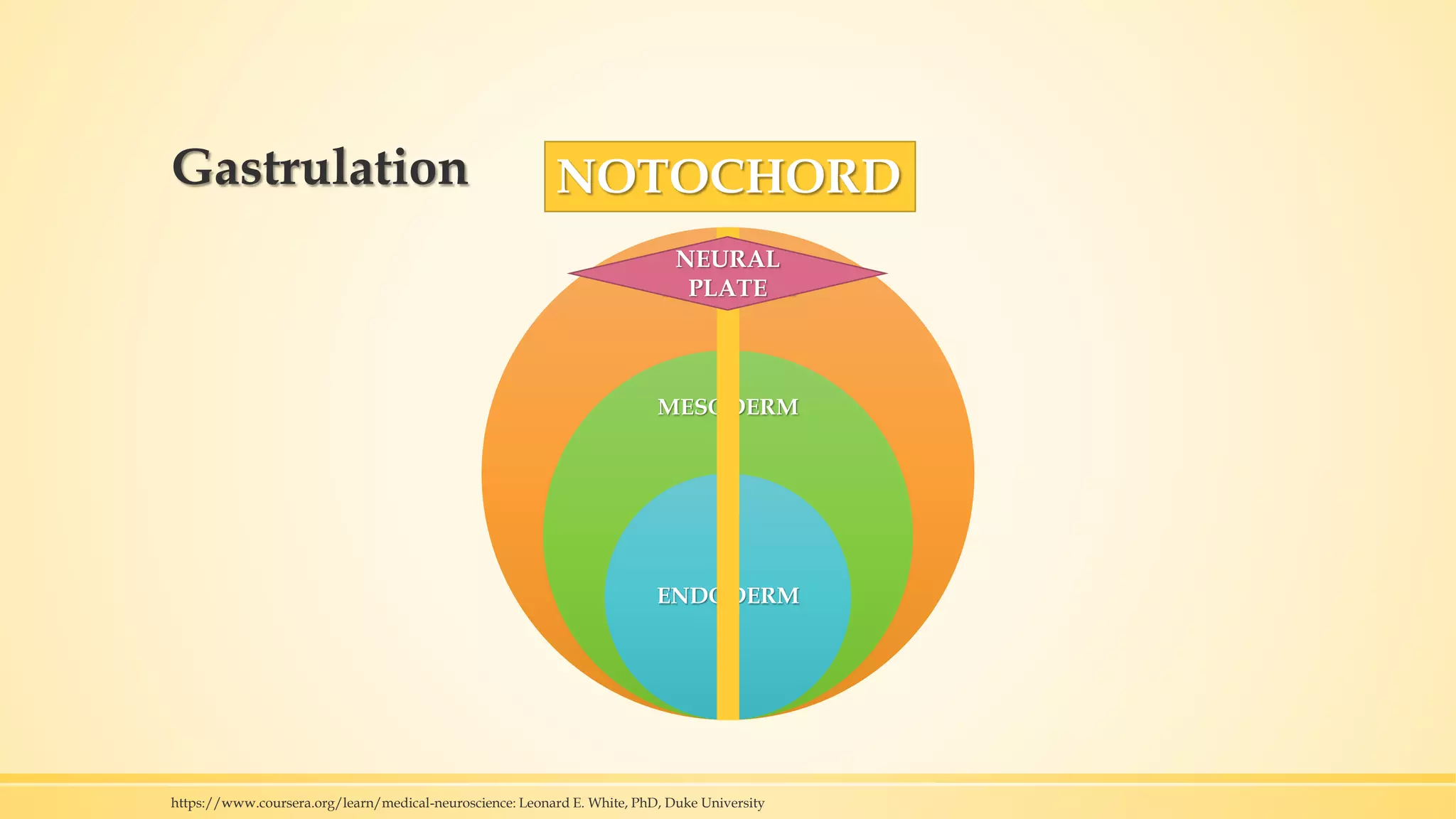
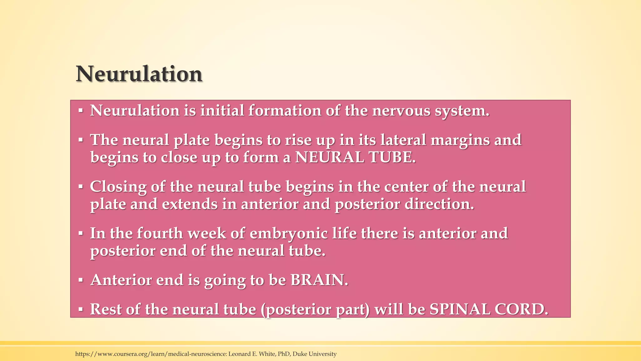
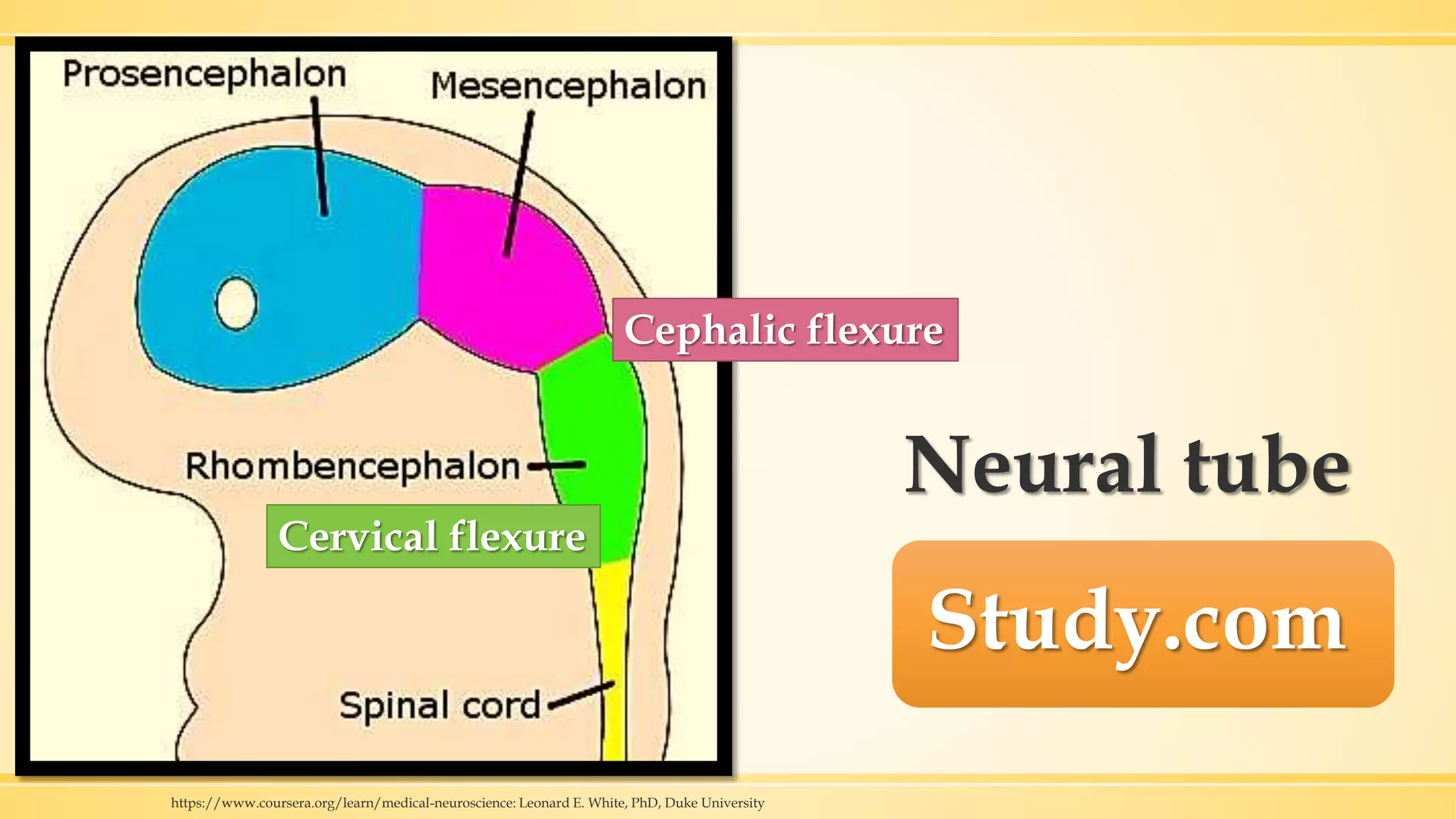
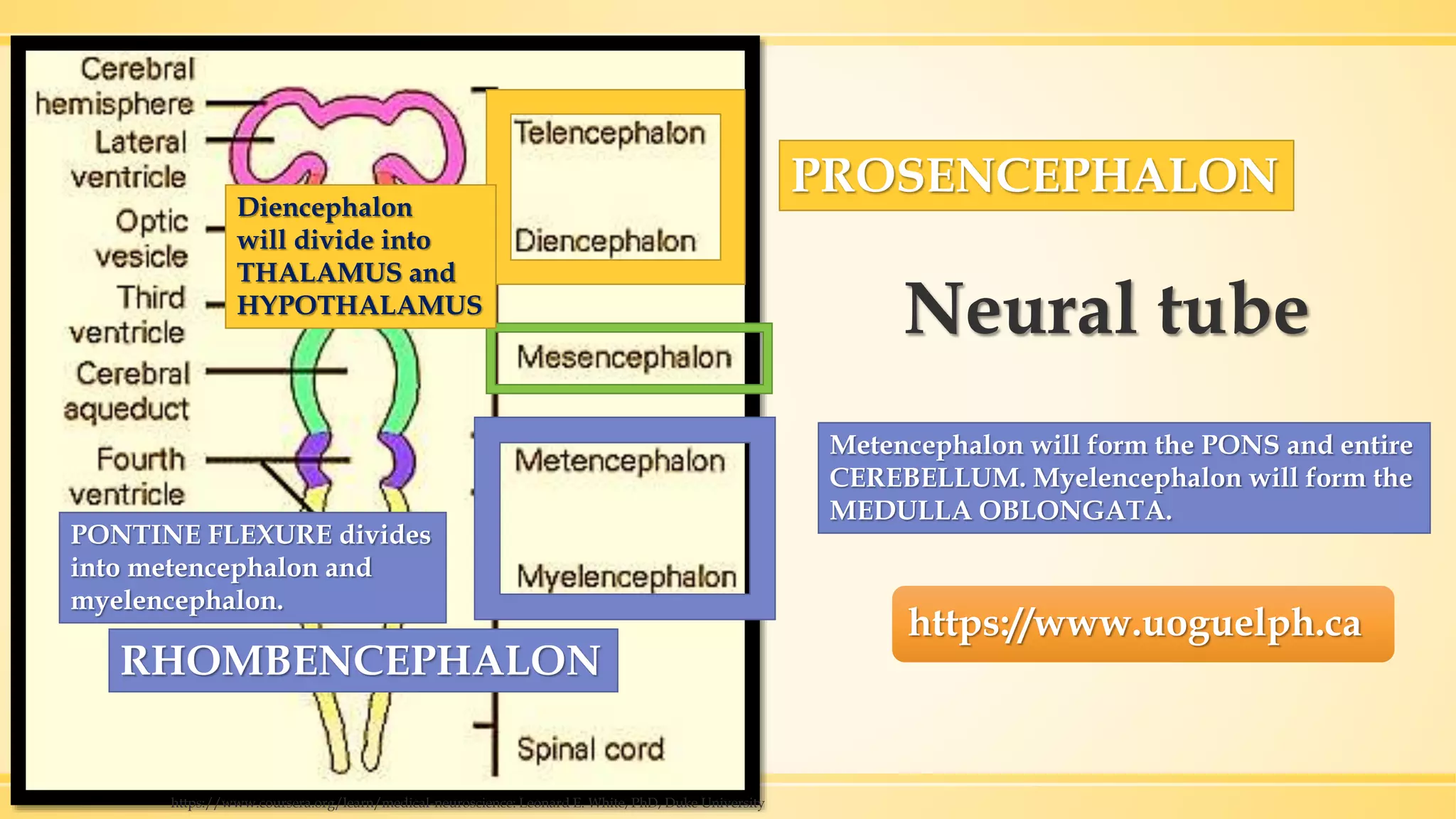

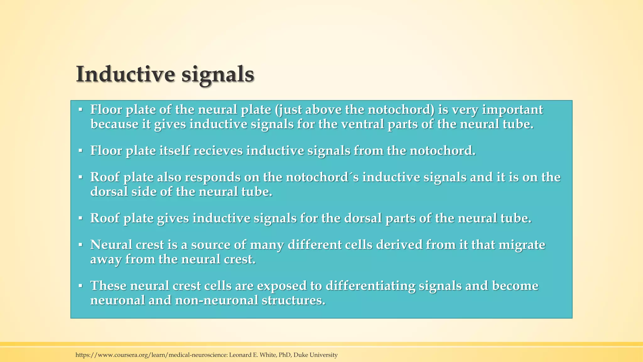
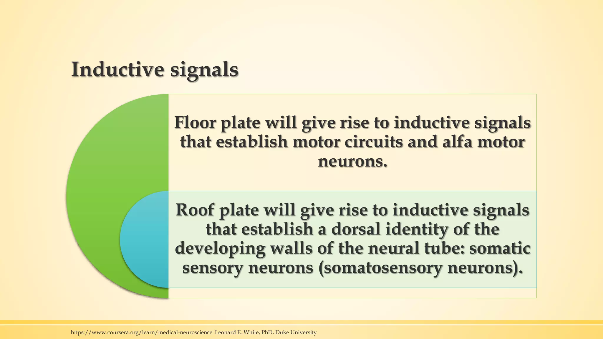
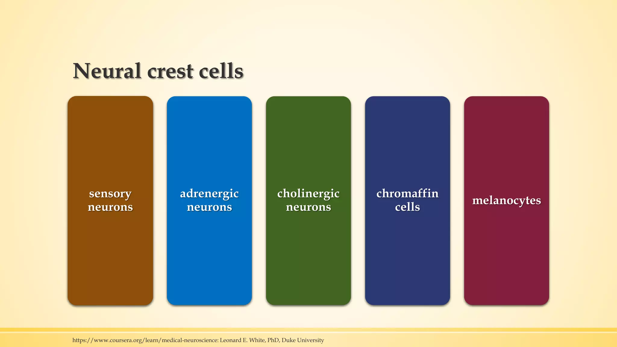
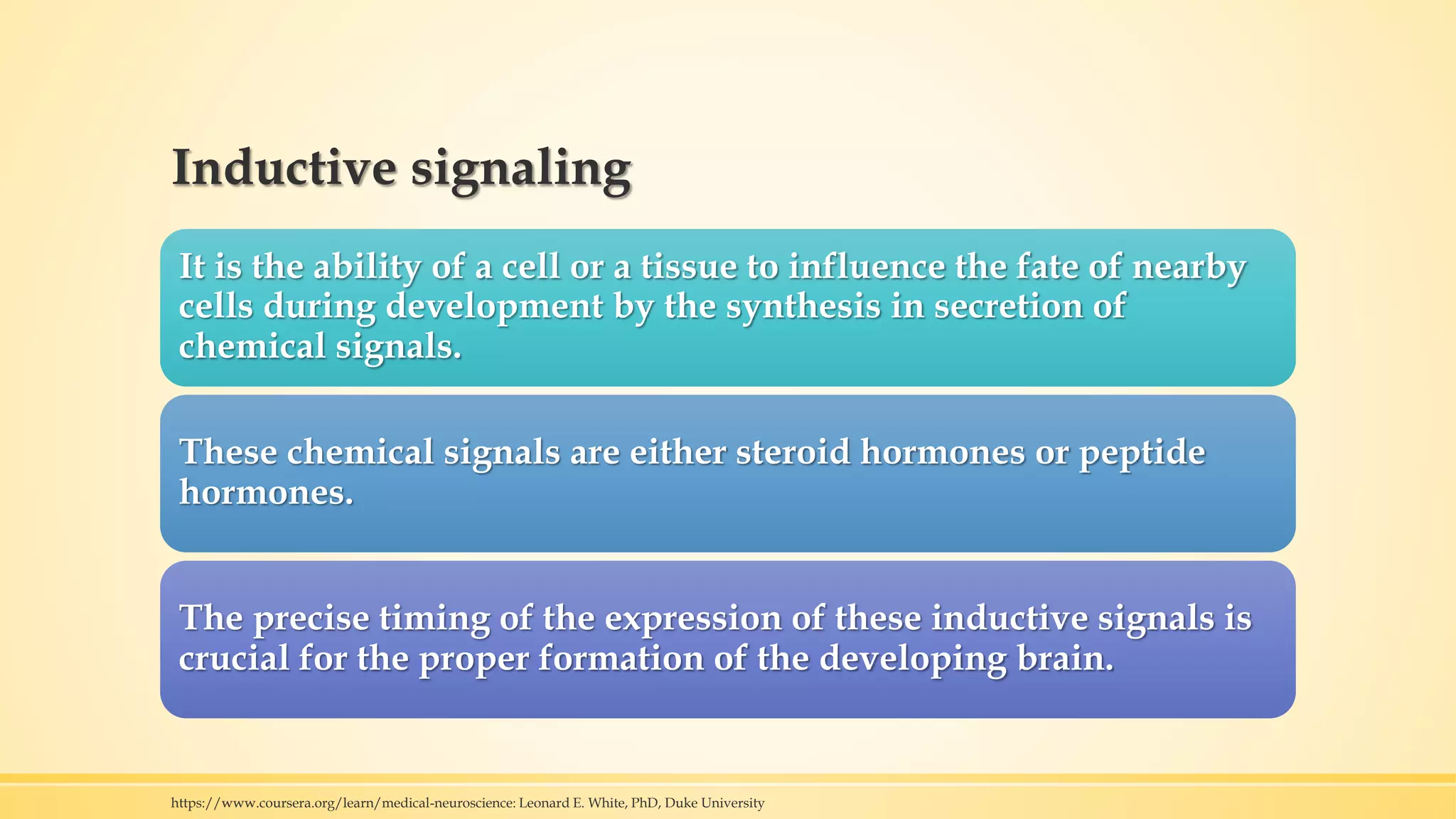
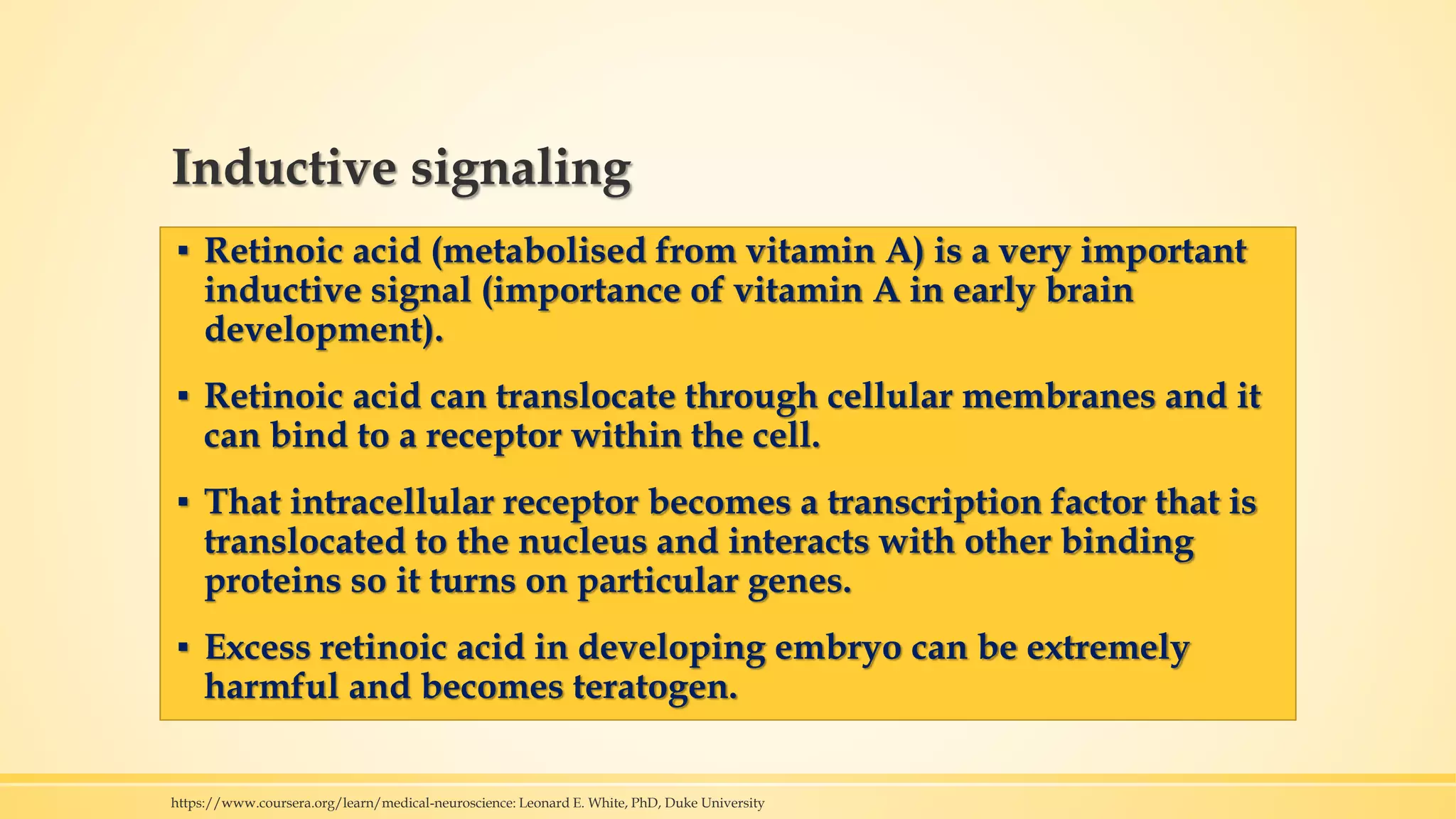
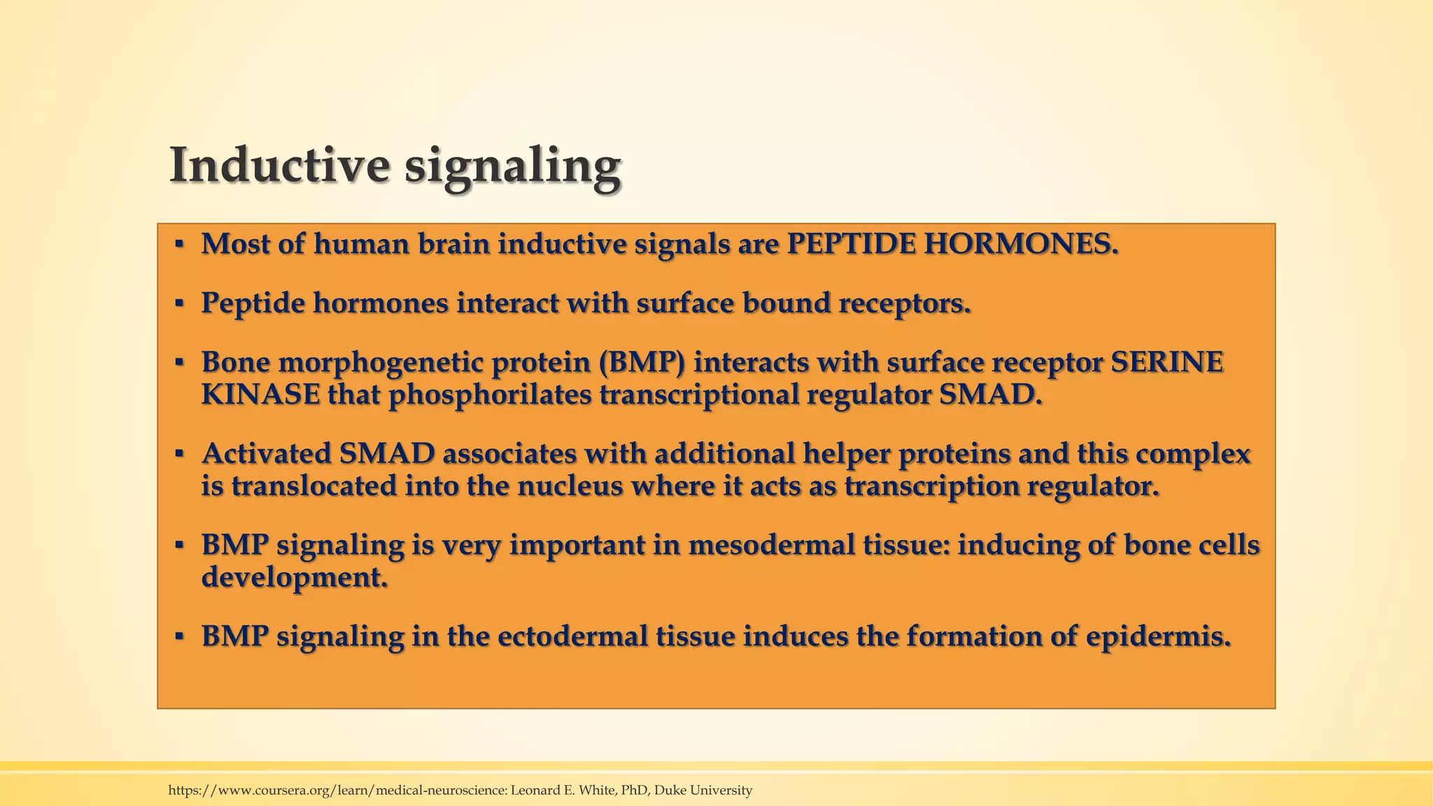
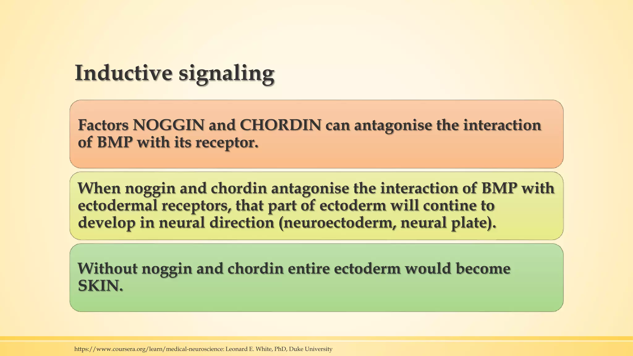
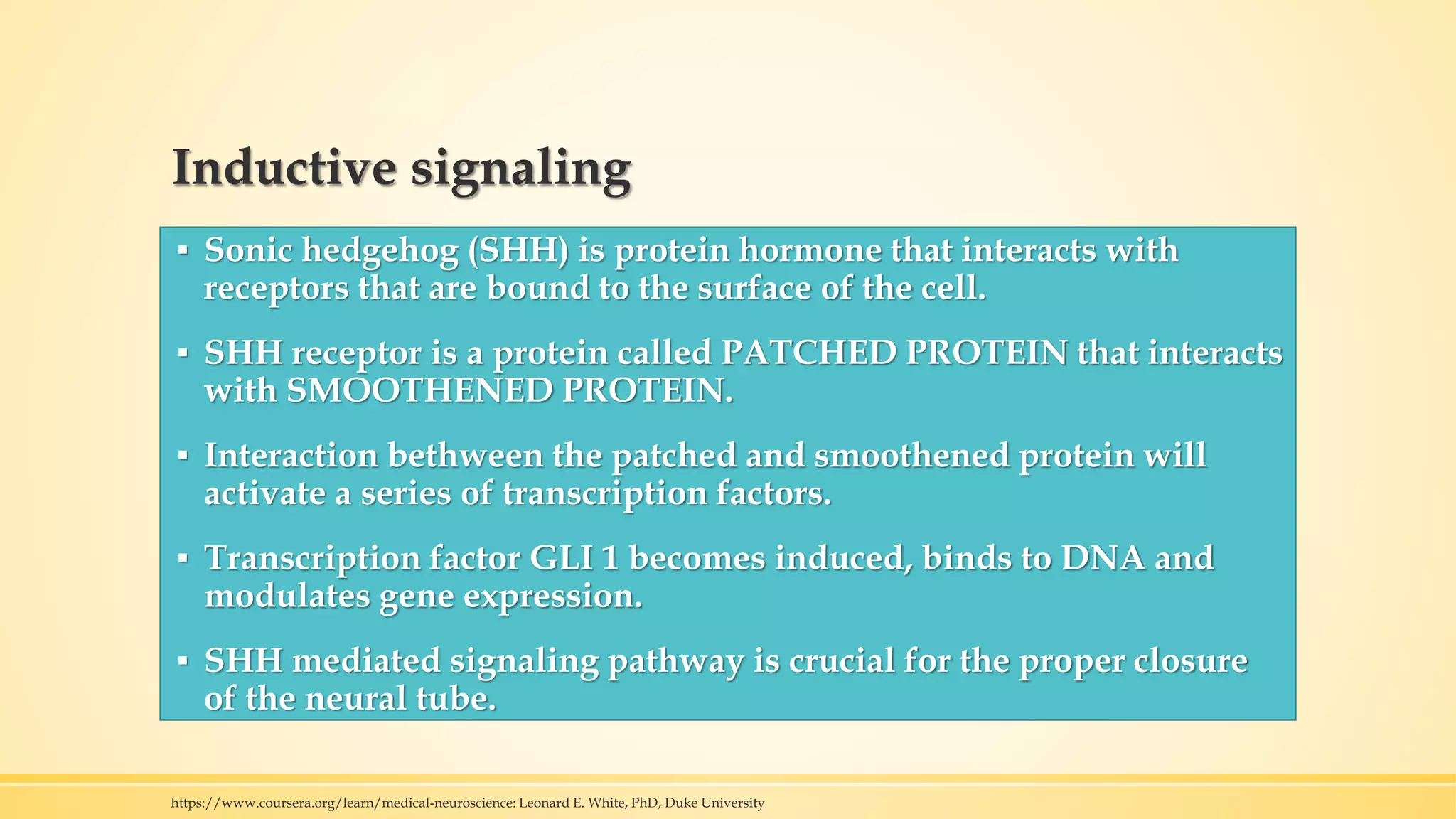
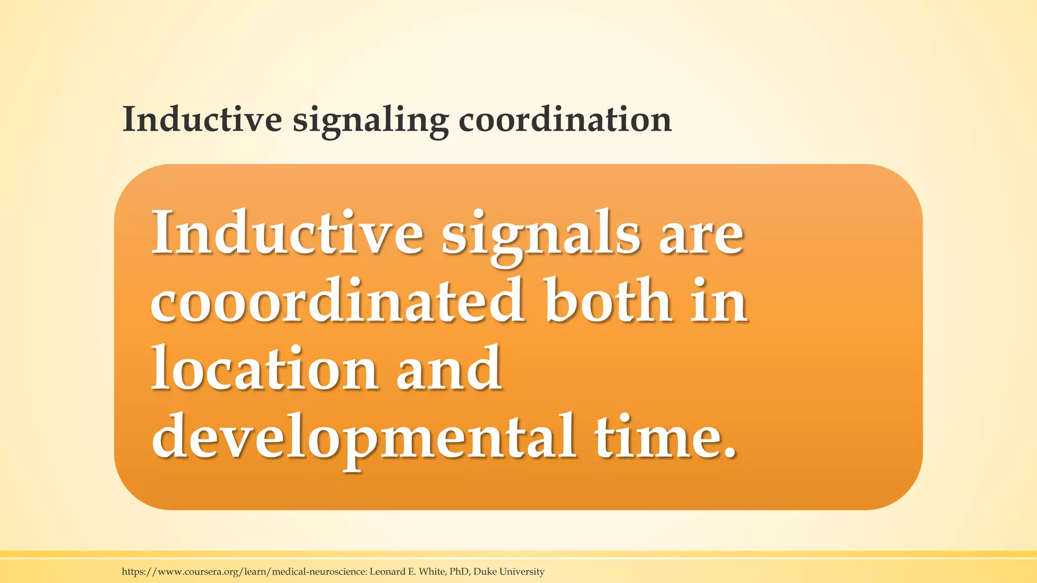
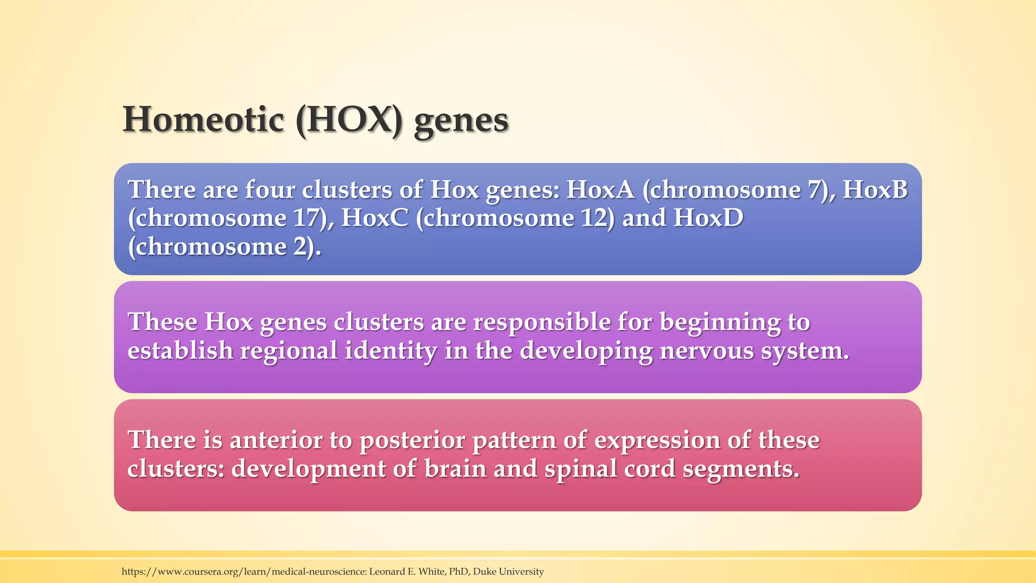
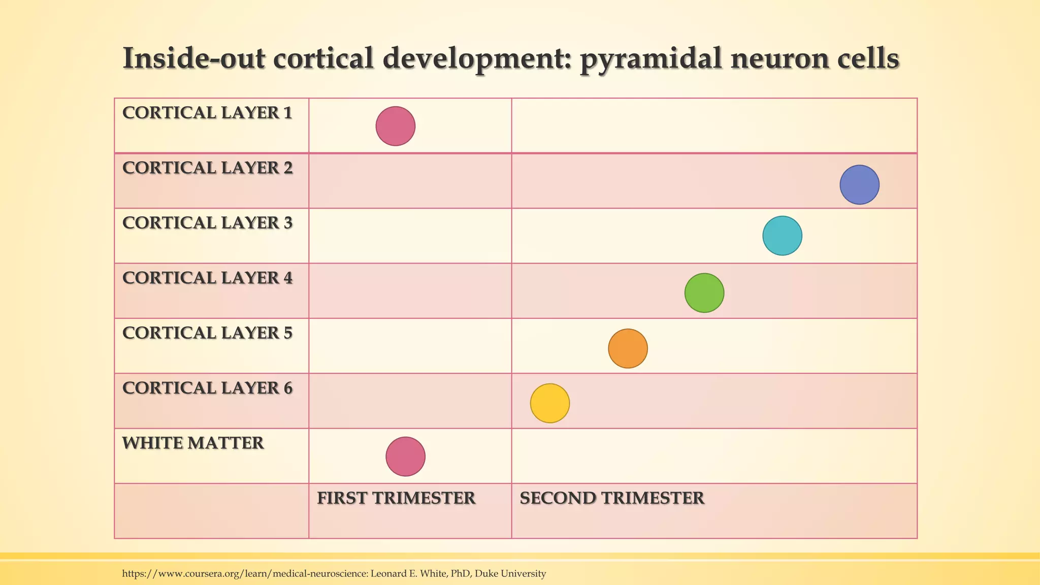
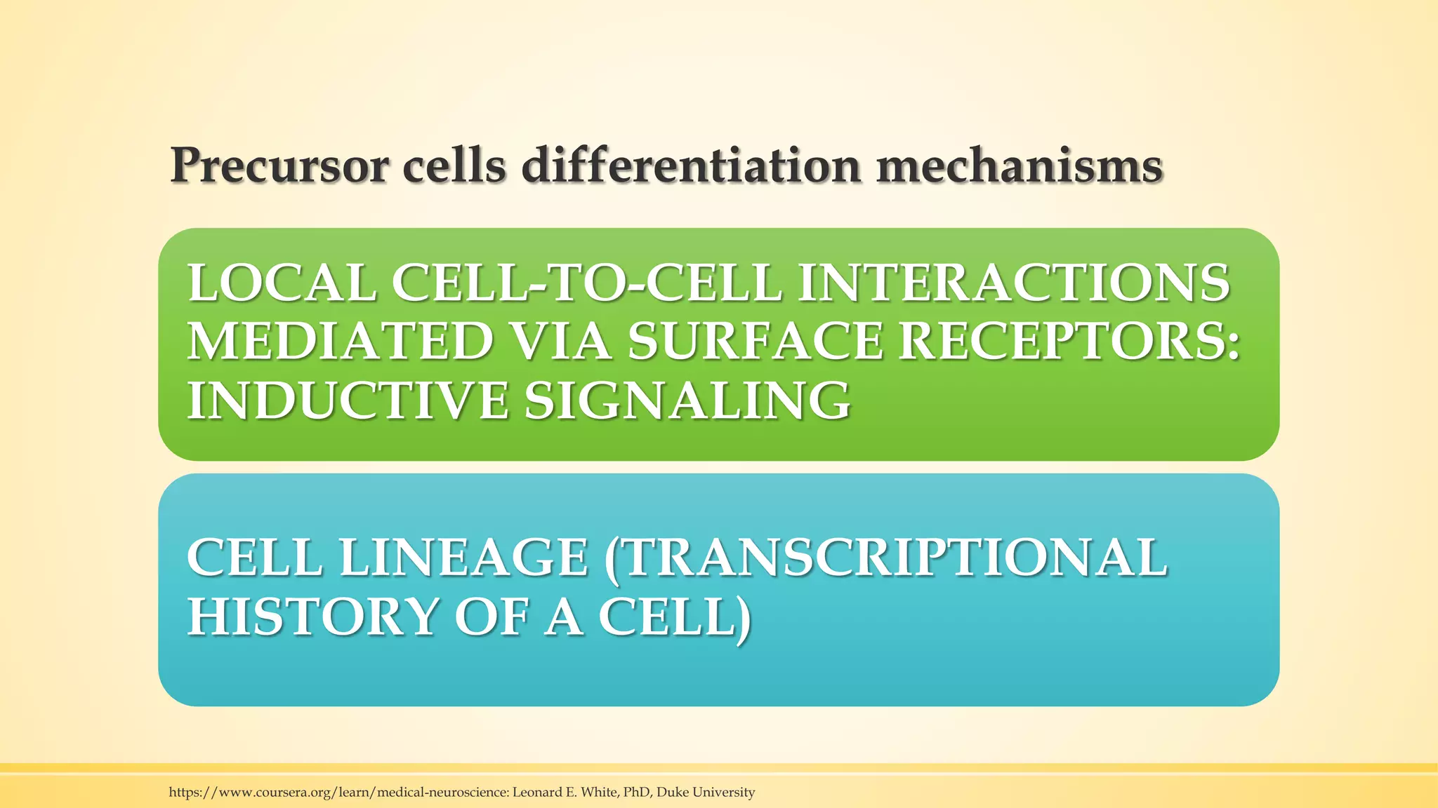
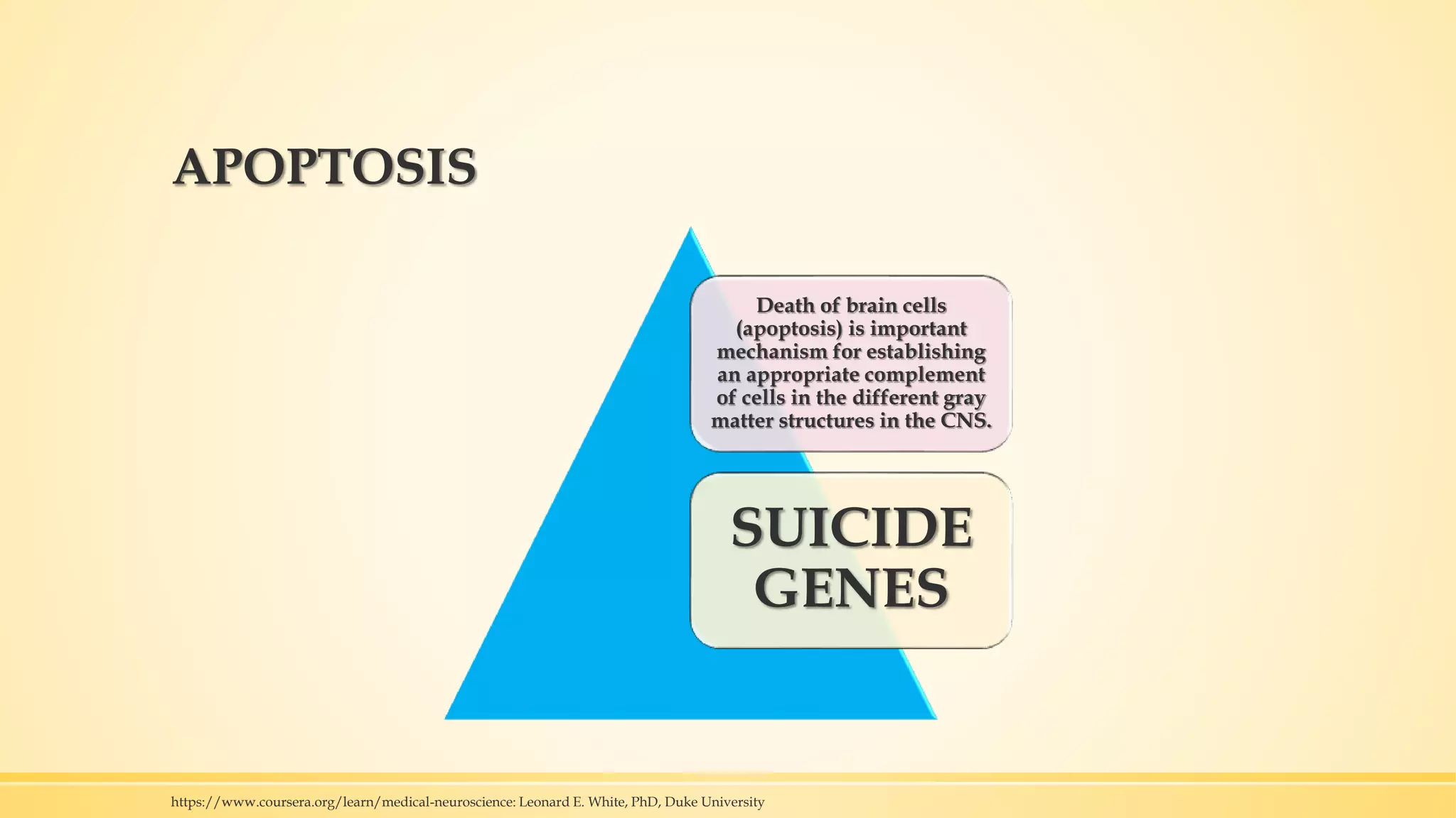


Early brain development is influenced by genetic, epigenetic, and environmental factors. Key processes include gastrulation, which forms the three germ layers; neurulation, which forms the neural tube; proliferation of neural stem cells; migration of neurons; and apoptosis which sculpt the brain. Precisely coordinated inductive signals involving molecules like BMP, SHH and retinoic acid guide cell differentiation and regional patterning in the developing brain.


