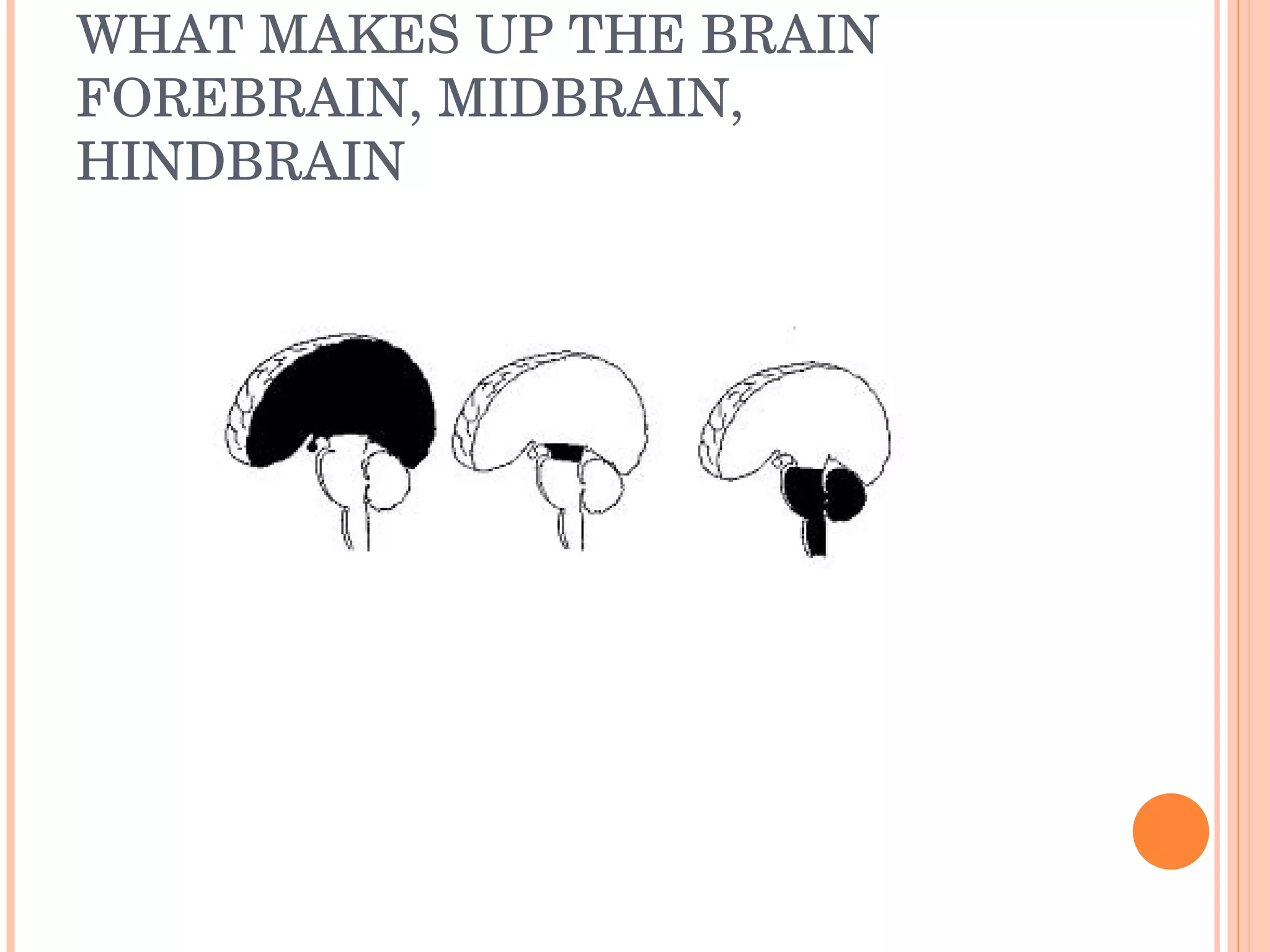
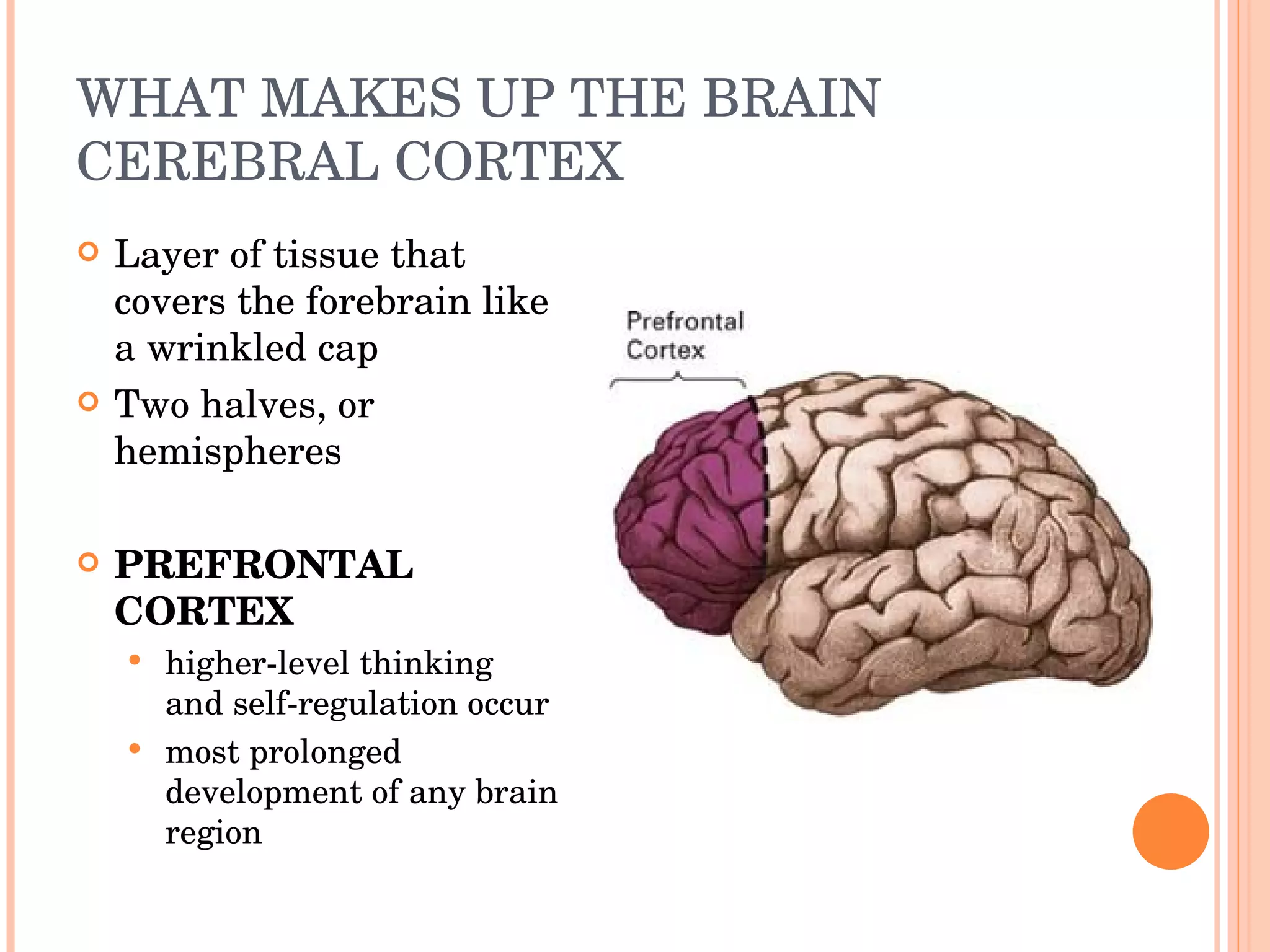
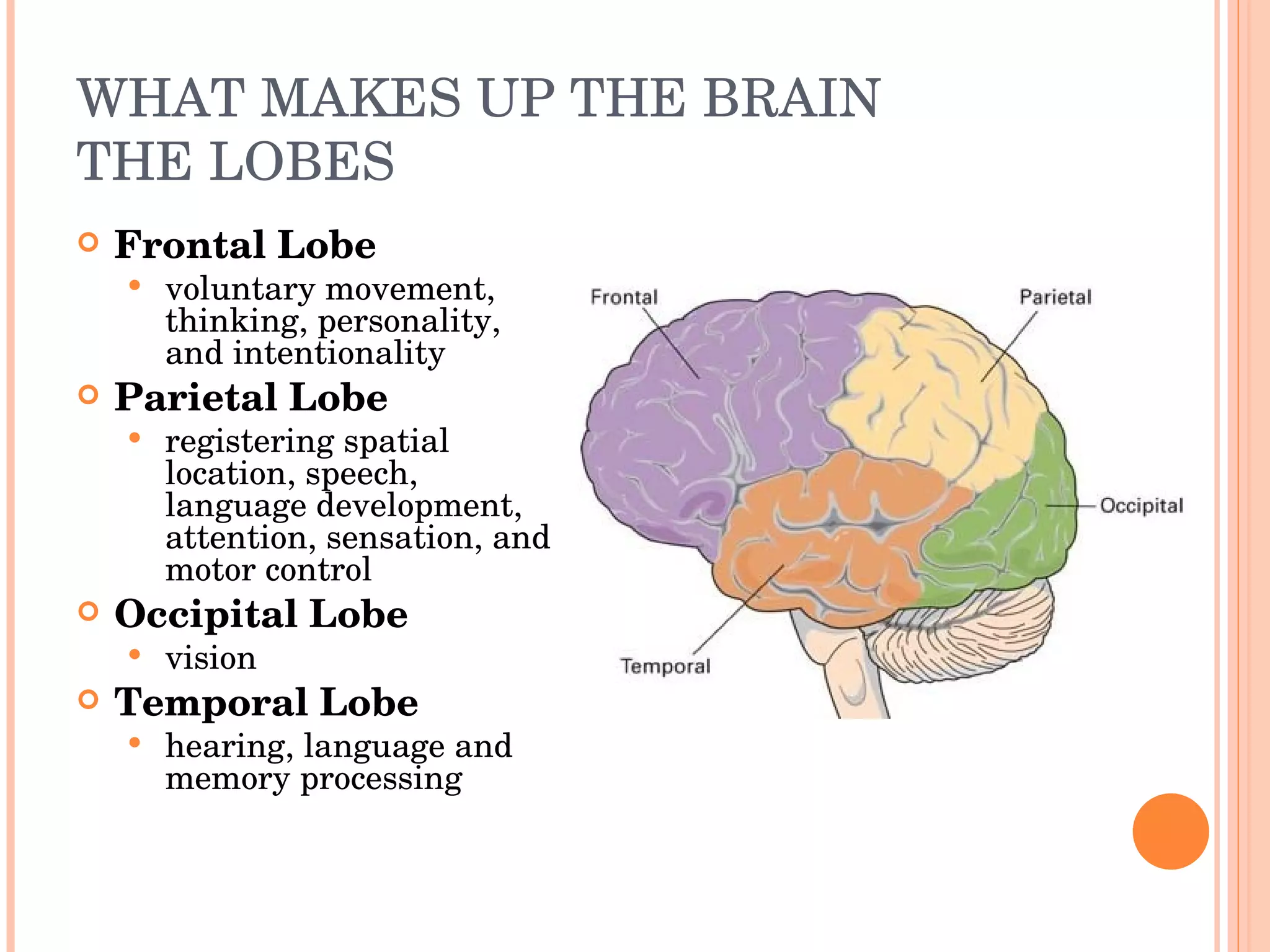
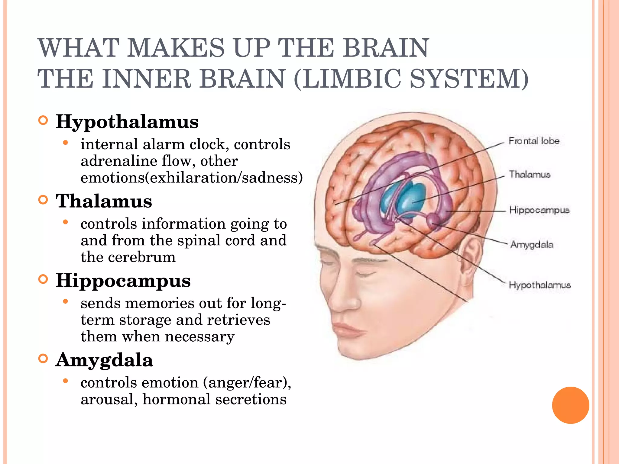
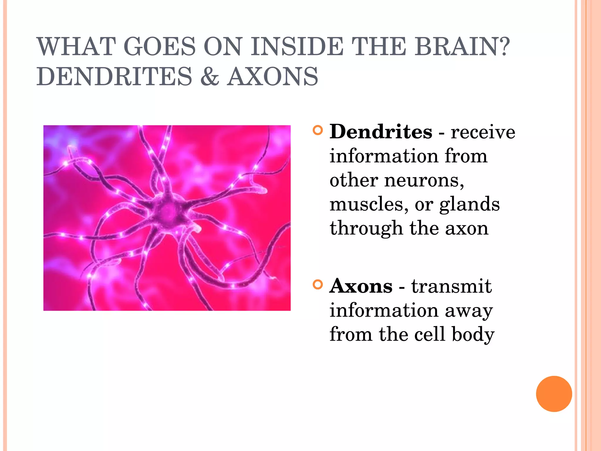
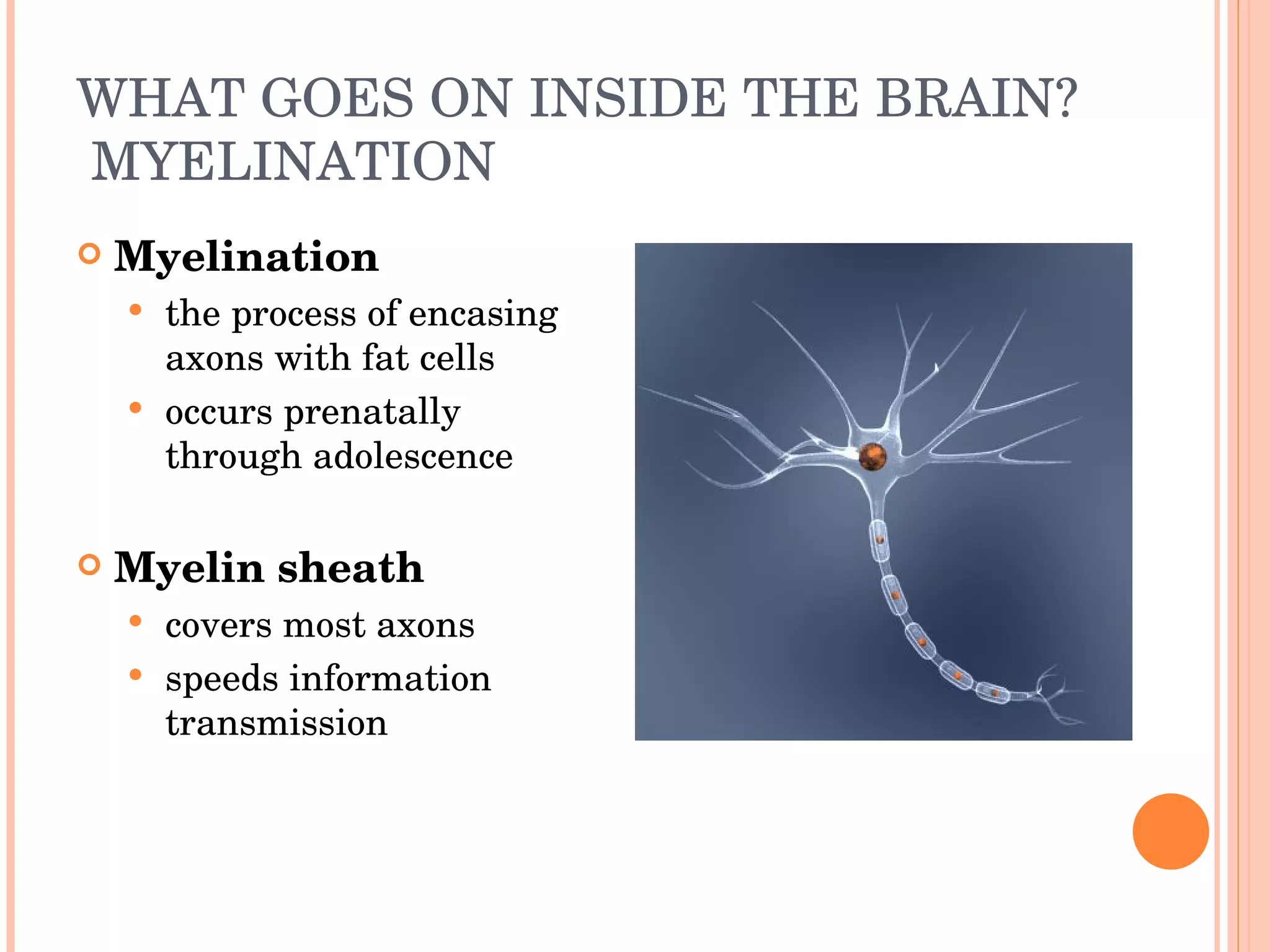
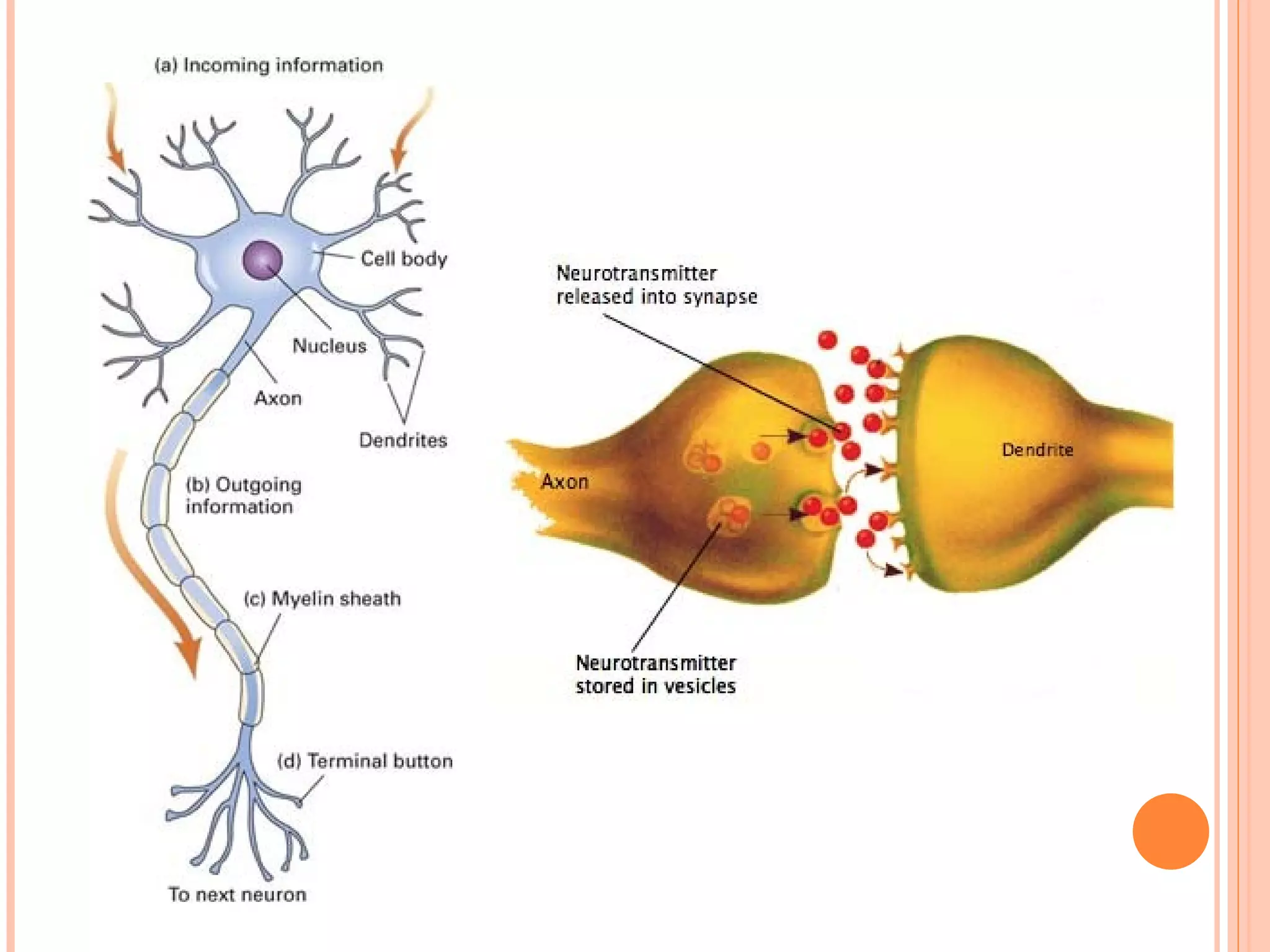
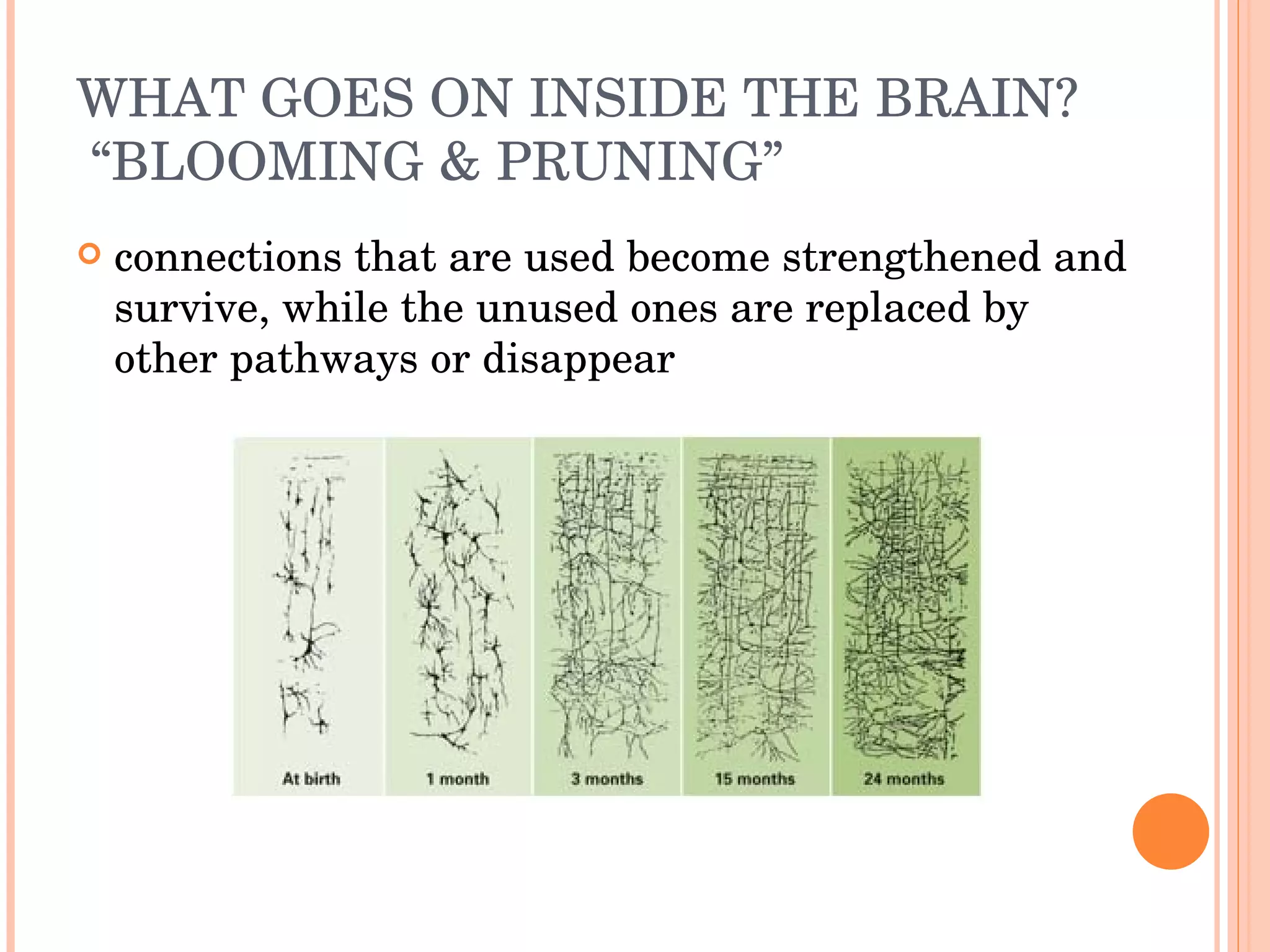
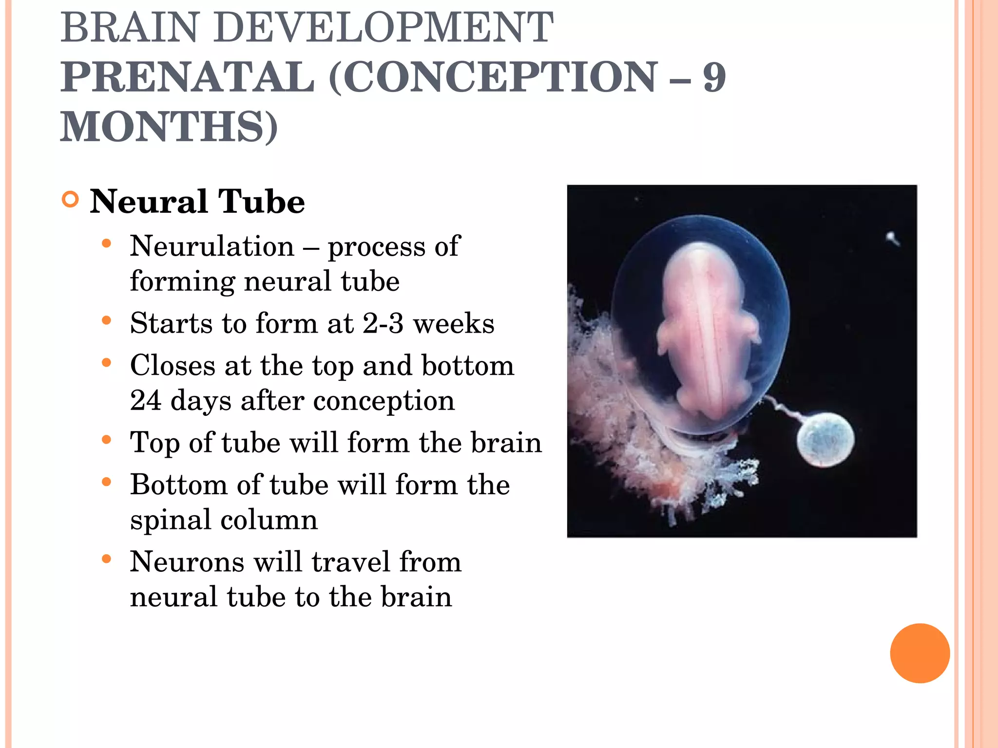
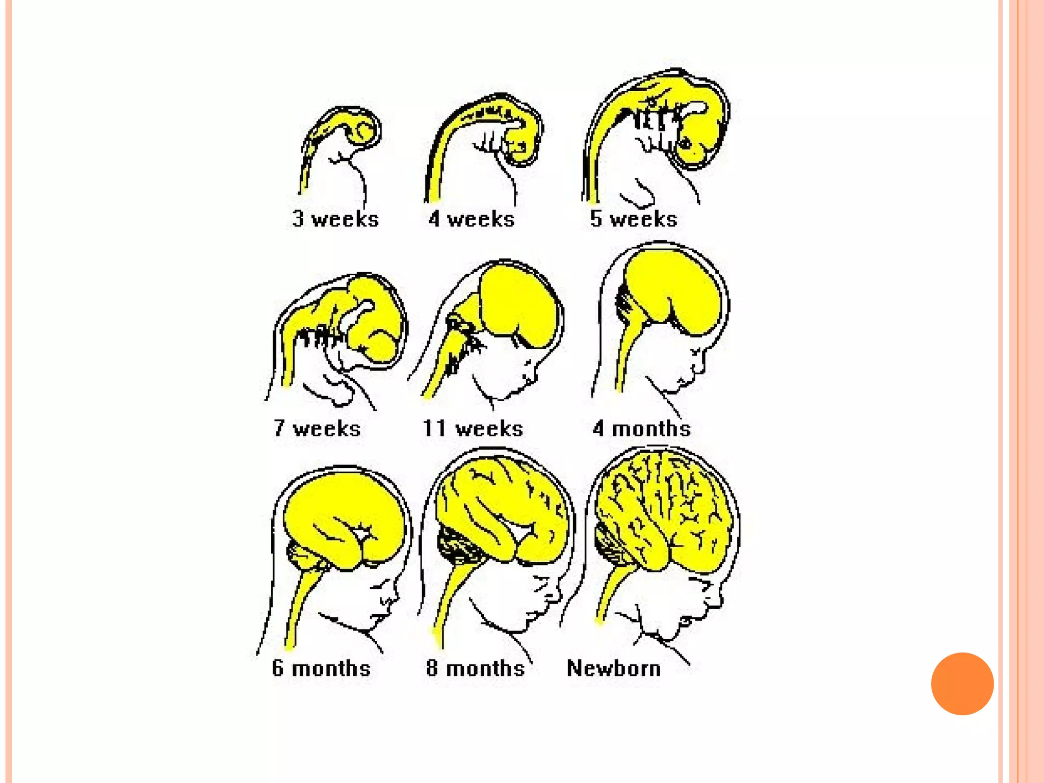
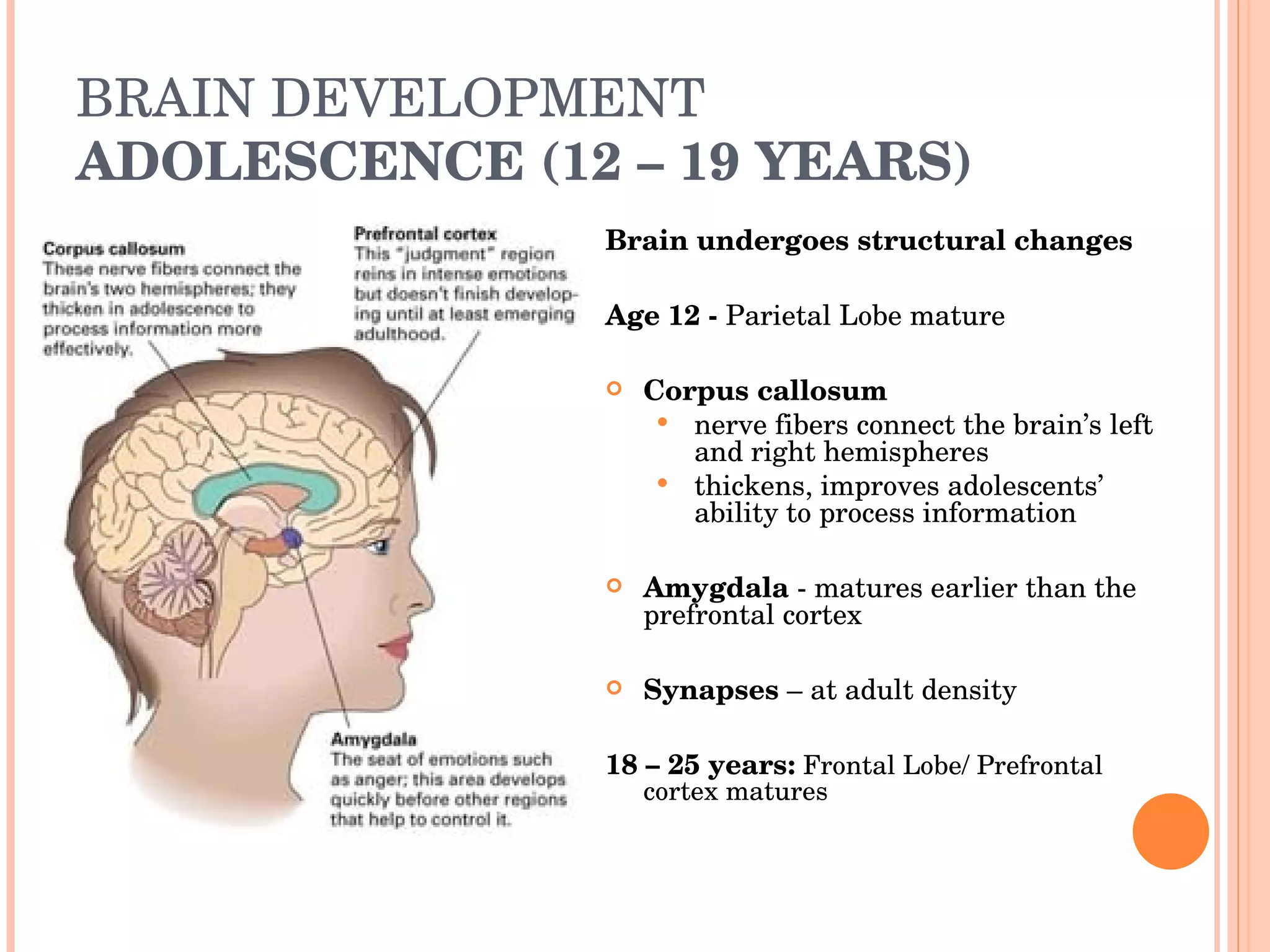
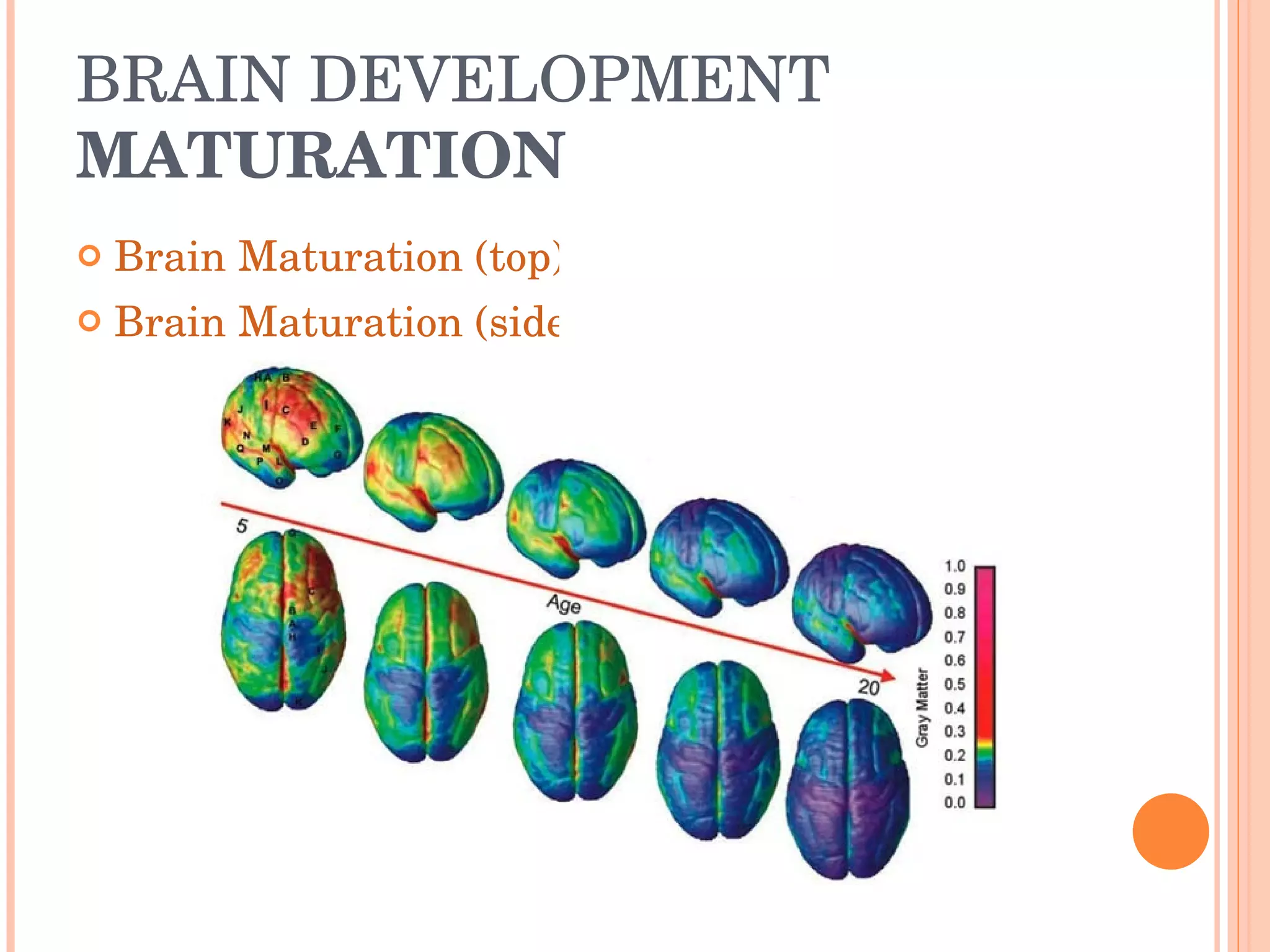
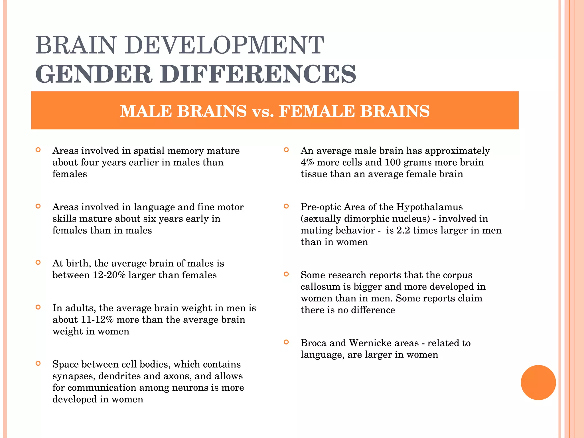
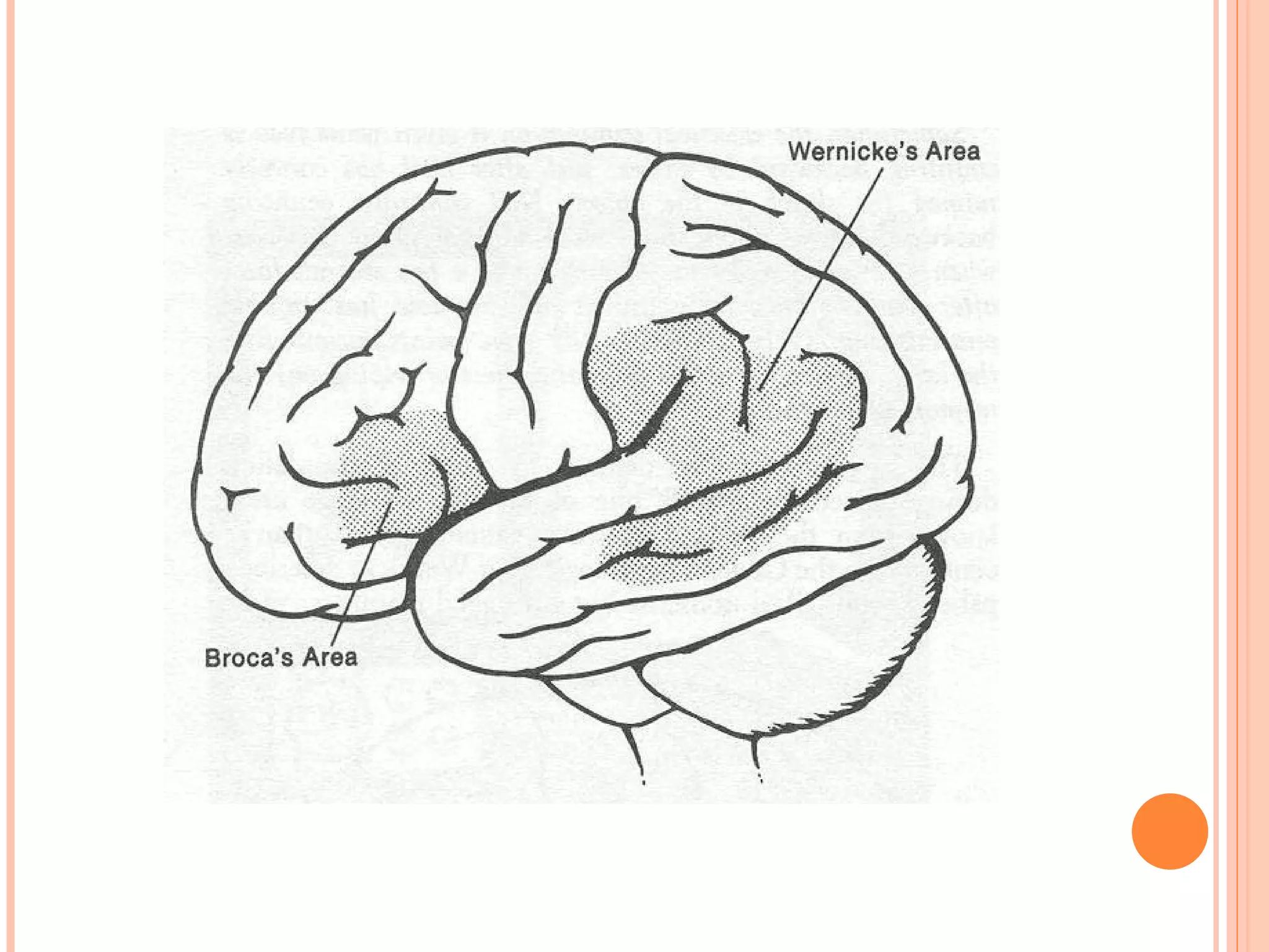
THE BRAIN Prenatal, Infancy, Childhood, Adolescence, Early Adulthood Development
WHAT MAKES UP THE BRAIN Forebrain, Midbrain, Hindbrain Cerebral cortex Prefrontal cortex Hemispheres Right Hemisphere/Left Hemisphere Lateralization The Lobes Frontal, Parietal, Occipital, Temporal The Inner Brain (Limbic System)
WHAT MAKES UP THE BRAIN FOREBRAIN, MIDBRAIN, HINDBRAIN FOREBRAIN the largest and most highly developed part consists of the cerebrum & the structures beneath MIDBRAIN uppermost part of the brainstem controls reflex actions, voluntary movements (eye movement) HINDBRAIN  upper part of the spinal cord the cerebellum controls body’s vital functions (respiration & heart rate) coordinates movement (hitting a tennis ball)
WHAT MAKES UP THE BRAIN FOREBRAIN, MIDBRAIN, HINDBRAIN
WHAT MAKES UP THE BRAIN CEREBRAL CORTEX Layer of tissue that covers the forebrain like a wrinkled cap Two halves, or hemispheres PREFRONTAL CORTEX higher-level thinking and self-regulation occur most prolonged development of any brain region
WHAT MAKES UP THE BRAIN HEMISPHERES Right Hemisphere specializes in humor and the use of metaphors, abstract reasoning skills, spatial reasoning, facial recognition, nonverbal memory Left Hemisphere specializes in speech and grammar, arithmetic, writing, complex motor functions, short-term verbal recall Lateralization  specialization of function in one hemisphere of the cerebral cortex or the other complex functions such as reading or performing music use both hemispheres
WHAT MAKES UP THE BRAIN THE LOBES Frontal Lobe voluntary movement, thinking, personality, and intentionality Parietal Lobe registering spatial location, speech, language development, attention, sensation, and motor control Occipital Lobe vision Temporal Lobe hearing, language and memory processing
WHAT MAKES UP THE BRAIN THE INNER BRAIN (LIMBIC SYSTEM) Hypothalamus internal alarm clock, controls adrenaline flow, other emotions(exhilaration/sadness) Thalamus controls information going to and from the spinal cord and the cerebrum Hippocampus sends memories out for long-term storage and retrieves them when necessary Amygdala controls emotion (anger/fear), arousal, hormonal secretions
WHAT GOES ON INSIDE THE BRAIN? NEURONS Nerve cells runs on own programming that follows genetic code handles information processing at the cellular level sends electrical and chemical signals to communicate with each other
WHAT GOES ON INSIDE THE BRAIN? DENDRITES & AXONS Dendrites  - receive information from other neurons, muscles, or glands through the axon Axons  - transmit information away from the cell body
WHAT GOES ON INSIDE THE BRAIN?  MYELINATION Myelination the process of encasing axons with fat cells  occurs prenatally through adolescence Myelin sheath covers most axons speeds information transmission
WHAT GOES ON INSIDE THE BRAIN? BUTTONS, NEUROTRANSMITTERS, SYNAPSES  & RECEPTORS Terminal Buttons releases chemicals called neurotransmitters into synapses Neurotransmitters Chemicals (i.e. inhibitory neurotransmitters such as serotonin, dopamine) Synapses  - tiny gaps between neurons’ fibers  Synaptogenesis  - the formation of synapses Receptors molecular structure that binds with substances
 
WHAT GOES ON INSIDE THE BRAIN?  “BLOOMING & PRUNING” connections that are used become strengthened and survive, while the unused ones are replaced by other pathways or disappear
BRAIN DEVELOPMENT  EVOLUTION OF THE BRAIN Evolutionary psychology emphasizes the importance of adaptation, reproduction, and “survival of the fittest” in shaping behavior Original assumption was that size of brain meant more intelligence – not true More reasonable approach was to compare evolution of brain regions Cerebrum (learning, perception, motivation) Brainstem (reflex activities)
BRAIN DEVELOPMENT  EVOLUTION OF THE BRAIN Three important points: The brain has increased in size Most of the increase in size has occurred in the cerebrum There is an increase in the number of convolutions (folds in cerebral surface). Increases the volume of the cerebral cortex An extended childhood period humans require time to develop a large brain and learn the complexity of human societies
BRAIN DEVELOPMENT PRENATAL (CONCEPTION – 9 MONTHS) Neural Tube Neurulation – process of forming neural tube Starts to form at 2-3 weeks Closes at the top and bottom 24 days after conception Top of tube will form the brain Bottom of tube will form the spinal column Neurons will travel from neural tube to the brain
BRAIN DEVELOPMENT PRENATAL (CONCEPTION – 9 MONTHS) Neurogenesis (5 th  week) the generation of new neurons 200-500,000 neurons are being generated every minute Neuronal migration (through 6 – 24 th  week) neurons move to specific locations to await instructions Travel along glia fibers connections between neurons begin (23 rd  week)  genes direct wiring patterns
BRAIN DEVELOPMENT PRENATAL (CONCEPTION – 9 MONTHS) Neuronal migration Neuron Traveling on glia fiber Neuron Migration Myelination Begins in spinal cord proceeds through subcortical regions (beneath the cortex) Cortical regions myelinate at different times – moves from posterior regions to anterior regions Frontal & Parietal Lobes are completed last
 
BRAIN DEVELOPMENT INFANCY (BIRTH – 2 YEARS OLD) At Birth   Contains approximately 100 billion+ neurons 25% of its adult weight 360 grams in females (approx. 12.7 oz.) 380 grams in males (approx. 13.4 oz.) Rapid growth begins in all areas Frontal lobes are immature Occipital & Temporal Lobe progression Sensory areas of Parietal Lobe mature
BRAIN DEVELOPMENT INFANCY (BIRTH – 2 YEARS OLD) Birth – 1 year old Weighs about 1000 grams at 1 year (approx. 2 lbs) 970 grams in males 940 grams in females Prefrontal cortex develops massively between 7-12 months Myelination – increase in brain’s weight 3-6 months - visual pathways complete
BRAIN DEVELOPMENT INFANCY (BIRTH – 2 YEARS OLD) Occipital Lobe (Vision) NEWBORN Occipital Lobe (Vision) 1 MONTH OLD
BRAIN DEVELOPMENT INFANCY (BIRTH – 2 YEARS OLD) Occipital Lobe (Vision) 2 MONTHS OLD Occipital Lobe (Vision) 3 MONTHS OLD
BRAIN DEVELOPMENT INFANCY (BIRTH – 2 YEARS OLD) Occipital Lobe (Vision) 6 MONTHS OLD
BRAIN DEVELOPMENT INFANCY (BIRTH – 2 YEARS OLD) Temporal Lobe  4 months: responds to every sound produced in all the languages 10 months: distinguish and produce the sounds of their own language no longer pays attention to the sounds of foreign languages  Lateralization Show greater electrical brain activity in the left hemisphere when listening to speech/sounds Amygdala  - matures over the course of the first 8 months Dendrite connections  Experiences determine how connections are made 8 to 9 months: form specific memories from their experiences “ Blooming and Pruning”  the more babies engage in physical activity or use language, more pathways will be strengthened 2 years old   75% of its adult weight 1,120 grams in males (2.5 lbs) 1,040 grams in females (2.3 lbs)
BRAIN DEVELOPMENT  EARLY CHILDHOOD (3 – 5 YEARS) Rapid, distinct spurts of growth (not as rapid as during infancy) Continuous increases in dendrite connections and myelination “ Blooming and Pruning” ‘ Experience is the sculptor’
BRAIN DEVELOPMENT  EARLY CHILDHOOD (3 – 5 YEARS) 3 years old Brain is ¾ of adult size Brain weight: 1,270 grams in males (approx. 2.8 lbs) 1,090 grams in females (approx. 2.4 lbs) 4 years old Parietal Lobe - Hand-eye coordination myelination is complete (gross/fine motor skills) Temporal Lobe - Auditory myelination is complete 5 years old 95% of its adult size Occipital Lobe - Acuity reaches adult levels
BRAIN DEVELOPMENT EARLY CHILDHOOD (3 – 5 YEARS)
BRAIN DEVELOPMENT EARLY CHILDHOOD (3 – 5 YEARS) Effects of Neglect and/or Abuse the frontal lobe develops improperly when deprived of affection and positive emotional experiences the lack of stimulation causes neurons in the brain to die off
BRAIN DEVELOPMENT MIDDLE/LATE CHILDHOOD (6 – 11 YEARS) Brain volume stabilizes Brain weight: Age 10 -11 1,440 grams in males (3.2 lbs) 1,260 grams in females (2.7 lbs) Age 6-7:  maturation of occipital lobe contrast sensitivity and peripheral vision reach adult levels Age 8:  maturation of temporal lobe   Cerebral Cortex - thickens in the temporal and frontal lobe areas improved language abilities such as reading Prefrontal Cortex - pathways and circuitry increases improved attention, reasoning, and cognitive control “ Blooming & Pruning” some brain areas increase in use while others decrease moves from broader areas to more focal, smaller areas
BRAIN DEVELOPMENT ADOLESCENCE (12 – 19 YEARS) Brain undergoes structural changes  Age 12 -  Parietal Lobe mature Corpus callosum nerve fibers connect the brain’s left and right hemispheres  thickens, improves adolescents’ ability to process information Amygdala  - matures earlier than the prefrontal cortex Synapses  – at adult density 18 – 25 years:  Frontal Lobe/ Prefrontal cortex matures
BRAIN DEVELOPMENT EARLY ADULTHOOD (20 – 30 YEARS) Anatomically, physiologically, functionally mature Brain weight:  1,450 grams in males (3.2 lbs) 1,310 grams in females (2.9 lbs) Brain continues to develop connections throughout adulthood Synapse formation is much slower than in childhood Synapses are formed based only on specific experiences in the adult's life
BRAIN DEVELOPMENT MATURATION Brain Maturation (top) Brain Maturation (side)
BRAIN DEVELOPMENT GENDER DIFFERENCES Areas involved in spatial memory mature about four years earlier in males than females Areas involved in language and fine motor skills mature about six years early in females than in males At birth, the average brain of males is between 12-20% larger than females In adults, the average brain weight in men is about 11-12% more than the average brain weight in women Space between cell bodies, which contains synapses, dendrites and axons, and allows for communication among neurons is more developed in women An average male brain has approximately 4% more cells and 100 grams more brain tissue than an average female brain Pre-optic Area of the Hypothalamus (sexually dimorphic nucleus) - involved in mating behavior -  is 2.2 times larger in men than in women Some research reports that the corpus callosum is bigger and more developed in women than in men. Some reports claim there is no difference Broca and Wernicke areas - related to language, are larger in women MALE BRAINS vs. FEMALE BRAINS


The Brain

  • 1.
    THE BRAIN Prenatal,Infancy, Childhood, Adolescence, Early Adulthood Development
  • 2.
    WHAT MAKES UPTHE BRAIN Forebrain, Midbrain, Hindbrain Cerebral cortex Prefrontal cortex Hemispheres Right Hemisphere/Left Hemisphere Lateralization The Lobes Frontal, Parietal, Occipital, Temporal The Inner Brain (Limbic System)
  • 3.
    WHAT MAKES UPTHE BRAIN FOREBRAIN, MIDBRAIN, HINDBRAIN FOREBRAIN the largest and most highly developed part consists of the cerebrum & the structures beneath MIDBRAIN uppermost part of the brainstem controls reflex actions, voluntary movements (eye movement) HINDBRAIN upper part of the spinal cord the cerebellum controls body’s vital functions (respiration & heart rate) coordinates movement (hitting a tennis ball)
  • 4.
    WHAT MAKES UPTHE BRAIN FOREBRAIN, MIDBRAIN, HINDBRAIN
  • 5.
    WHAT MAKES UPTHE BRAIN CEREBRAL CORTEX Layer of tissue that covers the forebrain like a wrinkled cap Two halves, or hemispheres PREFRONTAL CORTEX higher-level thinking and self-regulation occur most prolonged development of any brain region
  • 6.
    WHAT MAKES UPTHE BRAIN HEMISPHERES Right Hemisphere specializes in humor and the use of metaphors, abstract reasoning skills, spatial reasoning, facial recognition, nonverbal memory Left Hemisphere specializes in speech and grammar, arithmetic, writing, complex motor functions, short-term verbal recall Lateralization specialization of function in one hemisphere of the cerebral cortex or the other complex functions such as reading or performing music use both hemispheres
  • 7.
    WHAT MAKES UPTHE BRAIN THE LOBES Frontal Lobe voluntary movement, thinking, personality, and intentionality Parietal Lobe registering spatial location, speech, language development, attention, sensation, and motor control Occipital Lobe vision Temporal Lobe hearing, language and memory processing
  • 8.
    WHAT MAKES UPTHE BRAIN THE INNER BRAIN (LIMBIC SYSTEM) Hypothalamus internal alarm clock, controls adrenaline flow, other emotions(exhilaration/sadness) Thalamus controls information going to and from the spinal cord and the cerebrum Hippocampus sends memories out for long-term storage and retrieves them when necessary Amygdala controls emotion (anger/fear), arousal, hormonal secretions
  • 9.
    WHAT GOES ONINSIDE THE BRAIN? NEURONS Nerve cells runs on own programming that follows genetic code handles information processing at the cellular level sends electrical and chemical signals to communicate with each other
  • 10.
    WHAT GOES ONINSIDE THE BRAIN? DENDRITES & AXONS Dendrites - receive information from other neurons, muscles, or glands through the axon Axons - transmit information away from the cell body
  • 11.
    WHAT GOES ONINSIDE THE BRAIN? MYELINATION Myelination the process of encasing axons with fat cells occurs prenatally through adolescence Myelin sheath covers most axons speeds information transmission
  • 12.
    WHAT GOES ONINSIDE THE BRAIN? BUTTONS, NEUROTRANSMITTERS, SYNAPSES & RECEPTORS Terminal Buttons releases chemicals called neurotransmitters into synapses Neurotransmitters Chemicals (i.e. inhibitory neurotransmitters such as serotonin, dopamine) Synapses - tiny gaps between neurons’ fibers Synaptogenesis - the formation of synapses Receptors molecular structure that binds with substances
  • 13.
  • 14.
    WHAT GOES ONINSIDE THE BRAIN? “BLOOMING & PRUNING” connections that are used become strengthened and survive, while the unused ones are replaced by other pathways or disappear
  • 15.
    BRAIN DEVELOPMENT EVOLUTION OF THE BRAIN Evolutionary psychology emphasizes the importance of adaptation, reproduction, and “survival of the fittest” in shaping behavior Original assumption was that size of brain meant more intelligence – not true More reasonable approach was to compare evolution of brain regions Cerebrum (learning, perception, motivation) Brainstem (reflex activities)
  • 16.
    BRAIN DEVELOPMENT EVOLUTION OF THE BRAIN Three important points: The brain has increased in size Most of the increase in size has occurred in the cerebrum There is an increase in the number of convolutions (folds in cerebral surface). Increases the volume of the cerebral cortex An extended childhood period humans require time to develop a large brain and learn the complexity of human societies
  • 17.
    BRAIN DEVELOPMENT PRENATAL(CONCEPTION – 9 MONTHS) Neural Tube Neurulation – process of forming neural tube Starts to form at 2-3 weeks Closes at the top and bottom 24 days after conception Top of tube will form the brain Bottom of tube will form the spinal column Neurons will travel from neural tube to the brain
  • 18.
    BRAIN DEVELOPMENT PRENATAL(CONCEPTION – 9 MONTHS) Neurogenesis (5 th week) the generation of new neurons 200-500,000 neurons are being generated every minute Neuronal migration (through 6 – 24 th week) neurons move to specific locations to await instructions Travel along glia fibers connections between neurons begin (23 rd week) genes direct wiring patterns
  • 19.
    BRAIN DEVELOPMENT PRENATAL(CONCEPTION – 9 MONTHS) Neuronal migration Neuron Traveling on glia fiber Neuron Migration Myelination Begins in spinal cord proceeds through subcortical regions (beneath the cortex) Cortical regions myelinate at different times – moves from posterior regions to anterior regions Frontal & Parietal Lobes are completed last
  • 20.
  • 21.
    BRAIN DEVELOPMENT INFANCY(BIRTH – 2 YEARS OLD) At Birth Contains approximately 100 billion+ neurons 25% of its adult weight 360 grams in females (approx. 12.7 oz.) 380 grams in males (approx. 13.4 oz.) Rapid growth begins in all areas Frontal lobes are immature Occipital & Temporal Lobe progression Sensory areas of Parietal Lobe mature
  • 22.
    BRAIN DEVELOPMENT INFANCY(BIRTH – 2 YEARS OLD) Birth – 1 year old Weighs about 1000 grams at 1 year (approx. 2 lbs) 970 grams in males 940 grams in females Prefrontal cortex develops massively between 7-12 months Myelination – increase in brain’s weight 3-6 months - visual pathways complete
  • 23.
    BRAIN DEVELOPMENT INFANCY(BIRTH – 2 YEARS OLD) Occipital Lobe (Vision) NEWBORN Occipital Lobe (Vision) 1 MONTH OLD
  • 24.
    BRAIN DEVELOPMENT INFANCY(BIRTH – 2 YEARS OLD) Occipital Lobe (Vision) 2 MONTHS OLD Occipital Lobe (Vision) 3 MONTHS OLD
  • 25.
    BRAIN DEVELOPMENT INFANCY(BIRTH – 2 YEARS OLD) Occipital Lobe (Vision) 6 MONTHS OLD
  • 26.
    BRAIN DEVELOPMENT INFANCY(BIRTH – 2 YEARS OLD) Temporal Lobe 4 months: responds to every sound produced in all the languages 10 months: distinguish and produce the sounds of their own language no longer pays attention to the sounds of foreign languages Lateralization Show greater electrical brain activity in the left hemisphere when listening to speech/sounds Amygdala - matures over the course of the first 8 months Dendrite connections Experiences determine how connections are made 8 to 9 months: form specific memories from their experiences “ Blooming and Pruning” the more babies engage in physical activity or use language, more pathways will be strengthened 2 years old 75% of its adult weight 1,120 grams in males (2.5 lbs) 1,040 grams in females (2.3 lbs)
  • 27.
    BRAIN DEVELOPMENT EARLY CHILDHOOD (3 – 5 YEARS) Rapid, distinct spurts of growth (not as rapid as during infancy) Continuous increases in dendrite connections and myelination “ Blooming and Pruning” ‘ Experience is the sculptor’
  • 28.
    BRAIN DEVELOPMENT EARLY CHILDHOOD (3 – 5 YEARS) 3 years old Brain is ¾ of adult size Brain weight: 1,270 grams in males (approx. 2.8 lbs) 1,090 grams in females (approx. 2.4 lbs) 4 years old Parietal Lobe - Hand-eye coordination myelination is complete (gross/fine motor skills) Temporal Lobe - Auditory myelination is complete 5 years old 95% of its adult size Occipital Lobe - Acuity reaches adult levels
  • 29.
    BRAIN DEVELOPMENT EARLYCHILDHOOD (3 – 5 YEARS)
  • 30.
    BRAIN DEVELOPMENT EARLYCHILDHOOD (3 – 5 YEARS) Effects of Neglect and/or Abuse the frontal lobe develops improperly when deprived of affection and positive emotional experiences the lack of stimulation causes neurons in the brain to die off
  • 31.
    BRAIN DEVELOPMENT MIDDLE/LATECHILDHOOD (6 – 11 YEARS) Brain volume stabilizes Brain weight: Age 10 -11 1,440 grams in males (3.2 lbs) 1,260 grams in females (2.7 lbs) Age 6-7: maturation of occipital lobe contrast sensitivity and peripheral vision reach adult levels Age 8: maturation of temporal lobe   Cerebral Cortex - thickens in the temporal and frontal lobe areas improved language abilities such as reading Prefrontal Cortex - pathways and circuitry increases improved attention, reasoning, and cognitive control “ Blooming & Pruning” some brain areas increase in use while others decrease moves from broader areas to more focal, smaller areas
  • 32.
    BRAIN DEVELOPMENT ADOLESCENCE(12 – 19 YEARS) Brain undergoes structural changes Age 12 - Parietal Lobe mature Corpus callosum nerve fibers connect the brain’s left and right hemispheres thickens, improves adolescents’ ability to process information Amygdala - matures earlier than the prefrontal cortex Synapses – at adult density 18 – 25 years: Frontal Lobe/ Prefrontal cortex matures
  • 33.
    BRAIN DEVELOPMENT EARLYADULTHOOD (20 – 30 YEARS) Anatomically, physiologically, functionally mature Brain weight: 1,450 grams in males (3.2 lbs) 1,310 grams in females (2.9 lbs) Brain continues to develop connections throughout adulthood Synapse formation is much slower than in childhood Synapses are formed based only on specific experiences in the adult's life
  • 34.
    BRAIN DEVELOPMENT MATURATIONBrain Maturation (top) Brain Maturation (side)
  • 35.
    BRAIN DEVELOPMENT GENDERDIFFERENCES Areas involved in spatial memory mature about four years earlier in males than females Areas involved in language and fine motor skills mature about six years early in females than in males At birth, the average brain of males is between 12-20% larger than females In adults, the average brain weight in men is about 11-12% more than the average brain weight in women Space between cell bodies, which contains synapses, dendrites and axons, and allows for communication among neurons is more developed in women An average male brain has approximately 4% more cells and 100 grams more brain tissue than an average female brain Pre-optic Area of the Hypothalamus (sexually dimorphic nucleus) - involved in mating behavior - is 2.2 times larger in men than in women Some research reports that the corpus callosum is bigger and more developed in women than in men. Some reports claim there is no difference Broca and Wernicke areas - related to language, are larger in women MALE BRAINS vs. FEMALE BRAINS
  • 36.
  • 37.
  • 38.