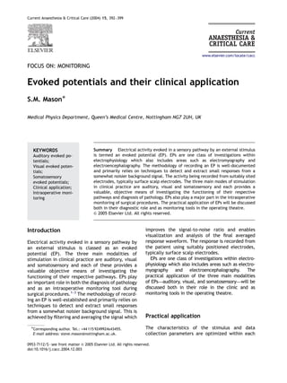
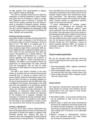
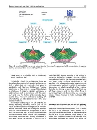
The document summarizes evoked potentials (EPs), which are electrical responses recorded from the brain or sensory pathways in response to stimulation. It discusses the three main types of EPs used clinically - auditory, visual, and somatosensory EPs. For each type, it describes the stimulation methods, recording techniques, clinical applications in diagnosis and monitoring, and how EPs provide objective assessment of sensory pathway function.
Auditory brainstem responses are generated by the
activity in structures of the ascending auditory
pathways that occurs during the first 8–10 ms
after a transient sound such as a click sound has
been applied to the ear.
Anesthesiology And Intraoperative Neurophysiological Monitoring Anurag Tewari MD
Anesthesiologists play a central role in optimizing IONM.
Intraoperative neuromonitoring (IONM) offers a near-real-time assessment of the functional integrity of the neuronal pathways during surgery. Evoked Potential signals may thus be regarded as surrogate markers of neuronal function and can be thought of as a repeated but limited neurological examination under general anesthesia. Optimization of anesthetic management contributes to the successful integration of IONM into perioperative care
Lower Extremity SSEP: Obligate peaks and recording montages following stimulation of the posterior tibial nerve. EP = Erb's. Obligate peaks and recording montages following stimulation of the posterior tibial nerve. T12 = 12th thoracic vertebra, CS = Cervical Spine, Fpz = center of frontal pole, CP = midpoint between ...
This presentation looks at intraoperative monitoring of auditory evoked potential, somato sensory evoked potential and motor evoked potential, procedure, pitfalls and utility.
EEG variants, are always to be recognized while interpreting the EEG one must be aware of these. Major and most common EEG is variants are discussed in the stated presentation.
Syed Irshad Murtaza.
Basic MEP techniques and understanding for Intraoperative neuromonitoring of the motors tracts during Brain and Spinal surgeries to prevent postoperative complications.
Artifacts in EEG - Recognition and differentiationRahul Kumar
This Presentation discusses the variously commonly seen artifacts in EEG, and how to recognize them. In EEG interpretation, it is often more important to identify an artifact than to identify true pathology. Once all the artifacts are ruled out, one is sure that what one is dealing with represents disease/abnormality
Guideline 11B: RECOMMENDED STANDARDS FOR INTRAOPERATIVE MONITORING OF SOMATOS...Anurag Tewari MD
Somatosensory evoked potentials (SSEPs) can be used intraoperatively to assess the function of the somatosensory pathways during surgical procedures in which the spinal cord, brainstem, or cerebrum is at risk and to localize the sensorimotor cortex
EEG - Montages, Equipment and Basic PhysicsRahul Kumar
This presentation discusses the 10-20 system of electrode placement, with its modifications. Also discussed are the Equipment Specifications, basic Physics and sources of interference
This presentation discusses the basic principles governing EEG Rhythm Generation, and discusses the various circuits that generate and maintain cerebral oscillations.
Auditory brainstem response (ABR)
Approximately 1 of every 1000 children is born deaf. Many more are born with less severe degrees of hearing impairment, while others may acquire hearing loss during early childhood.
combination of technological advances in ABR and otoacoustic emissions (OAE) testing methods are used for evaluation of hearing in newborns.
This slideshow describes unusual intraoperative motor evoked potentials observed from individuals with pre-existing spinal cord injury. These unusual motor evoked potentials demonstrated response thresholds, response amplitudes, response latencies, and response laterality differences from the anticipated motor evoked potentials.
Mental function examination is a part of Neurologic and Psychiatric examination as an emergency and as an outpatient clinic.
Detail Mental examination is required for cases of Dementia in various neurological diseases.
This set of slides are not for Psychiatric patients with disturbance of thought and mood.
ECochG is a variant of brainstem audio evoked response (ABR) where the recording electrode is placed as close as practical to the cochlea. We will use the abbreviation ECOG and ECochG interchangeably below. ECOG is preferable to us as it is shorter.
ECOG is intended to diagnose Meniere's disease, and particular, hydrops (swelling of the inner ear). ECOG may also be abnormal in perilymph fistula, and in superior canal dehiscence. The common feature connecting these illnesses is an imbalance in pressure between the endolymphatic and perilymphatic compartment of the inner ear.
ECOG can also be used to show that the cochlea is normal, in persons who are deaf. The cochlear microphonic of ECOG may be normal in auditory neuropathy (Santarelli and Arslan 2002) as well as other disorders in which the cochlea is preserved but the auditory nerve is damaged (Yokoyama, Nishida et al. 1999).
Finally, ECOG's have also been used to as a indicator of the temporary threshold shift that may follow noise injury (Nam et al, 2004).
EDIC is pleased to announce a webinar with Dr. R. Bruce Donoff, the Dean at Harvard Dental School. Dr. Donoff’s presentation will cover the risk factors for inferior alveolar and lingual nerve injury after third molar extraction, as well as the proper documentation and follow up of nerve injuries. Dr. Donoff will also discuss the potential for recovery from paresthesia after surgical intervention. The webinar will be held on May 10, 2011 at 7:00 PM.
Computer Aided Detection of Obstructive Sleep Apnea from EEG Signalssipij
Sleep Apnea is an anomaly in sleeping characterized by short pause in breathing. Failure to treat sleep
apnea leads to fatal complications in both psychological and physiological being of human.
Electroencephalogram (EEG) performs an important task in probing for sleep apnea through identifying
and recording the brain’s activities while sleeping. In this study, computer aided detection of sleep apnea
from EEG signals is developed to optimize and increase the prompt recognition and diagnosis of sleep
apnea in patients. The time domain, wavelets, and frequency domain of the EEG signals were computed,
and features were extracted from these domains. These features are inputted into two machine learning
algorithms: Support Vector Machine and K-Nearest Neighbors of different kernel functions and orders.
Evaluation metrics such as specificity, accuracy, and sensitivity are computed and analyzed for the
classifiers. The KNN classifier outperforms the SVM in classifying apnea from non-apnea events in
patients. The KNN order 3 shows the highest performance sensitivity of 85.92%, specificity of 80% and
accuracy of 82.69%.
Similar to Evoked potentials and their clinical application (20)
HOT NEW PRODUCT! BIG SALES FAST SHIPPING NOW FROM CHINA!! EU KU DB BK substit...GL Anaacs
Contact us if you are interested:
Email / Skype : kefaya1771@gmail.com
Threema: PXHY5PDH
New BATCH Ku !!! MUCH IN DEMAND FAST SALE EVERY BATCH HAPPY GOOD EFFECT BIG BATCH !
Contact me on Threema or skype to start big business!!
Hot-sale products:
NEW HOT EUTYLONE WHITE CRYSTAL!!
5cl-adba precursor (semi finished )
5cl-adba raw materials
ADBB precursor (semi finished )
ADBB raw materials
APVP powder
5fadb/4f-adb
Jwh018 / Jwh210
Eutylone crystal
Protonitazene (hydrochloride) CAS: 119276-01-6
Flubrotizolam CAS: 57801-95-3
Metonitazene CAS: 14680-51-4
Payment terms: Western Union,MoneyGram,Bitcoin or USDT.
Deliver Time: Usually 7-15days
Shipping method: FedEx, TNT, DHL,UPS etc.Our deliveries are 100% safe, fast, reliable and discreet.
Samples will be sent for your evaluation!If you are interested in, please contact me, let's talk details.
We specializes in exporting high quality Research chemical, medical intermediate, Pharmaceutical chemicals and so on. Products are exported to USA, Canada, France, Korea, Japan,Russia, Southeast Asia and other countries.
Pulmonary Thromboembolism - etilogy, types, medical- Surgical and nursing man...VarunMahajani
Disruption of blood supply to lung alveoli due to blockage of one or more pulmonary blood vessels is called as Pulmonary thromboembolism. In this presentation we will discuss its causes, types and its management in depth.
Acute scrotum is a general term referring to an emergency condition affecting the contents or the wall of the scrotum.
There are a number of conditions that present acutely, predominantly with pain and/or swelling
A careful and detailed history and examination, and in some cases, investigations allow differentiation between these diagnoses. A prompt diagnosis is essential as the patient may require urgent surgical intervention
Testicular torsion refers to twisting of the spermatic cord, causing ischaemia of the testicle.
Testicular torsion results from inadequate fixation of the testis to the tunica vaginalis producing ischemia from reduced arterial inflow and venous outflow obstruction.
The prevalence of testicular torsion in adult patients hospitalized with acute scrotal pain is approximately 25 to 50 percent
263778731218 Abortion Clinic /Pills In Harare ,sisternakatoto
263778731218 Abortion Clinic /Pills In Harare ,ABORTION WOMEN’S CLINIC +27730423979 IN women clinic we believe that every woman should be able to make choices in her pregnancy. Our job is to provide compassionate care, safety,affordable and confidential services. That’s why we have won the trust from all generations of women all over the world. we use non surgical method(Abortion pills) to terminate…Dr.LISA +27730423979women Clinic is committed to providing the highest quality of obstetrical and gynecological care to women of all ages. Our dedicated staff aim to treat each patient and her health concerns with compassion and respect.Our dedicated group ABORTION WOMEN’S CLINIC +27730423979 IN women clinic we believe that every woman should be able to make choices in her pregnancy. Our job is to provide compassionate care, safety,affordable and confidential services. That’s why we have won the trust from all generations of women all over the world. we use non surgical method(Abortion pills) to terminate…Dr.LISA +27730423979women Clinic is committed to providing the highest quality of obstetrical and gynecological care to women of all ages. Our dedicated staff aim to treat each patient and her health concerns with compassion and respect.Our dedicated group of receptionists, nurses, and physicians have worked together as a teamof receptionists, nurses, and physicians have worked together as a team wwww.lisywomensclinic.co.za/
Flu Vaccine Alert in Bangalore Karnatakaaddon Scans
As flu season approaches, health officials in Bangalore, Karnataka, are urging residents to get their flu vaccinations. The seasonal flu, while common, can lead to severe health complications, particularly for vulnerable populations such as young children, the elderly, and those with underlying health conditions.
Dr. Vidisha Kumari, a leading epidemiologist in Bangalore, emphasizes the importance of getting vaccinated. "The flu vaccine is our best defense against the influenza virus. It not only protects individuals but also helps prevent the spread of the virus in our communities," he says.
This year, the flu season is expected to coincide with a potential increase in other respiratory illnesses. The Karnataka Health Department has launched an awareness campaign highlighting the significance of flu vaccinations. They have set up multiple vaccination centers across Bangalore, making it convenient for residents to receive their shots.
To encourage widespread vaccination, the government is also collaborating with local schools, workplaces, and community centers to facilitate vaccination drives. Special attention is being given to ensuring that the vaccine is accessible to all, including marginalized communities who may have limited access to healthcare.
Residents are reminded that the flu vaccine is safe and effective. Common side effects are mild and may include soreness at the injection site, mild fever, or muscle aches. These side effects are generally short-lived and far less severe than the flu itself.
Healthcare providers are also stressing the importance of continuing COVID-19 precautions. Wearing masks, practicing good hand hygiene, and maintaining social distancing are still crucial, especially in crowded places.
Protect yourself and your loved ones by getting vaccinated. Together, we can help keep Bangalore healthy and safe this flu season. For more information on vaccination centers and schedules, residents can visit the Karnataka Health Department’s official website or follow their social media pages.
Stay informed, stay safe, and get your flu shot today!
The prostate is an exocrine gland of the male mammalian reproductive system
It is a walnut-sized gland that forms part of the male reproductive system and is located in front of the rectum and just below the urinary bladder
Function is to store and secrete a clear, slightly alkaline fluid that constitutes 10-30% of the volume of the seminal fluid that along with the spermatozoa, constitutes semen
A healthy human prostate measures (4cm-vertical, by 3cm-horizontal, 2cm ant-post ).
It surrounds the urethra just below the urinary bladder. It has anterior, median, posterior and two lateral lobes
It’s work is regulated by androgens which are responsible for male sex characteristics
Generalised disease of the prostate due to hormonal derangement which leads to non malignant enlargement of the gland (increase in the number of epithelial cells and stromal tissue)to cause compression of the urethra leading to symptoms (LUTS
New Drug Discovery and Development .....NEHA GUPTA
The "New Drug Discovery and Development" process involves the identification, design, testing, and manufacturing of novel pharmaceutical compounds with the aim of introducing new and improved treatments for various medical conditions. This comprehensive endeavor encompasses various stages, including target identification, preclinical studies, clinical trials, regulatory approval, and post-market surveillance. It involves multidisciplinary collaboration among scientists, researchers, clinicians, regulatory experts, and pharmaceutical companies to bring innovative therapies to market and address unmet medical needs.
These simplified slides by Dr. Sidra Arshad present an overview of the non-respiratory functions of the respiratory tract.
Learning objectives:
1. Enlist the non-respiratory functions of the respiratory tract
2. Briefly explain how these functions are carried out
3. Discuss the significance of dead space
4. Differentiate between minute ventilation and alveolar ventilation
5. Describe the cough and sneeze reflexes
Study Resources:
1. Chapter 39, Guyton and Hall Textbook of Medical Physiology, 14th edition
2. Chapter 34, Ganong’s Review of Medical Physiology, 26th edition
3. Chapter 17, Human Physiology by Lauralee Sherwood, 9th edition
4. Non-respiratory functions of the lungs https://academic.oup.com/bjaed/article/13/3/98/278874
micro teaching on communication m.sc nursing.pdfAnurag Sharma
Microteaching is a unique model of practice teaching. It is a viable instrument for the. desired change in the teaching behavior or the behavior potential which, in specified types of real. classroom situations, tends to facilitate the achievement of specified types of objectives.
For Better Surat #ℂall #Girl Service ❤85270-49040❤ Surat #ℂall #Girls
Evoked potentials and their clinical application
1. Current Anaesthesia & Critical Care (2004) 15, 392–399
FOCUS ON: MONITORING
Evoked potentials and their clinical application
S.M. MasonÃ
Medical Physics Department, Queen’s Medical Centre, Nottingham NG7 2UH, UK
Summary Electrical activity evoked in a sensory pathway by an external stimulus
is termed an evoked potential (EP). EPs are one class of investigations within
electrophysiology which also includes areas such as electromyography and
electroencephalography. The methodology of recording an EP is well-documented
and primarily relies on techniques to detect and extract small responses from a
somewhat noisier background signal. The activity being recorded from suitably sited
electrodes, typically surface scalp electrodes. The three main modes of stimulation
in clinical practice are auditory, visual and somatosensory and each provides a
valuable, objective means of investigating the functioning of their respective
pathways and diagnosis of pathology. EPs also play a major part in the intraoperative
monitoring of surgical procedures. The practical application of EPs will be discussed
both in their diagnostic role and as monitoring tools in the operating theatre.
& 2005 Elsevier Ltd. All rights reserved.
Introduction
Electrical activity evoked in a sensory pathway by
an external stimulus is classed as an evoked
potential (EP). The three main modalities of
stimulation in clinical practice are auditory, visual
and somatosensory and each of these provides a
valuable objective means of investigating the
functioning of their respective pathways. EPs play
an important role in both the diagnosis of pathology
and as an intraoperative monitoring tool during
surgical procedures.1–3
The methodology of record-
ing an EP is well-established and primarily relies on
techniques to detect and extract small responses
from a somewhat noisier background signal. This is
achieved by filtering and averaging the signal which
improves the signal-to-noise ratio and enables
visualization and analysis of the final averaged
response waveform. The response is recorded from
the patient using suitably positioned electrodes,
typically surface scalp electrodes.
EPs are one class of investigations within electro-
physiology which also includes areas such as electro-
myography and electroencephalography. The
practical application of the three main modalities
of EPs—auditory, visual, and somatosensory—will be
discussed both in their role in the clinic and as
monitoring tools in the operating theatre.
Practical application
The characteristics of the stimulus and data
collection parameters are optimized within each
ARTICLE IN PRESS
www.elsevier.com/locate/cacc
KEYWORDS
Auditory evoked po-
tentials;
Visual evoked poten-
tials;
Somatosensory
evoked potentials;
Clinical application;
Intraoperative moni-
toring
0953-7112/$ - see front matter & 2005 Elsevier Ltd. All rights reserved.
doi:10.1016/j.cacc.2004.12.003
ÃCorresponding author. Tel.: +44 115 9249924x43455.
E-mail address: steve.mason@nottingham.ac.uk.
2. modality so as to evoke an EP that is diagnostic at
different levels of the sensory pathway. The
majority of EPs are recorded from surface electro-
des attached to the scalp. However, some techni-
ques require more specialized electrodes such as an
electrode positioned on the front of the eye to
record retinal responses (electroretinography) or
an epidural electrode to record spinal cord re-
sponses during monitoring in spinal surgery. The
electrode forms the all important interface be-
tween the patient and the recording equipment
and is a key component in achieving good quality
EPs. For this reason it is essential to achieve a
stable, low impedance contact with the electrode
(typically less than 5 kO) which helps to reduce the
level of interference on the signal baseline,
particularly 50 Hz mains.
EPs range in amplitude from less than 1 mV for
surface recordings of the auditory brainstem
response (ABR) up to 200–300 mV for an electro-
retinogram. The majority of EPs are small when
compared to the level of background noise and
techniques are employed to enhance the signal to
noise ratio of the final recording of the response.
Firstly, the frequency content of the signal picked
up by the electrodes is controlled by a filter where
the lower and upper cut-off frequencies (band-
width) are set so that the response is passed
through but as much of the background noise is
excluded. Secondly, time synchronized signal aver-
aging is employed to enhance the signal-to-noise
ratio. A train of stimuli is presented and time
locked responses from individual stimuli add to-
gether, whereas background noise is generally
random and tends to cancel out.
EPs are often called objective investigations. How-
ever, it is important to remember that they are only
objective as far as the patient is concerned, because
the recorded waveform in the majority of clinical
applications still requires the skilled interpretation of
an experienced professional. Passive cooperation of
the patient is required in order to achieve a quiet
signal baseline with minimal contamination with
movement artefacts and myogenic activity. High levels
of noise can easily mask out a small amplitude
response. These recording conditions may be difficult
to achieve in a young child and may require the use of
sedation or even a general anaesthetic.
Interpretation of evoked potential
waveforms
The two main characteristics of the response
waveform that typically define where or not it lies
within the normal range are its amplitude and
latency with respect to the stimulus. The overall
response waveform often consists of a number of
different components, as shown in Fig. 1 for the
ABR. Measurement of the amplitude and latency of
these components is essential when using the ABR
as a diagnostic tool. The diagnostic ability of an EP
relies on the pathology affecting the relevant
sensory pathway and thereby disrupting the gen-
eration and transmission of response activity.
Auditory evoked potentials
It is possible to record a range of different auditory
EPs originating from progressively higher structures
along the pathway from cochlea to cortex as
described in Hall:1
1. Electrocochleography (ECochG): cochlear hair
cells and peripheral auditory nerve.
2. Auditory brainstem response (ABR): neural path-
ways in the brainstem from auditory nerve to the
inferior colliculus.
3. Middle latency response (MLR): upper brain-
stem, thalamus and primary auditory cortex.
4. Auditory cortical response (ACR): primary, sec-
ondary and associated cortical areas.
Auditory stimuli
There are three types of transient stimuli routinely
employed in the generation of these auditory EPs:
Click—a short duration, fast rise time stimulus
which has good synchronization properties for
generation of EPs in the cochlea and brainstem
pathways (ECochG and ABR). Acoustically this is a
wideband stimulus with poor frequency specifi-
city.
Tone pip—a slightly longer, slower stimulus than
the click, consisting of a few cycles of a pure
tone. The slower rise time of this stimulus
improves its acoustical frequency specificity but
this is achieved at the expense of degradation of
the EP from the cochlea and brainstem path-
ways. However, this stimulus is employed widely
with the ABR and ECochG but to a lesser extent
with the MLR and ACR.
Tone burst—a longer burst of a pure tone
with a relatively slow rise time. This stimulus
has excellent frequency specificity equivalent
to the stimuli used in subjective pure tone
ARTICLE IN PRESS
Evoked potentials and their clinical application 393
3. audiometry. It is an ideal stimulus for evoking
the ACR but cannot be used for ECochG and the
ABR as there is insufficient synchronization of
neural activity to enable identification of the
response.
In addition to these transient stimuli, an ampli-
tude and frequency modulated tonal stimulus can
be used to evoke an auditory steady-state response
(ASSR) from the auditory system as reviewed
by Jerger.4
The modulation frequency of the
stimulus determines primarily where the recorded
response originates from; 80 Hz predominantly
auditory brainstem and 40 Hz mainly middle latency
components.
Clinical application
Auditory EPs have a major diagnostic role in a wide
range of clinical applications in the disciplines of
audiology and neurology. The most widely studied
and applied EP is the ABR3,5
followed by the ACR
and ECochG and to a lesser extent the MLR. The
ABR is used for the objective assessment of hearing
in babies and young children when behavioural
testing is not reliable. It is also a valuable oto-
neurological tool for investigation of auditory nerve
tumours and brainstem pathology although over
recent years MRI has become the primary investiga-
tion in many of these cases. However, ABR is still
required in some patients due to long waiting lists
ARTICLE IN PRESS
ms
60dBnHL
STIM
1.9 3.0 4.1 5.2 5.8 5.9 7.6 9.2
1 2 3 4 5 6 7 8 9 10
CZ
+
CZ −
4.0 ms
vUNITS
COCHLEA
AUDITORY
CORTEX
MEDIAL
GENICULATE
BODY
INFERIOR
COLLICULUS
MIDBRAIN
LEVELNUCLEUS OF
LATERAL
LEMNISCUS
DORSAL
COCHLEAR
NUCLEUS
VENTRAL
COCHLEAR
NUCLEUS
ACOUSTIC NERVE
ORGAN OF
CORTI
OUTER
HAIR
CELLS
INNER
HAIR
CELL
SUPERIOR
OLIVARY
COMPLEX
PONTO-
MEDULLARY
I
II
III
IV
VI
VII
V
0.1
Figure 1 The characteristics and origins of the auditory brainstem response.
S.M. Mason394
4. for MRI, patients with claustrophobia or obesity,
and in specific types of pathology.
The ACR is a valuable clinical tool for objective
assessment of hearing thresholds in older children
and adults who are unwilling or unable to comply
with subjective tests of hearing. It assesses the
whole of the auditory pathway and the measure-
ment of threshold is frequency specific. However
the ACR is dependent on the subject being awake
and alert and this highlights the advantage of ABR
in young subjects as it is resistant to the effects of
sleep, sedation and anaesthesia.
Newborn hearing screening
Since 2002 newborn hearing screening programmes
(NHSP) have been introduced in the UK across an
increasing number of centres following a review by
Davis et al.14
The ABR is an integral part of a
hearing screening programme. It is used at the
early screening stage where automated ABR sys-
tems (AABR) provide a completely objective pass or
refer result for the screening test that does not
require interpretation by a skilled observer. This
objective result is determined by a computer
scoring algorithm that detects the presence or
absence of an ABR at a fixed screening level of
45 dBnHL. The AABR is a rare example of where the
subjectivity of interpretation has been removed
from the investigation. EPs are ripe for wider
application of these objective scoring techniques in
the future.
The ABR is also heavily involved in the early
follow-up of babies that are referred following the
screening test. In striving to achieve more fre-
quency specific objective information about hear-
ing loss there has been an increased emphasis on
the use of tone pip stimuli with the ABR rather than
a wideband click stimulus. Recordings of the ASSR
also offer the possibility of an alternative method
of measuring frequency specific thresholds. Re-
cently this technique has been bolstered by the
availability of commercial ASSR systems which
apply objective statistical methods for detection
of the presence of a response. Although this
technique of estimating hearing thresholds shows
considerable promise, its consistency and reliabil-
ity still needs to be established in routine clinical
use, particularly when testing very young babies.
Cochlear implantation
In the last 10–15 years cochlear implantation has
been increasingly used as a means of restoring some
perception of sounds and speech in patients with
profound hearing loss. Auditory EPs play an
essential role in the management of these patients
and particularly in young children.7,8
An example of
this is the ABR which can be evoked using electrical
stimulation presented to the auditory nerve by the
cochlear implant rather than conventional acous-
tical stimulation.9
The electrically evoked ABR
(EABR) can used to assist with tuning of the implant
which involves setting an appropriate dynamic
range for the electrical stimulus.
A recent development in cochlear implant
technology is a technique for recording the
electrically evoked, compound auditory nerve
action potential from the electrode array within
the cochlea. One electrode on the array is used as a
stimulating electrode and an adjacent electrode as
the recording site. Information is passed to and
from the implant using a radio frequency telemetry
link. On the Nucleus cochlear implant supplied by
Cochlear Europe Ltd. this technique is known as
neural response telemetry (NRT). An example of
recordings acquired using the NRT software is
shown in Fig. 2.
Visual evoked potentials
EPs can be recorded which represent electrical
activity originating from the retina, optic nerve and
visual cortex
1. Electrooculography (EOG): pigment epithelium
layer of the retina;
2. Electroretinography (ERG): rod and cone recep-
tors and the inner and outer nuclear layers; and
3. Visual evoked potential (VEP): response activity
from the visual cortex.
Visual stimuli
Two important characteristics associated with the
visual stimuli are contrast and luminance and are
incorporated in the main types of visual stimuli
used in clinical practice.
Plain flash of light—a low contrast stimulus that
can be presented at high and low levels of
luminance.
Pattern onset—a black and white checkerboard
stimulus that is flashed on and off the screen with
an overall constant luminance. The field size of
the stimulus is typically 201 with individual check
sizes ranging from 15 to 60 min of arc subtended
at the eye.
Pattern reversal—a checkerboard stimulus
where the individual black and white checks
are reversed on successive stimuli.
ARTICLE IN PRESS
Evoked potentials and their clinical application 395
5. ERG and VEPs are subdivided depending on the
characteristics of the stimulus presentation and
whether the investigations are carried out in the
dark adapted (scotopic) or light adapted eye
(phototopic). Recordings of the ERG and VEP are
standardized to the recommendations of the Inter-
national Society for the Clinical Electrophysiology
of Vision (ISCEV). For example, the standardized
ERG to a full field flash stimulus includes a range of
measurements that specifically investigate differ-
ential function of the rods and cones. The ERG can
also be evoked by a checkerboard pattern stimulus
(pattern ERG). The response components of the
PERG arise from more central structures and are
selectively affected in macular disease and gang-
lion cell dysfunction. The full field flash ERG and
the pattern ERG complement each other in the
differential diagnosis of peripheral retinal involve-
ment and more central dysfunction.
A more recent development of the ERG and the
VEP is the use of a multifocal stimulus where a two-
dimensional array of small stimulus elements is
used to evoke response activity from specific small
regions of the visual field. A detailed discussion of
the multifocal technique is described in Hood.10
A
typical recording of the multifocal ERG in a normal
subject is shown in Fig. 3.
Clinical application
Visual electrodiagnostic investigations of the func-
tioning of the retina, optic nerve pathways and
visual cortex will complement, and often supple-
ment, clinical examination.11,12
Typical examples
of their role, either individually or in combination,
are as follows:
Identification of the location and possible nature
of dysfunction along the visual pathway, for
example, a delayed response from the visual
cortex (VEP) due to inflammation of the optic
nerve (optic neuritis).
Involvement of particular retinal cell types such
as in cone dystrophy or more gross retinal
involvement as in cases of retinitis pigmentosa
where there is reduction in all components of the
ERG.
Involvement of a particular subpopulation of
visual nerve fibres or processing system such as
in cases of macular degeneration (e.g., Star-
gardt’s disease). The pattern ERG is a valuable
clinical tool in the assessment of macular
dysfunction.
In non-organic aetiology (e.g., hysterical am-
blyopia) the VEP evoked by different stimulus
ARTICLE IN PRESS
Figure 2 Recording of the electrically evoked compound auditory nerve action potential (ECAP) using the NRTsoftware.
In this example the electrical stimulus has been applied to electrode 10 of the 22-electrode array and the ECAP
response recorded from electrode 12.
S.M. Mason396
6. check sizes is a valuable tool to objectively
assess visual function.
Historically, visual electrodiagnostic investiga-
tions have been extensively studied and documen-
ted in adults but more recently their value in
paediatric work has been highlighted. Practical
application of these tests in a young child or baby is
a challenge. It requires special techniques to
address issues such as compliance with the proce-
dure and need for fixation of the stimulus by an
awake child. This is in contrast to auditory EPs
where testing can often be carried out with a child
asleep or sedated.
The multifocal techniques for ERG and VEP are
rapidly becoming essential clinical tools in the
visual electrodiagnostic investigation of patients.
Retinal functional losses due to regional disorders
in outer retinal layers can be described in detail
with the multifocal ERG. In macular disease there
are decreased or absent central ERG components
surrounded by normal ERG activity. In diseases of
the outer retina the pattern of distribution of
multifocal ERG activity is similar to the pattern of
the visual field defect. However this relationship is
dependent on whether the disease primarily affects
the outer retina (retinitis pigmentosa) or the
ganglion cells (glaucoma) or optic nerve (ischemic
optic neuropathy and optic neuritis). It is important
to measure not only the amplitude of the response
but also the timing as some diseases affect one
of these measures and not the other. There is
currently a big drive in documenting and comparing
disease-related findings in both multifocal ERG
and VEP.
Somatosensory evoked potentials (SSEP)
The most common form of stimulus used to evoke
an SSEP is an electrical pulse applied to a
peripheral afferent nerve such as the median nerve
at the wrist or the posterior tibial nerve in the
lower limb. The resultant EP can be recorded from
electrodes positioned at various sites along the
ARTICLE IN PRESS
Figure 3 A multifocal ERG in a normal subject showing the array of responses and a 3D representation of response
activity (courtesy of Colin Barber and Yaqin Wen).
Evoked potentials and their clinical application 397
7. pathway where the nerve becomes superficial or at
cortical level.
The amplitude of the SSEP varies consider-
ably across subjects, and the interpretation of
clinical diagnostic studies is based primarily
on component latencies rather than amplitude.
They have diagnostic role in peripheral neuro-
pathy and more central degenerative disorders
through measurement of the conduction time of
the afferent nerves. However, the develop-
ment and easier access of imaging such as MRI
have had an impact on the usage of SSEPs in
clinical practice and fewer studies are now per-
formed.
Intraoperative monitoring
Implementation of intraoperative monitoring re-
quires a somewhat different approach when com-
pared to recording EPs in the outpatient clinic. The
environment and practical procedure is more
challenging from the point of view of electrical
interference, lack of access to the patient during
the procedure, time pressures and a requirement
to provide immediate feedback of information on
which the surgeon can act if necessary. A team
approach is required involving the surgeon, anaes-
thetist, and the monitoring staff.
The use of EPs in intraoperative monitoring
of surgical procedures can provide valuable feed-
back about the status and potential compromise
of important neural pathways and vascular struc-
tures2,8,13
Monitoring the status of the auditory pathway
during cerebello-pontine angle (CPA) surgery
where the aim is to prevent avoidable post-
operative hearing deficit during tumour removal
Recording EPs related to monitoring of spinal
cord function during spinal surgery, such as
correction of scoliosis deformatives.
Monitoring cranial/vascular procedures such as
endartectomy and aneurysm surgery using soma-
tosensory EPs.
Monitoring the functional status of a cochlear
implant and stimulation of the auditory pathways
during implant surgery.
In addition to the surgical procedure there
are other factors that can affect the EP recorded
intraoperatively such as tissue temperature,
blood pressure and anaesthetic agents. These
need to be taken into consideration when report-
ing potential changes in the EP as a result of the
surgery. The most susceptible EPs to the effects
of anaesthesia are those arising from more central
structures and particularly cortical responses
whereas more peripheral responses are sparred.
In general the longer the latency of a response
component, the more synapses there are between
the stimulation site and the neural generator, the
greater the degree of effect of the anaesthetic
agent.
This susceptibility of the EP to anaesthetic agents
is exploited in a technique to monitor the depth of
anaesthesia. It has been demonstrated by Davies et
al.6
that components of the auditory MLR show
changes in latency during transitions between
consciousness and unconsciousness. Analysis of
these changes may provide an indicator of potential
awareness during anaesthesia.
Summary
EPs have a wide ranging role in assisting with the
diagnosis and management of patients both in the
clinic and during surgery. They can provide objec-
tive information about the functioning of sensory
pathways which is difficult to acquire using other
techniques. Some recordings are well-established
and form part of standard clinical practice whereas
others have an exciting future ahead such as
developments with multi-focal ERG and VEP,
electrical EPs associated with cochlear implanta-
tion, and the ASSR.
References
1. Hall III JW. Handbook of auditory evoked potentials. MA:
Allyn Bacon; 1992.
2. Misulis KE. Essentials of clinical neurophysiology. Boston:
Butterworth-Heinemann; 1997.
3. Mason SM. Electric response audiometry. In: McCormick B,
editor. Paediatric audiology 0–5 years. 2nd ed. London:
Whurr Publishers Ltd; 2004. p. 188–264.
4. Jerger J. The auditory steady state response: parts 1 and 2.
J Am Acad Audiol 2002;13:numbers 4 and 5.
5. Hood LJ. Clinical applications of the auditory brainstem
response. San Diego, CA: Singular Publishing; 1998.
6. Davies FW, Mantzaridis H, Kenny GN, Fisher AC. Middle
latency auditory evoked potentials during repeated transi-
tions from consciousness to unconsciousness. Anaesthesia
1996;51(2):107–13.
7. Cullington HE. Cochlear implants: objective measures.
London: Whurr Publishers; 2003.
8. Mason SM. Electrophysiological and objective tests. In:
McCormick B, Archbold S, editors. Cochlear implants
for young children. 2nd ed. London: Whurr Publishers;
2003. p. 162–216.
9. Mason SM. The electrically evoked auditory brainstem
response. In: Cullington H, editor. Cochlear implants:
ARTICLE IN PRESS
S.M. Mason398
8. objective measures. London: Whurr Publishers Ltd; 2003.
p. 130–59.
10. Hood DC. The multifocal technique: topographical ERG and
VEP responses. Documenta Ophthamologica: 100, No 2/3;
2000.
11. Halliday AM, editor. Evoked potentials in clinical testing.
2nd ed. London: Churchill Livingstone; 1993.
12. Fishman GA, Birch DG, Holder GE, Brigell MG. Electrophy-
siologic testing in disorders of the retina, optic nerve, and
visual pathway. 2nd ed. Mongraph published by American
Academy of Ophthalmology. 2001.
13. Jones SJ, Boyd S, Hetreed M, Smith NJ, editors. Handbook of
spinal cord monitoring. Dordrecht: Kluwer Academic Pub-
lishers; 1994.
14. Davis A, Bamford J, Wilson I, Ramkalawan T, Forshaw M,
Wright S. A critical review of the role of neonatal hearing
screening in the detection of congenital hearing impair-
ment. Health Technol Assess 1997;1(10):i–iv 1–176.
ARTICLE IN PRESS
Evoked potentials and their clinical application 399