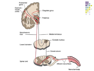
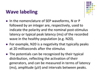
This document discusses somatosensory evoked potentials (SEPs), which are electrical signals generated in the nervous system in response to sensory stimuli. SEPs reflect the activation of neural structures along somatosensory pathways. They are recorded using electrodes on the scalp and spine in response to electrical stimulation of peripheral nerves. SEP waves are labeled according to their polarity and latency. Clinical uses of SEPs include evaluating peripheral nerves and central somatosensory pathways, localizing lesions, and monitoring patients in intensive care and during surgery. Abnormal SEPs can indicate disorders of the peripheral or central nervous system.
what is RNS and what the techniques to perform this test in the lab. Its significance in the evaluation and diagnosis of NMJ disorders like MG, LEMBS etc..
Basic MEP techniques and understanding for Intraoperative neuromonitoring of the motors tracts during Brain and Spinal surgeries to prevent postoperative complications.
Late response are the most helpful findings in some of the diseases affecting the peripheral nerves, (e.g GBS, Radiculopathies, ). How to assess these responses while performing Nerve Conduction Studies, is the most technical and theoretical consideration.... Here we go with the same things in the stated slides
This presentation describes the common conditions, anatomy and the ideal ways to do and perform nerve conduction studies in lower limbs. It is nicely depicted with self explanatory pictures.
This presentation discusses the basic principles governing EEG Rhythm Generation, and discusses the various circuits that generate and maintain cerebral oscillations.
This presentation is an introduction to the principles of Nerve Conduction Study and is entirely sourced from the book by David C Preston and Barbara E Shapiro: Electromyography and Neuromuscular disorders, 3rd Edition
This presentation looks at intraoperative monitoring of auditory evoked potential, somato sensory evoked potential and motor evoked potential, procedure, pitfalls and utility.
NCS are done by placing electrodes on the skin and stimulating the nerves through electrical impulses. To study motor nerves, electrodes are placed over a muscle that receives its innervation from the nerve you want to test (stimulate).
This presentation was provided by Steph Pollock of The American Psychological Association’s Journals Program, and Damita Snow, of The American Society of Civil Engineers (ASCE), for the initial session of NISO's 2024 Training Series "DEIA in the Scholarly Landscape." Session One: 'Setting Expectations: a DEIA Primer,' was held June 6, 2024.
Thinking of getting a dog? Be aware that breeds like Pit Bulls, Rottweilers, and German Shepherds can be loyal and dangerous. Proper training and socialization are crucial to preventing aggressive behaviors. Ensure safety by understanding their needs and always supervising interactions. Stay safe, and enjoy your furry friends!
The simplified electron and muon model, Oscillating Spacetime: The Foundation...RitikBhardwaj56
Discover the Simplified Electron and Muon Model: A New Wave-Based Approach to Understanding Particles delves into a groundbreaking theory that presents electrons and muons as rotating soliton waves within oscillating spacetime. Geared towards students, researchers, and science buffs, this book breaks down complex ideas into simple explanations. It covers topics such as electron waves, temporal dynamics, and the implications of this model on particle physics. With clear illustrations and easy-to-follow explanations, readers will gain a new outlook on the universe's fundamental nature.
Safalta Digital marketing institute in Noida, provide complete applications that encompass a huge range of virtual advertising and marketing additives, which includes search engine optimization, virtual communication advertising, pay-per-click on marketing, content material advertising, internet analytics, and greater. These university courses are designed for students who possess a comprehensive understanding of virtual marketing strategies and attributes.Safalta Digital Marketing Institute in Noida is a first choice for young individuals or students who are looking to start their careers in the field of digital advertising. The institute gives specialized courses designed and certification.
for beginners, providing thorough training in areas such as SEO, digital communication marketing, and PPC training in Noida. After finishing the program, students receive the certifications recognised by top different universitie, setting a strong foundation for a successful career in digital marketing.
June 3, 2024 Anti-Semitism Letter Sent to MIT President Kornbluth and MIT Cor...Levi Shapiro
Letter from the Congress of the United States regarding Anti-Semitism sent June 3rd to MIT President Sally Kornbluth, MIT Corp Chair, Mark Gorenberg
Dear Dr. Kornbluth and Mr. Gorenberg,
The US House of Representatives is deeply concerned by ongoing and pervasive acts of antisemitic
harassment and intimidation at the Massachusetts Institute of Technology (MIT). Failing to act decisively to ensure a safe learning environment for all students would be a grave dereliction of your responsibilities as President of MIT and Chair of the MIT Corporation.
This Congress will not stand idly by and allow an environment hostile to Jewish students to persist. The House believes that your institution is in violation of Title VI of the Civil Rights Act, and the inability or
unwillingness to rectify this violation through action requires accountability.
Postsecondary education is a unique opportunity for students to learn and have their ideas and beliefs challenged. However, universities receiving hundreds of millions of federal funds annually have denied
students that opportunity and have been hijacked to become venues for the promotion of terrorism, antisemitic harassment and intimidation, unlawful encampments, and in some cases, assaults and riots.
The House of Representatives will not countenance the use of federal funds to indoctrinate students into hateful, antisemitic, anti-American supporters of terrorism. Investigations into campus antisemitism by the Committee on Education and the Workforce and the Committee on Ways and Means have been expanded into a Congress-wide probe across all relevant jurisdictions to address this national crisis. The undersigned Committees will conduct oversight into the use of federal funds at MIT and its learning environment under authorities granted to each Committee.
• The Committee on Education and the Workforce has been investigating your institution since December 7, 2023. The Committee has broad jurisdiction over postsecondary education, including its compliance with Title VI of the Civil Rights Act, campus safety concerns over disruptions to the learning environment, and the awarding of federal student aid under the Higher Education Act.
• The Committee on Oversight and Accountability is investigating the sources of funding and other support flowing to groups espousing pro-Hamas propaganda and engaged in antisemitic harassment and intimidation of students. The Committee on Oversight and Accountability is the principal oversight committee of the US House of Representatives and has broad authority to investigate “any matter” at “any time” under House Rule X.
• The Committee on Ways and Means has been investigating several universities since November 15, 2023, when the Committee held a hearing entitled From Ivory Towers to Dark Corners: Investigating the Nexus Between Antisemitism, Tax-Exempt Universities, and Terror Financing. The Committee followed the hearing with letters to those institutions on January 10, 202
Biological screening of herbal drugs: Introduction and Need for
Phyto-Pharmacological Screening, New Strategies for evaluating
Natural Products, In vitro evaluation techniques for Antioxidants, Antimicrobial and Anticancer drugs. In vivo evaluation techniques
for Anti-inflammatory, Antiulcer, Anticancer, Wound healing, Antidiabetic, Hepatoprotective, Cardio protective, Diuretics and
Antifertility, Toxicity studies as per OECD guidelines
Macroeconomics- Movie Location
This will be used as part of your Personal Professional Portfolio once graded.
Objective:
Prepare a presentation or a paper using research, basic comparative analysis, data organization and application of economic information. You will make an informed assessment of an economic climate outside of the United States to accomplish an entertainment industry objective.
How to Build a Module in Odoo 17 Using the Scaffold MethodCeline George
Odoo provides an option for creating a module by using a single line command. By using this command the user can make a whole structure of a module. It is very easy for a beginner to make a module. There is no need to make each file manually. This slide will show how to create a module using the scaffold method.
A workshop hosted by the South African Journal of Science aimed at postgraduate students and early career researchers with little or no experience in writing and publishing journal articles.
Executive Directors Chat Leveraging AI for Diversity, Equity, and InclusionTechSoup
Let’s explore the intersection of technology and equity in the final session of our DEI series. Discover how AI tools, like ChatGPT, can be used to support and enhance your nonprofit's DEI initiatives. Participants will gain insights into practical AI applications and get tips for leveraging technology to advance their DEI goals.
2. Evoked potentials are the electrical signals
generated by the nervous system in response to
sensory stimuli.
Auditory, visual, and somatosensory stimuli are
used commonly for clinical evoked potential
studies.
Somatosensory evoked potentials (SEPs) consist
of a series of waves that reflect sequential
activation of neural structures along the
somatosensory pathways.
3. The somatosensory system sub serves five modalities:
mechanoreception, thermoreception, nociception,
proprioception and visceroception.
These sub modalities provide conscious perception of sensory
information from the skin, the musculo-skeletal system and
the viscera
In addition, somatosensory afferents are involved in
numerous motor and autonomous reflex pathways and
feedback loops with reflex centers in the spinal cord,
brainstem and forebrain.
Somatosensory afferents also provide a powerful excitatory
input to the ascending RAS that regulates sleep and
wakefulness.
4.
5. Methods of recording and
nomenclature
Stimulation
Recording
Wave labeling, generators and normal values
6. Stimulation
SEPs are usually evoked by bipolar transcutaneous
electrical stimulation applied on the skin over the
selected nerve.
Monophasic square-wave electrical pulses of 0.1–
0.2 ms should be delivered, preferably by a constant
current stimulator.
In routine recording, stimulus parameters include a
stimulus intensity able to produce a clear but
tolerable sensation.
Stimulation rate should be 3–5 Hz
7. The stimulation sites typically used for clinical
diagnostic SEP studies are the median nerve at the
wrist, the common peroneal nerve at the knee,
and/or the posterior tibial nerve at the ankle.
Recording of SEPs to stimulation of the ulnar nerve
at the wrist is useful for intraoperative monitoring
when the mid cervical spinal cord or parts of the
brachial plexus are at risk.
Recording electrodes are placed over the scalp,
spine, and peripheral nerves proximal to the
stimulation site
8. Recording
Recording electrodes are standard EEG disk
electrodes.
Skin-electrode impedance should be less than
5000 Ω.
The optimal condition to record clinically-
relevant SEP components is a high pass filter at
less than 3 Hz and a low-pass filter over 2000 Hz
9. Median nerve SEP
For standard clinical recordings at least four channels
designed to highlight one or more component each:
peripheral (Erb’s point) channel (N9), cervical channel (N13),
parietal channel (P14 and N20) and frontal channel (P14, P20
and N30).
Peripheral Erb’s point electrodes are designated as EP and
must be placed within the angle formed by the posterior
border of the clavicular head of the sternomastoid muscle
and the clavicle, 2–3 cm above the clavicle (Erb’s point).
The active electrode is ipsilateral to stimulation (EPi), and the
reference is either the contralateral EP electrode (EPc) or a
scalp electrode (generally Fz).
10.
11.
12. Tibial nerve SEP
For standard clinical recordings four channels designed to
highlight one component each: peripheral (N8), lumbar (N22),
subcortical (P30) and cortical (P39)
The peripheral recording electrode is placed in the popliteal
fossa (PF) 4–6 cm above the popliteal crease.
A reference electrode may be placed either on the medial
surface of the knee, over the medial femoral condyle or 3 cm
above the active electrode.
Lumbar electrodes should be placed on the skin overlying the
spinous processes of a lumbar vertebra, most often L1.
13.
14.
15. In the lower limb, posterior tibial SEPs are generally preferred
because of the following:
In clinical diagnostic use, they are larger and display less
intrasubject variability.
In intraoperative settings, they produce less patient
movement.
In intraoperative settings, electrodes at the ankle are more
accessible, and thus more easily replaced should they
malfunction, than those at the knee.
The peripheral nerve compound action potential (CAP) can be
recorded at the popliteal fossa and can be used to determine
whether the nerve is being adequately stimulated.
16. Wave labeling
In the nomenclature of SEP waveforms, N or P
followed by an integer are, respectively, used to
indicate the polarity and the nominal post-stimulus
latency or typical peak latency (ms) of the recorded
wave in the healthy population (e.g. N20).
For example, N20 is a negativity that typically peaks
at 20 milliseconds after the stimulus
The potentials can be recognized by their typical
distribution, reflecting the activation of their
generators, and can be measured in terms of latency
(ms), amplitude (μV) and intervals between peaks.
17. Clinical uses of SEPs
1. Evaluation of the peripheral nervous system and the large-
fiber sensory tracts in the CNS
2. Localization of the anatomic site of somatosensory pathway
lesions
3. Identification of impaired conduction caused by axonal loss
or demyelination
4. Confirmation of a nonorganic cause of sensory loss
5. To confirm the presence of normal conduction pathways in
patients with conversion disorder, malingering, or other
psychological disturbances.
18. Peripheral disorders
SEPs have been used to evaluate peripheral nerves that
cannot be studied by conventional nerve conduction studies,
and the proximal portions of peripheral nerves that are
otherwise inaccessible for study.
To detect proximal involvement in patients with Guillain–
Barre´ syndrome, although F-wave studies are more useful for
this purpose.
For recognition of a lesion in patients with such proximal
entrapment neuropathies as meralgia paresthetica.
To assess brachial plexus lesions, but their utility is less than
that of EMG.
19. Central nervous system
In patients with possible multiple sclerosis (MS) who do not
have clinical involvement of the central somatosensory
pathways, the tibial-derived SEP may be abnormal in about
one-third of cases.
However, multifocal involvement of central white matter
either clinically or electrophysiologically is not specific to
multiple sclerosis but may occur in patients with human HIV
infection, vitamin B12 or vitamin E deficiency ,neurosyphilis,
hereditary ataxic syndromes, hereditary spastic paraplegia ,
and other neurological disorders.
20. Evoked potential studies and MRI are complementary
techniques for detecting lesions in patients with MS, but at
the present time SEPs are not recommended for the
detection of subclinical lesions unless imaging facilities are
unavailable.
SEPs may be useful, however, to test the integrity of
pathways in MS patients with vague symptoms of uncertain
significance.
SEPs have also been used for monitoring disease progression
and evaluating novel therapeutic agents in patients with
suspected or definite MS
21. In patients with spinal injury, SEPs may be helpful in showing
the completeness of the lesion.
An incomplete lesion is suggested by preserved responses or
their early recovery after injury and thus a good prognosis.
In patients with spinal cord tumors or other structural lesions
involving the dorsal column, SEPs may be abnormal and help
to localize the lesion; but they are usually unnecessary
because imaging studies are more useful in this regard.
22. In patients with intractable pain being considered for spinal
cord stimulation, good functional status of the dorsal columns
is mandatory if a good clinical result is to be achieved.
Accordingly, the finding of abnormal preoperative SEPs may
be taken to reliably predict a lack of clinical effect and is thus
a contraindication to spinal cord stimulation
23. Clinical applications of SEPs in the ICU
Diagnosis
Diagnosis is usually not the primary aim of SEPs in ICU, except
in some circumstances:
the identification of a possible structural brainstem lesion in a
coma of unknown aetiology (when MRI is unavailable)
as a contributory tool for the diagnosis of de-efferented
states and psychogenic unresponsiveness
and together with other neurophysiologic and/or
angiographic tools, to confirm a clinically suspected brain
death.
Brain death is associated with the loss of all cortical and
subcortical SEP components, including P14, with preserved
sensory nerve action potential, spinal N13 and P13.
24. Prognosis
The prognostic value of SEPs differs in anoxic and
traumatic coma.
Briefly speaking, SEPs are the most powerful tool
to provide bad (but not good) news in brain
anoxia, and an excellent tool to provide good
(but not bad) news in head trauma.
25. Anoxic coma
The bilateral absence of N20 (with P14 preservation) more
than 12 (and, probably six) hours after coma onset in anoxic
coma has always been associated with death or permanent
vegetative state.
This makes SEPs the most powerful tool for an unfavorable
prognosis in anoxic coma.
Conversely, mildly altered SEPs (CA Level 1) do not allow
drawing any conclusion in terms of prognosis.
26. Head trauma
One major difference between brain anoxia and head trauma
is that, in the latter situation, the bilateral absence of N20 has
been associated with recovery in about 15% of cases.
The most likely explanation is that, in head trauma, a
transient N20 disappearance may be consecutive to focal
midbrain dysfunction due to edema.
Conversely, mildly altered SEPs in the absence of brainstem
dysfunction has been associated with a good recovery in
more than 80% (Level 2 CA) to 90% (Level 1 CA) of cases.
27. Use of SEPs for intraoperative monitoring
Like other neurophysiologic tools, SEPs can be
used in the operating room (OR) for three main
purposes:
to prevent neurological damage
to follow-up induced physiological changes
and to locate the central sulcus
28. Basic principles of SEP analysis for
intra-operative monitoring
Structure Tested Clinical applications
Peripheral nerve Large sensory fibres Prevention of peripheral-nerve
lesions in peripheral surgery
Spinal cord Posterior columns Scoliosis surgery
Thoracic and thoracoabdominal
aorta surgery
Brainstem Lemniscal pathways Brainstem surgery (in association
with BAEPs and motor EPs)
Thalamo cortical
pathways
Cerebral cortex MCA territory(median
nerve)
ACA territory(tibial
nerve)
Carotid endarterectomy
Intracranial aneurysm surgery
Cardiac surgery