The document discusses intraoperative neuromonitoring (IONM) using electromyography (EMG) to detect nerve root injuries during lumbosacral spine surgery. There is a 10% risk of new postoperative neurologic deficits from nerve root injuries during such surgeries. EMG monitoring of specific muscles innervated by lumbosacral nerve roots allows detection of acute nerve root irritation through visualizing abnormal motor unit potentials, alerting surgeons to prevent further injury. Technical considerations include using intramuscular EMG needles in at-risk muscles and direct nerve root stimulation to elicit compound muscle action potentials for assessing nerve function. Prolonged, high frequency motor unit potentials called neurotonic discharges
Transcranial Motor Evoked Potentials Monitoring per aACNS guidelinesAnurag Tewari MD
Motor evoked potentials (MEPs) are electrical signals recorded from neural tissue or
muscle following activation of central motor pathways. They complement other clinical
neurophysiology techniques, such as somatosensory evoked potentials (SEPs), in the assessment
of the nervous system, especially during intraoperative neurophysiologic monitoring (IONM).
Intraoperative neurophysiological monitoring (IONM) is the use of electrophysiological methods such as electroencephalography (EEG), electromyography (EMG), and evoked potentials to monitor the functional integrity of certain neural structures during surgery. The purpose this lecture is to Introduce you to the neurophysiological signals in intra operative neurophysiological signals.
The AIM of IONM is to reduce the risk to the patient of iatrogenic damage to the nervous system, and/or to provide functional guidance to the surgeon and anesthesiologist.
Basic MEP techniques and understanding for Intraoperative neuromonitoring of the motors tracts during Brain and Spinal surgeries to prevent postoperative complications.
This presentation looks at intraoperative monitoring of auditory evoked potential, somato sensory evoked potential and motor evoked potential, procedure, pitfalls and utility.
Lower Extremity SSEP: Obligate peaks and recording montages following stimulation of the posterior tibial nerve. EP = Erb's. Obligate peaks and recording montages following stimulation of the posterior tibial nerve. T12 = 12th thoracic vertebra, CS = Cervical Spine, Fpz = center of frontal pole, CP = midpoint between ...
Intraoperative electromyography (EMG) provides useful diagnostic and prognostic information during spine and peripheral nerve surgeries. The basic techniques include free-running EMG, stimulus-triggered EMG, and intraoperative nerve conduction studies. These techniques can be used to monitor nerve roots during spine surgeries, the facial nerve during cerebellopontine angle surgeries, and peripheral nerves during brachial plexus exploration and repair.
Anesthesiology And Intraoperative Neurophysiological Monitoring Anurag Tewari MD
Anesthesiologists play a central role in optimizing IONM.
Intraoperative neuromonitoring (IONM) offers a near-real-time assessment of the functional integrity of the neuronal pathways during surgery. Evoked Potential signals may thus be regarded as surrogate markers of neuronal function and can be thought of as a repeated but limited neurological examination under general anesthesia. Optimization of anesthetic management contributes to the successful integration of IONM into perioperative care
Intraoperative neurophysiological monitoring (IONM) is the use of electrophysiological methods such as electroencephalography (EEG), electromyography (EMG), and evoked potentials to monitor the functional integrity of certain neural structures during surgery. The purpose this lecture is to Introduce you to the neurophysiological signals in intra operative neurophysiological signals.
The AIM of IONM is to reduce the risk to the patient of iatrogenic damage to the nervous system, and/or to provide functional guidance to the surgeon and anesthesiologist.
This presentation discusses the basic principles governing EEG Rhythm Generation, and discusses the various circuits that generate and maintain cerebral oscillations.
In extracranial surgeries, as in
carotid endarterectomies (CEAs), EEG may be
employed to monitor cortex directly at risk
for ischemia.When
looking for evidence of significant cerebral
hypoperfusion, as during carotid endarterectomy,
typical criteria indicating the need for
carotid shunting are 50% loss of overall amplitude,
50% loss of alpha and beta activity, or a
doubling of low-frequency activity
High-intensity LEDs are embedded in the flash stimulation pad
The small disc shape and silicone properties of the pad make it both flexible and lightweight
Illuminance can be set up to 20,000 lux, and different light emission times and cycles can be chosen.
A common system for placing electrodes is the “10-20 International System” which is based on measurements of head size (Jasper, 1958).
The mid-occipital electrode location (OZ) is on the midline.
The distance above the inion calculated as 10 % of the distance between the inion and nasion, which is 3-4 cm in most adults
Lateral occipital electrodes are a similar distance off the midline.
To have reliable VEPs, Intraoperatively, the following factors are important
Maintaining normal intraoperative physiological/hemodynamic parameters
Use of TIVA instead of inhalational anesthesia
Better stimulus delivery methods
Recording intraoperative ERG to ensure good retinal stimulation and
Employing optimal recording parameters
Artifacts in EEG - Recognition and differentiationRahul Kumar
This Presentation discusses the variously commonly seen artifacts in EEG, and how to recognize them. In EEG interpretation, it is often more important to identify an artifact than to identify true pathology. Once all the artifacts are ruled out, one is sure that what one is dealing with represents disease/abnormality
Guideline 11B: RECOMMENDED STANDARDS FOR INTRAOPERATIVE MONITORING OF SOMATOS...Anurag Tewari MD
Somatosensory evoked potentials (SSEPs) can be used intraoperatively to assess the function of the somatosensory pathways during surgical procedures in which the spinal cord, brainstem, or cerebrum is at risk and to localize the sensorimotor cortex
ANAESTHETIC CONSIDERATION IN MACROGLOSSIA DUE TO LYMPHANGIOMA OF TONGUEAnurag Tewari MD
Successful airway management of an infant or child with macroglossia prerequisites recognition of a potential airway problem. We describe our experience with a debilitated 13-year-old girl who presented with severe macroglossia, secondary to lymphangioma of the tongue. Along with the social discomfort she had inability to speak, eat or drink properly and exposure-induced dryness. Such patients are a challenge for the anaesthesiologists due to the anticipated difficult intubation associated with the oral mucosa occupying lesion. It also becomes pertinent to rule out any of the associated congenital anomalies. The importance of a thorough preoperative evaluation and attention to difficult intubation and maintenance of airway is emphasized. We endeavor to review the available literature regarding patient's perioperative management of such patients.
Keywords: Airway management, Anesthesia, Lymphangioma, Macroglossia, Difficult airway,
ANESTHETIC CONSIDERATIONS FOR STEREOTACTIC ELECTROENCEPHALOGRAPHY (SEEG) IMP...Anurag Tewari MD
The refractory seizures have significant impact on the quality of life and increase long term neurologic and non-neurologic complications. Implantation of Stereotactic Electroencephalography (SEEG) leads is one of the newer surgical techniques intended to localize seizure foci with higher accuracy than the conventional methods. Most of the commonly utilized anesthetic agents depress EEG waveforms affecting intra operative monitoring during these surgeries. Hence, the anesthetic goals include a stable induction and maintenance with agents which have minimal effect on EEG. This article discusses the peri-operative considerations of multiple anti-epileptic medications, recent advances in anesthetic management, and important post-operative concerns.
Keywords: Anesthesia, epilepsy surgery, intra-operative EEG, intra operative monitoring, refractory seizures, SEEG, seizure foci, stereotactic electroencephalography
CNIM Questions related to Mathematics and Formulas Anurag Tewari MD
There are a few questions in CNIM exam that would require you to use your knowledge of simple mathematics to derive to an answer. Here are a few representative questions. Please do read more and practice as many questions as you can.
Intraoperative neurophysiological monitoring team's communiqué with anesthesia professionals.
Background and Aims: Intraoperative neurophysiological monitoring (IONM) is the standard of care during many spinal, vascular, and intracranial surgeries. High-quality perioperative care requires the communication and cooperation of several multidisciplinary teams. One of these multidisciplinary services is intraoperative neuromonitoring (IONM), while other teams represent anesthesia and surgery. Few studies have investigated the IONM team's objective communication with anesthesia providers. We conducted a retrospective review of IONM-related quality assurance data to identify how changes in the evoked potentials observed during the surgery were communicated within our IONM-anesthesia team and determined the resulting qualitative outcomes.
Material and Methods: Quality assurance records of 3,112 patients who underwent surgical procedures with IONM (from 2010 to 2015) were reviewed. We examined communications regarding perioperative evoked potential or electroencephalography (EEG) fluctuations that prompted neurophysiologists to alert/notify the anesthesia team to consider alteration of anesthetic depth/drug regimen or patient positioning and analyzed the outcomes of these interventions.
Results: Of the total of 1280 (41.13%) communications issued, there were 347 notifications and 11 alerts made by the neurophysiologist to the anesthesia team for various types of neuro/orthopedic surgeries. Prompt communication led to resolution of 90% of alerts and 80% of notifications after corrective measures were executed by the anesthesiologists. Notifications mainly related to limb malpositioning and extravasation of intravenous fluid.
Conclusion: Based on our institutions' protocol and algorithm for intervention during IONM-supported surgeries, our findings of resolution in alerts and notifications indicate that successful communications between the two teams could potentially lead to improved anesthetic care and patient safety.
Every anesthesiologist worth their salt is guilty of administering a wrong drug at least once in their career. Most of the time the consequences have been harmless (albeit not without feeling of guilt or remorse), but in some cases they have caused an undesired iatrogenic morbidity and/or mortality. The high duress milieu of an operation theater (OT), intensive care unit (ICU) or emergency room (ER) predisposes flawed actions. Pediatric population in OT, ICU, or ER is at considerable hazard for medication blunders. Once injected into the blood stream, a drug cannot be retrieved, only countered. A time for change in the field of anesthesiology is inevitable. As indicated previously, medical errors are prevalent within this field and current safety protocol has not been changed in over 60 years. Not only will the implementation of a device like VEINROM increase practitioner's accountability, update patient records in real time and improve the overall health care system, it will most importantly save lives. It is an obligation for standards committee members and medical device manufacturers to implement safeguards that prevent human error. The Institute of medicine estimates that at least 1.5 million Americans are injured each year as a result of EDA, costing the US healthcare field more than 3.5 billion USD annually. The global health care system is in the process of implementing improved standards and regulations that require syringes to be pre-filled by outside pharmacies rather than medical practitioners during the pre-operation period. To support this claim, Transparency Market Research estimates that the global pre-filled syringe market will grow by a 13.3% compound annual rate, reaching a market value of 4.98 billion USD by the year 2019 . These trends point to an estimated 3 billion USD in profit opportunity within the next 7 years.
It is our moral and Hippocratic duty to continue risk management processes that decrease the probability of iatrogenic morbidities. For a device such as VEINROM, the time is right and future, bright. Medical device innovation is continuous and safety measures are continually updated. VEINROM is the next step in making the art of anesthesia safer for all involved.
Filters in Intraoperative Neurophysiological Monitoring Anurag Tewari MD
Intraoperative neurophysiological monitoring (IONM) is the use of electrophysiological methods such as electroencephalography (EEG), electromyography (EMG), and evoked potentials to monitor the functional integrity of certain neural structures during surgery. The purpose this lecture is to Introduce you to the use of FILTERS in neurophysiological signals in intra operative neurophysiological signals.
The AIM of IONM is to reduce the risk to the patient of iatrogenic damage to the nervous system, and/or to provide functional guidance to the surgeon and anesthesiologist.
Intraoperative neurophysiological monitoring (IONM) is the use of electrophysiological methods such as electroencephalography (EEG), electromyography (EMG), and evoked potentials to monitor the functional integrity of certain neural structures during surgery. The purpose this lecture is to Introduce you to the neurophysiological signals in intra operative neurophysiological signals.
The AIM of IONM is to reduce the risk to the patient of iatrogenic damage to the nervous system, and/or to provide functional guidance to the surgeon and anesthesiologist.
Electronics and Intra Operative Neurophysiological MonitoringAnurag Tewari MD
Basic information about how the fundamentals of electronics and how they are important for intra-operative neuro-physiological monitoring on day to day basis. First chapter to read before you start IONM
For intraoperative monitoring, it is most
important to know how the various nuclei of the
ascending auditory pathways are connected and
how these nuclei together with the fiber tracts
that connect them produce electrical activity
when the ear is stimulated with transient sounds.
Auditory brainstem responses are generated by the
activity in structures of the ascending auditory
pathways that occurs during the first 8–10 ms
after a transient sound such as a click sound has
been applied to the ear.
Improved transcranial motor evoked potentials after craniovertebral decompres...Anurag Tewari MD
Surgical strategies towards the treatment of patients with symptomatic Chiari II malformations
(CIIM) are favorable. Despite immediate evaluation and treatment with CSF shunt revision
surgery, a significant population of CIIM patients requires hindbrain decompression. There is
growing evidence for the utility of intraoperative electrophysiological studies, particularly
combinatorial assessment with SSEPS and Tc-MEPs in spinal surgeries for brainstem
compression and myelopathy, but scarce in the pediatric CIIM and myelodysplasia literature.
Here, we report our use of a departmental IONM safety checklist and its efficacy in two cases of
infants presenting with progressive brainstem dysfunction and long-tract signs CIIM hindbrain
decompression.
Ozempic: Preoperative Management of Patients on GLP-1 Receptor Agonists Saeid Safari
Preoperative Management of Patients on GLP-1 Receptor Agonists like Ozempic and Semiglutide
ASA GUIDELINE
NYSORA Guideline
2 Case Reports of Gastric Ultrasound
HOT NEW PRODUCT! BIG SALES FAST SHIPPING NOW FROM CHINA!! EU KU DB BK substit...GL Anaacs
Contact us if you are interested:
Email / Skype : kefaya1771@gmail.com
Threema: PXHY5PDH
New BATCH Ku !!! MUCH IN DEMAND FAST SALE EVERY BATCH HAPPY GOOD EFFECT BIG BATCH !
Contact me on Threema or skype to start big business!!
Hot-sale products:
NEW HOT EUTYLONE WHITE CRYSTAL!!
5cl-adba precursor (semi finished )
5cl-adba raw materials
ADBB precursor (semi finished )
ADBB raw materials
APVP powder
5fadb/4f-adb
Jwh018 / Jwh210
Eutylone crystal
Protonitazene (hydrochloride) CAS: 119276-01-6
Flubrotizolam CAS: 57801-95-3
Metonitazene CAS: 14680-51-4
Payment terms: Western Union,MoneyGram,Bitcoin or USDT.
Deliver Time: Usually 7-15days
Shipping method: FedEx, TNT, DHL,UPS etc.Our deliveries are 100% safe, fast, reliable and discreet.
Samples will be sent for your evaluation!If you are interested in, please contact me, let's talk details.
We specializes in exporting high quality Research chemical, medical intermediate, Pharmaceutical chemicals and so on. Products are exported to USA, Canada, France, Korea, Japan,Russia, Southeast Asia and other countries.
New Directions in Targeted Therapeutic Approaches for Older Adults With Mantl...i3 Health
i3 Health is pleased to make the speaker slides from this activity available for use as a non-accredited self-study or teaching resource.
This slide deck presented by Dr. Kami Maddocks, Professor-Clinical in the Division of Hematology and
Associate Division Director for Ambulatory Operations
The Ohio State University Comprehensive Cancer Center, will provide insight into new directions in targeted therapeutic approaches for older adults with mantle cell lymphoma.
STATEMENT OF NEED
Mantle cell lymphoma (MCL) is a rare, aggressive B-cell non-Hodgkin lymphoma (NHL) accounting for 5% to 7% of all lymphomas. Its prognosis ranges from indolent disease that does not require treatment for years to very aggressive disease, which is associated with poor survival (Silkenstedt et al, 2021). Typically, MCL is diagnosed at advanced stage and in older patients who cannot tolerate intensive therapy (NCCN, 2022). Although recent advances have slightly increased remission rates, recurrence and relapse remain very common, leading to a median overall survival between 3 and 6 years (LLS, 2021). Though there are several effective options, progress is still needed towards establishing an accepted frontline approach for MCL (Castellino et al, 2022). Treatment selection and management of MCL are complicated by the heterogeneity of prognosis, advanced age and comorbidities of patients, and lack of an established standard approach for treatment, making it vital that clinicians be familiar with the latest research and advances in this area. In this activity chaired by Michael Wang, MD, Professor in the Department of Lymphoma & Myeloma at MD Anderson Cancer Center, expert faculty will discuss prognostic factors informing treatment, the promising results of recent trials in new therapeutic approaches, and the implications of treatment resistance in therapeutic selection for MCL.
Target Audience
Hematology/oncology fellows, attending faculty, and other health care professionals involved in the treatment of patients with mantle cell lymphoma (MCL).
Learning Objectives
1.) Identify clinical and biological prognostic factors that can guide treatment decision making for older adults with MCL
2.) Evaluate emerging data on targeted therapeutic approaches for treatment-naive and relapsed/refractory MCL and their applicability to older adults
3.) Assess mechanisms of resistance to targeted therapies for MCL and their implications for treatment selection
ARTIFICIAL INTELLIGENCE IN HEALTHCARE.pdfAnujkumaranit
Artificial intelligence (AI) refers to the simulation of human intelligence processes by machines, especially computer systems. It encompasses tasks such as learning, reasoning, problem-solving, perception, and language understanding. AI technologies are revolutionizing various fields, from healthcare to finance, by enabling machines to perform tasks that typically require human intelligence.
Pulmonary Thromboembolism - etilogy, types, medical- Surgical and nursing man...VarunMahajani
Disruption of blood supply to lung alveoli due to blockage of one or more pulmonary blood vessels is called as Pulmonary thromboembolism. In this presentation we will discuss its causes, types and its management in depth.
Lung Cancer: Artificial Intelligence, Synergetics, Complex System Analysis, S...Oleg Kshivets
RESULTS: Overall life span (LS) was 2252.1±1742.5 days and cumulative 5-year survival (5YS) reached 73.2%, 10 years – 64.8%, 20 years – 42.5%. 513 LCP lived more than 5 years (LS=3124.6±1525.6 days), 148 LCP – more than 10 years (LS=5054.4±1504.1 days).199 LCP died because of LC (LS=562.7±374.5 days). 5YS of LCP after bi/lobectomies was significantly superior in comparison with LCP after pneumonectomies (78.1% vs.63.7%, P=0.00001 by log-rank test). AT significantly improved 5YS (66.3% vs. 34.8%) (P=0.00000 by log-rank test) only for LCP with N1-2. Cox modeling displayed that 5YS of LCP significantly depended on: phase transition (PT) early-invasive LC in terms of synergetics, PT N0—N12, cell ratio factors (ratio between cancer cells- CC and blood cells subpopulations), G1-3, histology, glucose, AT, blood cell circuit, prothrombin index, heparin tolerance, recalcification time (P=0.000-0.038). Neural networks, genetic algorithm selection and bootstrap simulation revealed relationships between 5YS and PT early-invasive LC (rank=1), PT N0—N12 (rank=2), thrombocytes/CC (3), erythrocytes/CC (4), eosinophils/CC (5), healthy cells/CC (6), lymphocytes/CC (7), segmented neutrophils/CC (8), stick neutrophils/CC (9), monocytes/CC (10); leucocytes/CC (11). Correct prediction of 5YS was 100% by neural networks computing (area under ROC curve=1.0; error=0.0).
CONCLUSIONS: 5YS of LCP after radical procedures significantly depended on: 1) PT early-invasive cancer; 2) PT N0--N12; 3) cell ratio factors; 4) blood cell circuit; 5) biochemical factors; 6) hemostasis system; 7) AT; 8) LC characteristics; 9) LC cell dynamics; 10) surgery type: lobectomy/pneumonectomy; 11) anthropometric data. Optimal diagnosis and treatment strategies for LC are: 1) screening and early detection of LC; 2) availability of experienced thoracic surgeons because of complexity of radical procedures; 3) aggressive en block surgery and adequate lymph node dissection for completeness; 4) precise prediction; 5) adjuvant chemoimmunoradiotherapy for LCP with unfavorable prognosis.
The prostate is an exocrine gland of the male mammalian reproductive system
It is a walnut-sized gland that forms part of the male reproductive system and is located in front of the rectum and just below the urinary bladder
Function is to store and secrete a clear, slightly alkaline fluid that constitutes 10-30% of the volume of the seminal fluid that along with the spermatozoa, constitutes semen
A healthy human prostate measures (4cm-vertical, by 3cm-horizontal, 2cm ant-post ).
It surrounds the urethra just below the urinary bladder. It has anterior, median, posterior and two lateral lobes
It’s work is regulated by androgens which are responsible for male sex characteristics
Generalised disease of the prostate due to hormonal derangement which leads to non malignant enlargement of the gland (increase in the number of epithelial cells and stromal tissue)to cause compression of the urethra leading to symptoms (LUTS
micro teaching on communication m.sc nursing.pdfAnurag Sharma
Microteaching is a unique model of practice teaching. It is a viable instrument for the. desired change in the teaching behavior or the behavior potential which, in specified types of real. classroom situations, tends to facilitate the achievement of specified types of objectives.
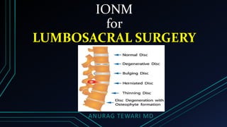
2. IONM for Lumbosacral Surgery
The adult spinal cord ends opposite the lower border of the L1 vertebra
Introduction
Anurag Tewari MD
3. IONM for Lumbosacral Surgery
• Below that, the spinal canal contains only individual lumbosacral nerve roots descending
toward their respective neural foramina, collectively known as the cauda equina
Introduction
Anurag Tewari MD
4. IONM for Lumbosacral Surgery
• There is a 10% risk of new postoperative neurologic deficit from Nerve Root
Injury during spinal fusion for degenerative lumbosacral spine disease
Introduction
Anurag Tewari MD
10-fold greater than the risk of spinal cord injury from scoliosis surgery
5. IONM for Lumbosacral Surgery
• Correction of spinal deformity during lumbosacral surgery can injure Nerve Roots by
• PROBING
• STRETCHING
• DRILLING
• MISPLACED HARDWARE
Introduction
Anurag Tewari MD
6. IONM for Lumbosacral Surgery
The most common deficit is a new FOOT DROP from surgical injury to
the L5 nerve root, although findings can include any pattern of
• Dermatomal numbness
• Radicular weakness
• Sphincter dysfunction
Introduction
Anurag Tewari MD
9. IONM for Lumbosacral Surgery
• Nerve root injuries are more frequent during revision than primary surgery and
in cases where multiple levels are fused
• Hence, the purpose of IONM with EMG during these lumbosacral spine cases is:
• DETECT early and potentially reversible surgical nerve root irritation
• ALERT the surgeon
• To prevent more significant injury and postoperative deficit(s)
Introduction
Anurag Tewari MD
RESULTS:
The overall incidence of lumbar nerve root palsy was 2.9% with a 1.4%
incidence in primary and 3.8% incidence in revision surgery
https://www.ncbi.nlm.nih.gov/pubmed/16025033
10. NERVE ROOT MONITORING USING FREE-RUNNING EMG
• Mixed nerve SSEPs and TcMEPs are most often used for spinal cord monitoring
but
THEY ARE NOT SENSITIVE FOR DETECTING NERVE ROOT INJURIES
WHY?
• SSEPs represent the summation of neural signals that enter the spinal cord
through multiple segments, and because of central amplification, they can
remain completely unchanged after an individual nerve root lesion
Background
Anurag Tewari MD
11. NERVE ROOT MONITORING USING FREE-RUNNING EMG
• TcMEPs recorded from an epidural electrode will obviously not be affected by a
more caudal nerve root injury
• TcMEPs recorded from a single distal limb muscle throughout surgery will not
detect nerve root injuries affecting other myotomes
There have been numerous well-documented cases of patients with acute radiculopathy from spine surgery despite
normal SSEP and TcMEP monitoring
Background
Anurag Tewari MD
12. NERVE ROOT MONITORING USING FREE-RUNNING EMG
• Dermatomal somatosensory evoked potentials (D-SSEPs) can detect individual
nerve root injuries but are technically difficult to resolve in the operating room
• They need to be averaged many times
• Resulting a slow turnaround time
• They test only dorsal root (sensory) function
Background
Anurag Tewari MD
13. NERVE ROOT MONITORING USING FREE-RUNNING EMG
• For these reasons, EMG has become the technique most often used to monitor
nerve root function during lumbosacral spine surgery
• EMG is more sensitive than SSEPs and D-SSEPs for detecting nerve root dysfunction, as it
• Provides immediate results without averaging
• Gives instantaneous feedback to the surgeon during critical phases of the procedure
• Can be monitored from multiple channels simultaneously
• (i.e., Multiple myotomes and nerve roots)
Background
Anurag Tewari MD
14. TECHNICAL CONSIDERATIONS for Lumbosacral Surgery
• EMG is recorded from limb muscles using paired intramuscular needle or wire electrodes
Types of electrodes
Recording Electrodes and Parameters
Anurag Tewari MD
Hooked wire electrodes can be used for deeper muscles such as iliacus or rectus abdominus
15. TECHNICAL CONSIDERATIONS for Lumbosacral Surgery
Typical recording parameters for intraoperative EMG are
Recording Electrodes and Parameters
Anurag Tewari MD
Free Running EMG Stimulus-Triggered EMG
Filters 20–10,000 Hz 10–10,000 Hz
Gain 50 μV/div 50 μV/div
Time Base 100 ms/div 10 ms/div
16. TECHNICAL CONSIDERATIONS for Lumbosacral Surgery
• Intraoperative CMAP responses are recorded from intramuscular needle electrodes and
submaximal stimulation and are highly complicated polyphasic responses with
• Variable onset Latencies and response Amplitudes
Recording Electrodes and Parameters
Anurag Tewari MD
CMAP responses from intraoperative stimulus-triggered EMG
CMAP response in tibialis anterior from stimulation of an L5 pedicle
screw at 5.3 mA, suggesting pedicular breach
CMAP responses recorded after direct stimulation of exposed cauda
equina (as a positive control) at 4 mA
17. TECHNICAL CONSIDERATIONS for Lumbosacral Surgery
• In effect they are all or nothing responses, although the stimulus threshold can
provide some quantitative information regarding nerve function
• A monopolar stimulating electrode is generally preferable during lumbosacral
spine surgeries to avoid current shunting from fluid within the operative field
Stimulating electrodes and parameters
Anurag Tewari MD
18. TECHNICAL CONSIDERATIONS for Lumbosacral Surgery
• The stimulator is a sterile handheld electrode handed to the surgeon during the
procedure
• The surgeon inserts the stimulator into pedicle holes and touches the
stimulator against pedicle screws or any other structure in the operative field
Stimulating electrodes and parameters
Anurag Tewari MD
19. TECHNICAL CONSIDERATIONS for Lumbosacral Surgery
• Although the surgeon holds the stimulating electrode, the IONM technologist
controls the electrical stimulator and will need to be sure that it is active when
the surgeon wants to stimulate
• Stimulation can be with single or recurrent shocks, with a stimulus duration of
0.2 ms and an initial “searching” stimulus intensity of 7 mA
Stimulating electrodes and parameters
Anurag Tewari MD
20. TECHNICAL CONSIDERATIONS for Lumbosacral Surgery
• Recurrent stimuli are delivered at approximately 2 Hz, usually 2.3 Hz to avoid an
exact multiple of 60 and potential artifact, with a stimulus intensity range of 0
to 25 mA
• We notify the surgeon if a CMAP response is seen at the searching stimulus
intensity and then progressively reduce the stimulus intensity and determine
the precise stimulus threshold
Stimulating electrodes and parameters
Anurag Tewari MD
21. TECHNICAL CONSIDERATIONS for Lumbosacral Surgery
It is important to Discuss The Case With The Surgeon Ahead Of Time to decide
which muscles to monitor, as there are a limited number of available recording
channels and it may not be possible to relocate recording EMG electrodes after the
patent has been prepared and draped for the procedure
Muscle Selection
Anurag Tewari MD
22. TECHNICAL CONSIDERATIONS for Lumbosacral Surgery
• Intramuscular EMG electrodes are usually inserted into the anesthetized
patient without the assistance of voluntary activation to confirm satisfactory
needle placement, and it is important to use muscles that are easily identified
using surface landmarks to assure accurate needle placement
• It is helpful to have an EMG anatomy atlas readily available in the operating
room to check the innervation or location of unfamiliar muscles
Muscle Selection
Anurag Tewari MD
http://teleemg.sectorlink.org/new/atlas.htm
EMG Muscle Identification PDF
23. NERVE ROOT MONITORING USING FREE-RUNNING EMG
• Continuous free-running EMG is monitored from muscles innervated by nerve roots
considered to be at risk for injury during surgery
Technique
Anurag Tewari MD
24. IONM for Lumbosacral Surgery
Nerve roots at
risk
Appropriate muscle for EMG monitoring
T7–12 External oblique and Rectus abdominus
L1–2 Iliacus
L2–4 Vastus medialis
L4–5 Tibialis anterior
S1–2 Medial gastrocnemius
S3–5 Anal and urethral sphincter
Muscles suitable for Intraoperative Electromyographic Monitoring of Thoraco-lumbosacral
Nerve Root Segments during Lumbar Fusion
Anurag Tewari MD
25. NERVE ROOT MONITORING USING FREE-RUNNING EMG
• Blunt mechanical trauma to nerve roots causes a depolarization that will be conducted
down the nerve, across the neuromuscular junction, and evoke identifiable motor unit
potentials in the monitored muscle
Technique
Anurag Tewari MD
EMG monitoring should be quiescent
under normal conditions
Blunt mechanical nerve root irritation
activates the motor nerve fibers, is
transmitted down the nerve and across
the neuromuscular junction, and
evokes recordable motor unit
potentials in the monitored muscle
26. NERVE ROOT MONITORING USING FREE-RUNNING EMG
• Minor nerve root manipulation will result in a short burst of motor unit potentials
Technique
Anurag Tewari MD
A. Minor nerve root manipulation evokes a short burst of motor unit
potentials
B and C. More severe blunt mechanical nerve root injury or retraction
evokes prolonged trains of high-frequency motor unit potentials, known as
neurotonic discharges
D. Rarely, myokymic discharges can be seen, but usually only after very severe
nerve injury
In each case the time base shown is 100 ms/div and the display sensitivity is 50 μV/div.
27. NERVE ROOT MONITORING USING FREE-RUNNING EMG
• More severe mechanical nerve root injury or retraction will cause prolonged trains of high-
frequency motor unit potentials, often referred to as neurotonic discharges
Technique
Anurag Tewari MD
28. NERVE ROOT MONITORING USING FREE-RUNNING EMG
• Repetitive grouped motor unit potentials, MYOKYMIC DISCHARGES, are seen less frequently
and usually indicate very Severe Nerve Injury
• Identification of this abnormal EMG activity can be used to alert the surgeon of inadvertent
trauma to nerve roots
• Allowing evasive action in an effort to prevent more severe or irreversible injury
Technique
Anurag Tewari MD
29. NERVE ROOT MONITORING USING FREE-RUNNING EMG
The ANAL and/or URETHRAL SPHINCTER muscles should be monitored in cases where the
conus medullaris or lower sacral nerve root are at particular risk for iatrogenic injury
Technique
Anurag Tewari MD
30. IONM for Lumbosacral Surgery
• EMG monitoring has a high sensitivity for detecting mechanical nerve root manipulation
but
• Has a low specificity and positive predictive value for postoperative nerve root injury
• Significant EMG activity occurs in 80% of monitored lumbosacral spine surgeries
• Although less that 10% of these develop a new persistent postoperative neurologic deficit
Outcome Data
Anurag Tewari MD
https://www.ncbi.nlm.nih.gov/pubmed/15014279
31. IONM for Lumbosacral Surgery
• SSEP monitoring has a lower sensitivity, but a higher specificity and positive predictive value
• However, SENSITIVITY is preferable to specificity for IONM
• As it is more important to detect a mild degree of nerve root irritation and alert the surgeon in time to take
evasive action, than it is to correctly predict an irreversible postoperative deficit
Outcome Data
Anurag Tewari MD
Sensitivity and Specificity of EMG vs. SEP Monitoring for Detecting 14 New Postoperative
Deficits in 213 Monitored Lumbar Spine Cases
32. IONM for Lumbosacral Surgery
• There are many published anecdotal case reports touting the benefits of intraoperative EMG
monitoring during lumbar spine surgery, there is a paucity of prospective outcome data
• Some studies have shown reduced incidence of postoperative C5 radiculopathy after
cervical decompression using EMG monitoring from the deltoid muscle, and cases detected
during surgery with EMG monitoring were felt to have been milder, with a more rapid
recovery because the surgeon was alerted immediately
Outcome Data
Anurag Tewari MD
Study EMG Monitoring No EMG Monitoring
Jimenez, 2005 1/116 (0.9%) 4/55 (7.3%)
Fan, 2002 2/68 (2.9%) 6/132 (4.5%)
33. IONM for Lumbosacral Surgery
• Although EMG monitoring is sensitive for blunt mechanical irritation and injury to motor
nerves, it may remain QUIESCENT DURING SHARP NERVE TRANSECTION
• Even after acute nerve transection the distal nerve stump can still be activated by
mechanical irritation and electrical stimulation
• The presence of these ongoing evoked EMG responses may be mistakenly interpreted as evidence of nerve
root continuity
Precautions
Anurag Tewari MD
34. IONM for Lumbosacral Surgery
• Mechanical trauma is less likely to evoke neurotonic discharges from abnormal motor nerves
• And EMG monitoring may miss additional intraoperative injury to nerve roots already partially injured by
preexisting lumbosacral radiculopathy
Precautions
Anurag Tewari MD
35. IONM for Lumbosacral Surgery
• Neurotonic discharges must be differentiated from other EMG findings that can occur during surgery
without nerve injury to avoid false alarms
In patients with preexisting radiculopathy and denervation, Spontaneous Fibrillation Potentials may be
seen throughout surgery
Precautions
Anurag Tewari MD
Spontaneous fibrillation potentials may been seen throughout surgery in patients
with preexisting radiculopathy and denervation
Voluntary motor unit potentials can be seen under conditions
of light anesthesia
Electrocautery artifact seen in EMGNeedle electrode movement artifact
36. IONM for Lumbosacral Surgery
• Spontaneous fibrillation potentials can be distinguished from neurotonic discharges because
they occur continuously (even before critical phases of surgery) and fire regularly at slow
rates (less than 15 Hz)
• Unlike neurotonic discharges, fibrillations are action potentials of single muscle fibers and
not motor units, so they are simple tri-phasic or biphasic spikes
Precautions
Anurag Tewari MD
37. IONM for Lumbosacral Surgery
• Voluntary motor unit potentials can be seen under conditions of light anesthesia
• These are semi-rhythmic motor unit potentials that usually occur simultaneously in muscles
from multiple bilateral myotomes
• Electrocautery artifact is easy to recognize but precludes concurrent EMG monitoring
Precautions
Anurag Tewari MD
38. IONM for Lumbosacral Surgery
• As nerve activation from mechanical irritation is transmitted across the
neuromuscular junction in order to evoke identifiable myogenic responses
• EMG cannot be monitored in the presence of total pharmacologic
neuromuscular blockade
Precautions
Anurag Tewari MD
39. STIMULUS-TRIGGERED EMG MONITORING TO VERIFY CORRECT PLACEMENT OF PEDICLE SCREWS
• Many spinal instrumentation systems use pedicle screws as a point of fixation
• Holes are drilled blindly through the narrow pedicles into the vertebral body to
facilitate pedicle screw placement
Background
Anurag Tewari MD
40. STIMULUS-TRIGGERED EMG MONITORING TO VERIFY CORRECT PLACEMENT OF PEDICLE SCREWS
• Clinical studies have detected postoperative symptoms from irritation or injury
to the adjacent nerve roots by misplaced pedicle screws in 5% to 10% cases
• Intraoperative fluoroscopy and radiographs have been used in an attempt to
verify correct pedicle screw placement during surgery but are not always
reliable, perhaps because of limited available projections
Background
Anurag Tewari MD
41. STIMULUS-TRIGGERED EMG MONITORING TO VERIFY CORRECT PLACEMENT OF PEDICLE SCREWS
• Computed tomography is more definitive, but is not available in real
time in OR
• Direct inspection and palpation of the medial wall of the
instrumented pedicle by the surgeon is the “gold standard” test for
misplaced screws during surgery
• but doing this for every screw may necessitate unnecessary laminectomies
and disproportionate operative times
Background
Anurag Tewari MD
42. STIMULUS-TRIGGERED EMG MONITORING TO VERIFY CORRECT PLACEMENT OF PEDICLE SCREWS
• Fortunately, stimulus-triggered EMG can quickly verify correct pedicle screw
placement in real time during surgery
Background
Anurag Tewari MD
43. STIMULUS-TRIGGERED EMG MONITORING TO VERIFY CORRECT PLACEMENT OF PEDICLE SCREWS
• Holes or screws that are correctly positioned within the pedicle wall are
separated from the adjacent nerve roots by a cortical bony layer with high
impedance to the passage of electrical current
Background
Anurag Tewari MD
44. STIMULUS-TRIGGERED EMG MONITORING TO VERIFY CORRECT PLACEMENT OF PEDICLE SCREWS
• However, a hole or screw that has perforated the bony pedicle wall will lie
directly against adjacent nerve roots
• Direct electrical stimulation of such misplaced holes and screws activates the
adjacent nerve roots, evoking CMAP responses in muscles from the appropriate
myotomes at lower stimulus intensities than those that lie completely
contained within the pedicle wall
Background
Anurag Tewari MD
45. STIMULUS-TRIGGERED EMG MONITORING TO VERIFY CORRECT PLACEMENT OF PEDICLE SCREWS
• Stimulus thresholds of less than 4 to 6 mA are suggestive of cortical bony perforation by
pedicular instrumentation when the adjacent nerve root is healthy
Background
Anurag Tewari MD
Structure Stimulus Threshold (mA)
Normal nerve root (0.2–5.7)
Chronically compressed nerve root 6.3–20
Normal hole (16.5–44.3)
Normal screw 24 (12.1–35.9)
Misplaced hole (1–6)
Misplaced screw (1–6)
Stimulus Thresholds for Normal Healthy Nerve Roots, Chronically Compressed Nerve Roots, and Normal and Misplaced Pedicle Holes and Screws
Maguire J, Wallace S, Madiga R, et al. Evaluation of intrapedicular screw position using intraoperative evoked electromyography. Spine 1995;20:1068–1074.
Raynor B, Lenke LG, Bridwell K, et al. Correlation between low triggered EMG thresholds and lumbar pedicle screw malposition: analysis of 4587 screws. Presented at the Scoliosis Research Society, 2005.
46. STIMULUS-TRIGGERED EMG MONITORING TO VERIFY CORRECT PLACEMENT OF PEDICLE SCREWS
• Holes and screws are tested using a monopolar stimulating electrode with a
remote anode needle electrode placed in the surgical wound and stimulus-
triggered EMG recordings made from appropriate limb muscles
• The stimulator is inserted directly into the pedicle hole after drilling
Technique
Anurag Tewari MD
47. STIMULUS-TRIGGERED EMG MONITORING TO VERIFY CORRECT PLACEMENT OF PEDICLE SCREWS
• It is preferable to have the stimulator halfway into the hole, so that the
uninsulated tip of the electrode is inside the pedicle close to the adjacent nerve
root and not deep inside the vertebral body
• Screws are tested by touching the stimulator against the exposed shank or port
of the screw, taking care to avoid any intervening soft tissue
Technique
Anurag Tewari MD
48. STIMULUS-TRIGGERED EMG MONITORING TO VERIFY CORRECT PLACEMENT OF PEDICLE SCREWS
• It is important to test each pedicle hole and screw individually as each is drilled
and inserted
• A misplaced hole should be redirected and then retested before it is
instrumented
Technique
Anurag Tewari MD
Some particular brands of pedicle screw have an unusually high electrical resistance based on their metallic composition, so just testing the screw (and not the preceding hole) may give rise to a false-negative result
49. STIMULUS-TRIGGERED EMG MONITORING TO VERIFY CORRECT PLACEMENT OF PEDICLE SCREWS
• To save time, each hole and screw can be initially tested at a searching stimulus intensity of 7
to 10 mA
• If no CMAP response is identified at this intensity level, then the hole or screw is considered
safe
• If a CMAP response is identified at the initial searching intensity, the stimulating current is
progressively reduced, using recurrent stimulation at 2 Hz, in order to determine the precise
stimulus threshold required to evoke the response and hence the probability of pedicular
perforation
Technique
Anurag Tewari MD
50. STIMULUS-TRIGGERED EMG MONITORING TO VERIFY CORRECT PLACEMENT OF PEDICLE SCREWS
• Pedicle screws or holes with stimulus thresholds of less than 4 mA should be
removed and/or redirected
• Pedicle screws with stimulus thresholds between 4 and 6 mA are considered
borderline and should probably be more closely inspected by the surgeon
• The very same technique of stimulus-triggered EMG using a handheld stimulator in the
operative field can also be used to identify viable nerve roots during dissection of intra-
spinal dumbbell and foraminal tumors and spinal surgery for tethered cord release
Technique
Anurag Tewari MD
51. STIMULUS-TRIGGERED EMG MONITORING TO VERIFY CORRECT PLACEMENT OF PEDICLE SCREWS
• Based on a stimulus threshold of 6 mA or less, the sensitivity of stimulus-triggered EMG for
identifying misplaced pedicle screws and hole is more than 90%,
• significantly exceeding the sensitivity of intraoperative radiography, which is only 63%
• The probability of a pedicle screw breach increases with decreasing stimulus threshold
Outcome Data
Anurag Tewari MD
Stimulus Threshold and Pedicle Screw Malposition (Confirmed by Palpation and/or Radiographs) for 4,587 Screws From Raynor et al.
52. STIMULUS-TRIGGERED EMG MONITORING TO VERIFY CORRECT PLACEMENT OF PEDICLE SCREWS
• It is important to recognize that the stimulus thresholds mentioned above refer
to pedicular instrumentation at levels where the bone and adjacent nerve roots
are normal and healthy
• False-positive stimulus-triggered EMGs can be seen in patients with advanced
osteoporosis, presumably because of thin cortical bone with a lower than
expected impedance to the passage of the electrical current
Precautions
Anurag Tewari MD
53. STIMULUS-TRIGGERED EMG MONITORING TO VERIFY CORRECT PLACEMENT OF PEDICLE SCREWS
• More importantly, false-negative results may be seen in patients with preexisting
radiculopathy
• The stimulus intensities required to evoke CMAP responses from direct electrical stimulation
of nerve roots are higher in the presence of preexisting axonotmetic radiculopathy,
sometimes as high as 10 to 20 mA
Precautions
Anurag Tewari MD
Stimulus Thresholds for Normal Healthy Nerve Roots, Chronically Compressed Nerve Roots, and Normal and Misplaced Pedicle Holes and Screws
54. STIMULUS-TRIGGERED EMG MONITORING TO VERIFY CORRECT PLACEMENT OF PEDICLE SCREWS
• Thus the failure to evoke a CMAP response from stimulation of a hole or screw
adjacent to a chronically compressed nerve root at the usual searching stimulus
intensity of 7 mA will not necessarily exclude a pedicular wall breach at that
level, with the potential for further iatrogenic nerve root injury
Precautions
Anurag Tewari MD
55. STIMULUS-TRIGGERED EMG MONITORING TO VERIFY CORRECT PLACEMENT OF PEDICLE SCREWS
• Clearly, pedicular instrumentation must be tested at higher stimulus intensities
when the adjacent nerve root is known to be axonotmetic
• Axonotmetic nerve root injury should be suspected in the presence of chronic
preoperative radicular motor deficits or abnormal preoperative
electromyographic studies
Precautions
Anurag Tewari MD
56. STIMULUS-TRIGGERED EMG MONITORING TO VERIFY CORRECT PLACEMENT OF PEDICLE SCREWS
• When possible it is best to first determine the electrical thresholds required to
evoke CMAP responses in the appropriate muscles from direct stimulation of
the exposed chronically compressed nerve root
• And then test the pedicular instrumentation at that level at an appropriately
higher intensity (perhaps 5 mA higher) in order to exclude a bony cortical
breach
• If the nerve root can not be directly tested, for example if there is no
laminectomy at that level, then it is probably best to empirically test the holes
and screws at a higher searching intensity such as 20 mA
Precautions
Anurag Tewari MD
57. STIMULUS-TRIGGERED EMG MONITORING TO VERIFY CORRECT PLACEMENT OF PEDICLE SCREWS
• EMG cannot be monitored in the presence of total pharmacologic neuromuscular blockade
• because nerve activation from electrical stimulation must be transmitted across the
neuromuscular junction in order to evoke identifiable CMAP responses
In the presence of partial neuromuscular blockade, a physiologically decremental
response may suggest a falsely high threshold and lead to a false-negative result
• Under these circumstances, it is better to use single shocks at an initial stimulus intensity
where a positive response is likely to be significant (e.g., 4 mA)
Precautions
Anurag Tewari MD
58. MONITORING NEUROMUSCULAR BLOCKADE
• Nerve activation, whether from mechanical irritation or electrical stimulation,
must be transmitted across the neuromuscular junction in order to evoke
identifiable myogenic responses
• However, muscle relaxation is a critical component of balanced anesthesia
Background
Anurag Tewari MD
59. MONITORING NEUROMUSCULAR BLOCKADE
• While adequate surgical relaxation may be achieved using inhalation anesthetic
agents alone, patients are more likely to move in the absence of specific
pharmacologic neuromuscular blockade, and this may compromise patient
safety
• Fortunately, EMG can be successfully monitored in the operating room under
conditions of partial (as much as 50% to 75%) neuromuscular blockade using
carefully titrated doses of short-acting agents such as Rocuronium
Background
Anurag Tewari MD
60. MONITORING NEUROMUSCULAR BLOCKADE
• Neuromuscular blockade has been traditionally monitored by the
anesthesiologist during surgery using the number of visible hand twitches after
a train-of-four peripheral stimulations to the ulnar nerve at the forearm
• However, unfamiliarity with peripheral nerve function and anatomy may result
in inaccurate electrode placement and misinterpretation of direct muscle
activation as twitches by the anesthesiologist, underestimating the degree of
pharmacologic neuromuscular blockade
Background
Anurag Tewari MD
61. MONITORING NEUROMUSCULAR BLOCKADE
• Muscles from the cranial, cervical, and lumbosacral myotomes have different
susceptibilities to the effects of neuromuscular blocking agents
• In addition, chronically compressed nerves may have enhanced sensitivity to
the effects of neuromuscular blockade
• For these reasons, neuromuscular transmission should be monitored using
repetitive nerve stimulation from a muscle belonging to the appropriate
myotome for the nerves or roots considered at risk from surgery
Background
Anurag Tewari MD
62. MONITORING NEUROMUSCULAR BLOCKADE
• It is more usual to monitor neuromuscular transmission in anesthetized
patients using repetitive nerve stimulation
• Less than 75% neuromuscular blockade corresponds to a decrement of less
than 100% over four successive supramaximal repetitive nerve stimulations
Technique
Anurag Tewari MD
63. MONITORING NEUROMUSCULAR BLOCKADE
• It is easy to monitor repetitive nerve stimulation from the tibialis anterior
muscles during lumbar spine surgery because this muscle is routinely used for
EMG monitoring during these cases
• The deep peroneal nerve can be stimulated transcutaneously using needle or
surface electrodes at the fibula head
Technique
Anurag Tewari MD
64. MONITORING NEUROMUSCULAR BLOCKADE
• Less than four identifiable CMAP responses indicates pharmacologic neuromuscular
blockade exceeding 75%, which might lower the sensitivity of EMG monitoring for the
detection of intraoperative nerve root injury
• As long as four CMAP responses are identifiable from supramaximal repetitive nerve
stimulation at 2 Hz, the degree of neuromuscular blockade is sufficiently low to allow EMG
monitoring
Technique
Anurag Tewari MD
65. TECHNICAL CONSIDERATIONS for Lumbosacral Surgery
• Many patients undergoing lumbosacral spine surgeries will already have
preoperative weakness
• Their preoperative EMG reports may show evidence of chronic motor axonal
loss from lumbar radiculopathy in the form of low amplitude CMAP responses
or denervation on needle EMG examination
• These patients have preexisting axonotmetic lumbar nerve root injuries,
necessitating special precautions to exclude pedicular breach with stimulus
triggered EMG at those levels
Preoperative Studies
Anurag Tewari MD
66. TECHNICAL CONSIDERATIONS for Lumbosacral Surgery
• Neuromuscular blockade must be carefully monitored and documented
throughout surgery
• The expected and preferred result is quiescent EMG monitoring, but quiescent
EMG is only meaningful when the IONMtechnologist is able to demonstrate
less than 75% pharmacologic blockade at the same time
Neuromuscular Blockade
Anurag Tewari MD
67. TECHNICAL CONSIDERATIONS for Lumbosacral Surgery
• Otherwise the false-negative absence of EMG activity may give the surgeon a
false sense of security and give rise to a surgical outcome that is worse than it
would have been with no monitoring at all
• Hence, the surgeon should be notified right away whenever excessive
pharmacologic neuromuscular blockade is detected; he or she will then likely
order reversal if approaching a critical phase of the procedure
Neuromuscular Blockade
Anurag Tewari MD
68. TECHNICAL CONSIDERATIONS for Lumbosacral Surgery
• Only one ground electrode should be attached to the patient, preferably a large adhesive
surface electrode, which has a higher contact area than a needle and therefore receives a
reduced current density in the event of current leakage,
• Thus minimizing the risk of a skin burn
• All electrical devices attached to the patient should share the same ground circuit as the
EMG machine
• The EMG machine should be turned on before any electrodes are attached to the patient
and turned off only after all electrodes have been disconnected so as to avoid power surges
that could be transmitted to the patient
Patient Safety
Anurag Tewari MD
69. TECHNICAL CONSIDERATIONS for Lumbosacral Surgery
• There have been skin burns at the site of EMG needle electrode insertion because of current
shunting from faulty electrocautery units during surgery; and the following steps should be
taken to avoid this:
• Ground pads from all electrocautery units must be properly positioned to ensure a large
area of contact with the patient’s skin and placed as close to the surgical site as possible
• Each electrocautery unit should contain a built-in ground monitoring system to warn the
surgeon in case of circuit discontinuity with a potential for current shunting through other
electrodes attached to the patient
• Needle electrodes should not be placed in close proximity to electrocautery ground pads
and, if possible, should not be placed within the predicted pathway between an active
electrocautery electrode and its ground pad
• Finally, electrocautery units, EMG monitoring devices, and all operating room electrical
circuits should be regularly checked for current leakage
Patient Safety
Anurag Tewari MD